ANS 206 practical 1
 Call Kai
 Learn
Learn Practice Test
 Spaced Repetition
 Match
Match Flashcards
 Knowt Play
1/168

There's no tags or description
Looks like no tags are added yet.
Name | Mastery | Learn | Test | Matching | Spaced | Call with Kai | Chat |
|---|
No analytics yet
Send a link to your students to track their progress
169 Terms

Label 1-4

5 functions of the skeletal system
Support: the hard framework of the skeletal system helps to support and anchor the soft organs
Protection: the skeletal system surrounds the organs, for example the brain and spinal cord, and provides protection
Movement: muscles are able to attach to the skeletal system, this allows bones to be used as levers
Storage: minerals and lipids are stored within bone material
Blood cell formation: bone marrow is responsible for blood cell production
Compact Bone
hard layer that covers most bones and forms the shaft of long bones. Compact bone contains both bone cells and blood vessels.
Cancellous (or Spongy) bone
located at the ends of long bones and is composed of spicules arranged in a porous network. The small, open spaces are then filled with marrow.
Medullary Cavity
space that is found inside the cortex of a long bone. In a young animal the medullary cavity is filled with red marrow, this will eventually be replaced by fatty yellow marrow as the animal ages.
Bone types
long
Short
Flat
Irregular
Sesamoid
Long bone anatomy
Epiphysis: this refers to the ends of the long bone. Each long bone has a proximal epiphysis and a distal epiphysis.
Proximal epiphysis - end closest to the body
Distal epiphysis - end furthest away from the body
Diaphysis: the shaft of the long bone, found between the two epiphyses and surrounds the medullary cavity.
Articular Cartilage: thin layer of hyaline cartilage that covers the end of the bone that articulates, cushions and allows for smooth movement.
Epiphyseal Plate: layer of cartilage that allows for the growth of the bone, this is the only area where a bone can lengthen. Separates the diaphysis and epiphysis in an immature bone.
Functions of long bones & examples
act as levers
aid in support, locomotion, and prehension
The best examples of long bones within the bones are the bones in the extremities such as the humerus, radius, and ulna in the forelimb and the femur, tibia, and fibula in the hindlimb.
Functions of short bones & examples
Short bones have a cube-like structure with no single marrow cavity. Instead, these bones are filled with spongy bone filled with marrow spaces.
absorb concussion and shock
In carpus & tarsus (found in complex joints where they can also provide a variety of movements)
Functions of flat bones & examples
There is an inner framework of spongy bone, called diploe, sandwiched between two plates of compact bone. These two plates are called the lamina externa and the lamina interna.
provide protection to vital organs such as the brain (skull), the heart and lungs (scapula and ribs).
The scapula provides large areas for attachment of muscles.
Functions of irregular bones & examples
unpaired bones found on the median plane that have an odd shape
provides protection, support, and muscular attachment.
Ex: vertebrae & pelvis.
Functions of sesamoid bones & examples
small, independent
Generally, sesamoid bones are found embedded in a tendon where they help to reduce friction, increase leverage, or change the direction of pull.
Ex: patella (or kneecap) and the distal sesamoid bone.
Three small bones found within soft organs:
Os penis - bone in the penis of dogs
Os cordis - bone in the heart of cattle
Os rostri - bone in the snout of pigs
Axial Skeletal System
Skull
Vertebral Column
Sternum
Ribs
These bones are important for protection, support, and carrying other body parts.
Appendicular skeletal System
Thoracic limbs (forelimb)
Pelvic limbs (hindlimb)
Shoulder
Pelvis
The two skeletal systems articulate at one point, this is the…?
sacroiliac joint or SI. This is where the sacrum of the vertebral column articulates with the ilium.
Vertebrae labeling
Horse vertebral formula
C7 T18 L6 S5 CL15-20
Pig vertebral formula
C7 T14-15 L6-7 S4 CL20-23
Dog vertebral formula
C7 T13 L7 S3 CL20-23
Appendicular skeleton
Shoulder - pectoral girdle (scapula)
Forelimbs - humerus, radius, ulna, carpals, metacarpals, phalanges
Pelvis - pelvic girdle (ilium, ischium, pubis)
Hindlimbs - femur, tibia, fibula, tarsals, metatarsals, phalanges
Forelimb & label
The forelimb is made up of the:
Scapula, shoulder blade
relatively flat, triangular bone
has a spine that extends to the dorsal border and divides the scapula unevenly into two portions
the distal end of the spine forms the acromion process
it is attached to the body via muscle, ligaments, etc. we call this a synsarcosis - there is no true joint
Humerus
a long bone that has a shaft and two extremities
proximal end has a rounded head that articulates with the scapula to form the shoulder joint, or scapulohumeral joint
Humerus + scapula = shoulder joint
The distal end of the humerus forms the elbow joint with the proximal ends of the ulna and radius
Radius and Ulna (forearm)
Radius is larger, proximally it forms the elbow with the humerus and distally articulates with the carpus
Ulna forms the olecranon process (point of elbow); in horses the ulna and radius is fused; ox, sheep, goats, dogs, cats have a complete ulna
Carpus - often referred to as the knee, this region corresponds to your wrist. The carpus consists of two rows of small bones called carpal bones.
Metacarpus - immediately distal to the carpus, in the horse it is often referred to as the cannon bone. Two small splint bones are found next to the metacarpus, one on the medial side and one on the lateral side. Metacarpus + Proximal Phalanx = Metacarpophalangeal
Digits -
Made up of three phalanges: proximal phalanx, middle phalanx, distal phalanx. In horses, these three bones are often referred to as the long pastern, short pastern, and coffin bone.
Proximal phalanx + middle phalanx = fetlock joint or proximal interphalangeal - two sesamoid bones are located here called the proximal sesamoid bones
Middle phalanx + coffin bone = pastern or distal interphalangeal
The distal sesamoid bone, or navicular bone, is located between the middle and distal phalanges

Pelvic girdle/Os Coxae & label
the fusion of 3 different bones:
Ilium - the largest and most dorsal portion
Tuber Sacrale - close to the sacroiliac joint, where the sacrum and ilium articulates
Tuber Coxae - point of hip, or hook bone in cattle
Wing - flat portion between the tuber coxae and tuber sacrale
Fracture of the tuber coxae in horses is called knock-down hip
Ischium - forms the caudal part of the floor of the pelvic cavity
Ischial tuber - pin bone in cattle
Pubis - smallest, forms the cranial part of the floor

Hindlimb & label
The bones of the hindlimb include:
Femur - thigh bone
head articulates with the acetabulum of the Ox Coxae to form the hip joint
distally, the bone has two condyles that articulate with the patella and the tibia and fibula
The tibia and fibula are fused in horses
the tibia is the larger bone
Femur + tibia + patella = stifle, corresponds to the knee joint in people
Tarsus or hock joint
consists of multiple small bones called tarsal bones
The two largest bones of the hock are the talus and calcaneus, the calcaneus process makes up the point of hock
The metatarsus (cannon) and digits are identical to the forelimb

Masseter muscle
involved in mastication, or chewing; broad muscle in the cheek of the horse

Trapezius (what does it act on & what are the two portions?)
Acts on the Shoulder Girdle
This muscle acts on the scapula
Trapezius - swings the scapula forward
It has two portions: cervical and thoracic

List the extensors acting on the shoulder joint
Brachiocephalicus (head to arm) and the Supraspinatus

List the flexors acting on the shoulder joint
the Latissimus Dorsi and the Infraspinatus.

List the adductor and the abductor of the shoulder joint
adductor: Pectoralis
abductor: deltoideus

Triceps brachii
Muscle that extends elbow joint

Biceps brachii
Flexes elbow joint

Hamstrings (in relation to the hip joint)
extensors of the hip joint
Made up of 3 muscles!
Bicep femoris, Semitendinosus, Semimembranosus

Quadriceps (in reference to the hip joint)
Flexors of the hip joint
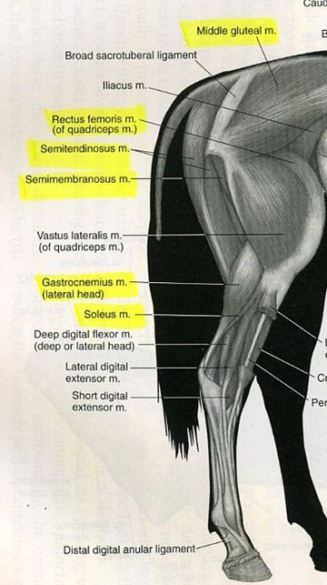
Gluteal muscle
Abductor of the hip joint

Quadriceps (in relation to the stifle joint)
Extensors of the stifle joint
Hamstrings & gastrocnemius (in relation to the stifle joint)
Flexors of the stifle

Gastrocnemius (In relation to the hock joint)
Extensor of the hock

Latissimus dorsi
Strong flexor of the shoulder
Will advance the trunk if the limb is fixed

external abdominal oblique
the most superficial muscle
helps to flex the vertebral column, support the organs of digestion and reproduction

Role of diaphragm and intercostals
Involved in respiration
define extension, flexion, adduction, & abduction
Extension: Straightening or increasing the angle
Flexion: Bending or decreasing the angle
Adduction: Moving towards the midline
Abduction: Moving away from the midline
What is the endocrine system
made up of a group of ductless glands that secrete hormones
What is a hormone?
A hormone is a chemical substance that is produced by a ductless gland, this substance is then released into the blood stream and carried to other parts of the body where they will produce a specific regulatory effect.
three ways hormones are secreted
Endocrine route
Exocrine route
Paracrine route
Endocrine route
the hormone will be transported via the blood stream to the target organ
Exocrine route
the hormone is secreted into ducts then it travels to outside the body where it interacts with a target organ. For example, hormones will be secreted into the lumen of the digestive tract.
Paracrine route
the hormone will be secreted into the space between cells and the interstitial fluid will then carry it to the target organ; these hormones often effect neighboring cells
Chemical structures of hormones
Classic hormones can be grouped based on their chemical structure as either peptides (protein), amines (modifications of tyrosine), or steroids (cholesterol).
Where are peptide hormones and amines stored?
stored in secretory granules by endocrine cells
Can steroid hormones be stored?
NO, must be synthesized
Hormones NOT Secreted by Endocrine Glands
Gastric
Secretin
Erythropoietin
Gastric
released from the wall of the stomach; stimulates the release of gastric juices from gastric glands for digestion to being
Secretin
released from the wall of the small intestine; stimulates the secretion of intestinal/pancreatic juices to continue digestion in duodenum
Erythropoietin
released by the kidney; stimulates bone marrow
Target organ
Only certain populations of cells will respond to an individual hormone
target organ identifies the tissue whose cells will be affected by a given hormone.
Cells within target organs can recognize and respond to a given hormone because they contain specific receptors capable of binding with the hormone.
Hormone Receptor
A receptor for a specific hormone must be present for a cell to respond to the hormone
Receptors are a complex of protein structures designed to bind a hormone to elicit its cellular effects
usually found in the cell membrane or the nucleus.
Term for the increase/decrease of the number of receptors on target cells
An increase in the number of receptors on target cells is called up-regulationand a decrease is termed down-regulation.
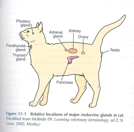
Endocrine organs/glands that only exist to produce hormones
Pituitary Gland
Thyroid Gland
Parathyroid Gland
Adrenal Glands
what are “releasing hormones” & what synthesizes & releases them?
hypothalamus
released & stimulate the release of various hormones from the adenohypophysis (or anterior pituitary).
The main hormones produced and released by the hypothalamus are:
Corticotropin Releasing Hormone or CRH
Gonadotropin Releasing Hormone or GnRH
Growth Hormone Releasing Hormone or GHRH
Thyrotropin Releasing Hormone or TRH
also produces Oxytocin and Antidiuretic Hormone, but these are then stored and released by the neurohypophysis (or posterior pituitary)
What is the hypothalamus considered?
considered to be the crucial interface between the endocrine and nervous systems.
What does the Pituitary gland contain
contains hormones that generally affect the function of many organs in a variety of systems.
What are the two histologically distinct areas of the pituitary gland?
Anterior lobe (or adenohypophysis) is made up of epithelial tissue
Posterior lobe (or neurohypophysis) is made up of neural tissue
What does the anterior pituitary do?
produces a few different hormones:
ACTH
FSH and LH
GH
TSH
PRL
What does the posterior pituitary do?
produces NO hormones, it only stores and releases:
Oxytocin
ADH
hypothalamopituitary axis
structure that allows the hypothalamus and pituitary to interact
How do the hypothalamus and pituitary connect? And how is this structure formed?
a stalk called the infundibulum
There are cell bodies of neurons in the hypothalamus, their axons then form this infundibulum.
What is the hypothalamohypophysial portal system
unique system of arterioles and capillaries associated with ghe infundibulum
Blood from a capillary network in the hypothalamus is able to flow through portal vessels into the glandular (anterior) portion of the pituitary.
Anterior Pituitary Hormones
Adrenocorticotrophic Hormone (ACTH)
LH & FSH
Growth hormone (GH)
Adrenocorticotrophic Hormone (ACTH)
Hormone of the anterior pituitary
Stimulates release of hormones from adrenal gland
Corticosteroids (from adrenal cortex) are associated with "stress responses" and the fight or flight response
Most organs have receptors for corticosteroids
LH & FSH
Involved with the regulation of male and female reproductive processes (have receptors)
Growth Hormone (GH)
hormone of anterior pituitary
Most organs, tissues, and cells within the body can respond to growth hormone
Stimulates changes in target organs that support growth (mitosis, increased mobilization of nutrients, etc.)
Posterior Pituitary Hormones
antidiuretic hormone (ADH
oxytocin
Antidiuretic Hormone (ADH)
Involved with kidney function (water retention)
Oxytocin
Involved with smooth muscle contraction (uterus, myoepithelial cells in mammary gland)
What hormones does the thyroid gland release?
Thyroid Hormones - T3 and T4
Calcitonin - peptide hormone

T3 and T4
tyrosine based, contain iodine
T4 - Thyroxine
T3 - Triiodothyronine
Increases basal metabolic rate - measure of oxygen use in resting conditions
Almost all tissues have receptors
Ratio of T4 to T3 is 20:1
Calcitonin
peptide hormone
Helps control calcium levels
Acts to decrease blood calcium by inhibiting osteoclasts
What does Parathyroid Gland release & how many do animals hv
Most domestic animals have 2 pairs associated with the thyroid
Secretes PTH
increases osteocytes and osteoclasts

Parathyroid hormone (PTH)
Increases blood calcium and lowers blood phosphorus
Stimulates release of calcium and phosphorus from bone
Adrenal gland anatomy
divided up into the Adrenal Cortex (outer region)
& Adrenal Medulla (inner region)

The Adrenal Cortex produces:
Glucocorticoids (cortisol and derivatives)
Enhances the production (or conversion) of glucose
Mineralocorticoids (aldosterone)
Regulates sodium and potassium levels in the blood
The Adrenal Medulla produces:
Catecholamines (epinephrine and norepinephrine)
Has a variety of functions in different organs but mostly prepares the animal for fight or flight
three main hormones produced by the Pancreas
Insulin
Glucagon
Somatostatin

Insulin
Produced by beta cells
Decreases blood glucose
Describe the Control of Blood Glucose Levels & the types of diabetes
As you consume food, glucose is released into the bloodstream → blood glucose concentration to rise
insulin is released → lowers blood glucose by allowing cells to uptake glucose. The uptake of glucose provides energy to the cells.
Extra glucose will be stored as Glycogen in the liver, if blood sugar concentration drops too low, Glucagon causes the release of Glycogen which can be converted into glucose via the process of Glycogenesis.
Sometimes, there can be issues in the regulation of blood glucose levels, when this happens we have either Type 1 diabetes or Type 2 diabetes.
Type 1: the pancreas is unable to produce insulin;
blood glucose levels too high
there is a malfunction of the beta cells of the pancreas
Type 2: insulin insensitivity
Insulin is produced in adequate amounts by the beta cells of the pancreas, but the body no longer responds to the insulin
When the body no longer responds to the insulin, blood glucose levels become too high

Somatostatin
Slows the mobilization & production of nutrients
Somatostatin
Slows the mobilization & production of nutrients
The masses of endocrine tissue in the Pancreas are called the…
Pancreatic Islets (or Islets of Langerhans)
Pineal Gland location
located dorsocaudally in the Diencephalon
Pinealocytes
Cells of the pineal gland
neuronal & secretory
supported by neuroglia
receive input from axons
Pinealocytes manufacture an enzyme that converts serotonin to melatonin (hormone).
Melatonin
Pinealocytes manufacture an enzyme that converts serotonin to melatonin (hormone)
regulates the sleep-wake cycle (circadian rhythm). Melatonin peaks during darkness.
Hormones are regulated by two main methods:
negative feedback & positive feedback
Negative Feedback Regulation
Rising levels of a hormone cause a biologic response to inhibit the further release
Blood glucose levels

Positive Feedback Regulation
less frequent than negative feedback
Hormones cause a biologic response that produces an increase in the release of a hormone
childbirth

What is the nervous system?
a network of billions of nerve cells linked together in a highly organized fashion to form the rapid control center of the body.
Functions of the nervous system
Regulating the movement of body parts
Regulating the secretion from glands
Collecting information about external and internal environments
Maintaining a state of consciousness
Stimulating behavior for survival
In order for the nervous system to perform these functions it requires rapid transmission of information.
The nervous system has two major divisions:
Central
Peripheral