Lecture 16 - Enzyme Mechanisms II
 Call Kai
 Learn
 Practice Test
 Spaced Repetition
 Match
 Flashcards
 Knowt Play
1/56

There's no tags or description
Looks like no tags are added yet.
57 Terms
Lecture 16 objectives
Understand what lysozyme does and know its reaction
know how a serine protease works
understand how different enzymes choose their substrate
understand what affinity labeling of an enzyme is

Lysozyme function
Digests bacterial cell walls by hydrolyzing β(1→4) glycosidic bonds between N-acetylmuramic acid (NAM) and N-acetylglucosamine (NAG)

NAG vs. NAM
NAG = N-acetylglucosamine
NAM = N-acetylmuramic acid (NAG with a lactic acid ether, CH3CHCOO-, on C3)
They alternate in the bacterial cell wall polysaccharide.
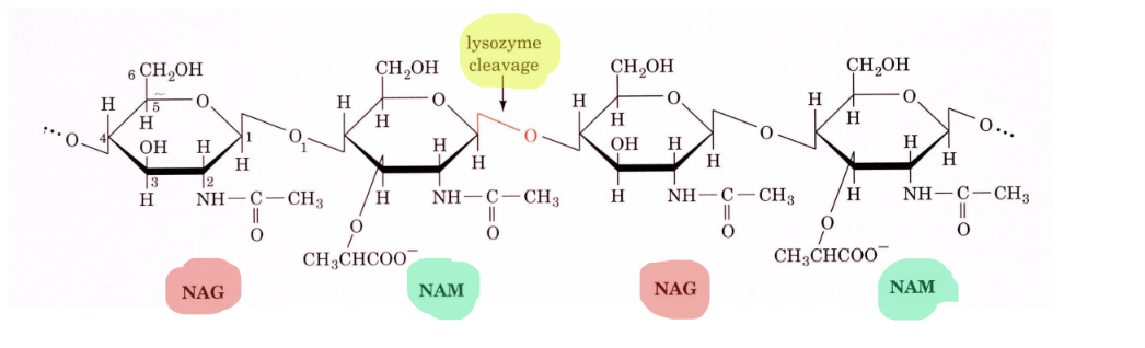
Exact bond lysozyme cleaves
The β(1→4) bond from C1 of NAM to O4 of NAG

Lysozyme structure
Hen egg white (HEW) lysozyme is a small globular protein of 129 residues, cross-linked by 4 disulfide bonds, with a deep substrate-binding cleft

Lysozyme catalytic residues
Glu 35 and Asp 52, which sit on either side of the D-E glycosidic bond
VERY IMPORTANT for CLEAVAGE

Lysozyme-substrate interactions
The substrate is held in the cleft by many hydrogen bonds with residues such as Asp 101, Trp 62, Trp 63, Ala 107, Asn 59, Gln 57, Asn 44, Glu 35, Phe 34, Asn 37, and Arg 114. The D ring is held in a half-chair conformation.

Table: lysozyme kcat for oligosaccharide substrates
As NAG increases, kcat increases

Lesson from the lysozyme kcat table
Rate rises sharply with chain length, especially from 4 to 5 to 6 sugars. Full activity needs all six subsites (A-F) filled, and alternating NAG-NAM is the best substrate.

H2(18)O labeling experiment with lysozyme
When hydrolysis is done in H2(18)O, configuration at C1 is retained.
Phillips mechanism step 1
Lysozyme binds a hexasaccharide unit of the bacterial cell wall
The D residue is distorted toward the half-chair conformation.
Phillips mechanism step 2
Glu 35 transfers its proton to O1 of the D ring (general acid catalysis)
The C1-O1 bond is cleaved, generating an oxonium ion at C1.
Phillips mechanism step 3
Asp 52 stabilizes the oxonium ion through charge-charge (electrostatic) interactions
The slide says the carboxylate cannot form a covalent bond because distances are too great, but also notes an SN2 path with transient formation of a C-O bond to the enzyme.
Phillips mechanism step 4
The E ring group is released from the enzyme, giving a glycosyl-enzyme intermediate
Water adds to reverse the chemistry and reprotonate Glu 35.
“resetting the enzyme”

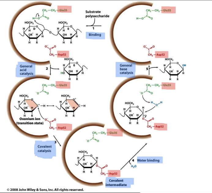
Lysozyme reaction schematic (5 steps)
(1) Binding of the substrate polysaccharide
(2) general acid catalysis by Glu 35 releases the E ring and forms the oxonium ion (transition state)
(3) covalent catalysis, where Asp 52 forms a covalent intermediate with C1
(4) water binding
(5) general base catalysis, where Glu 35- activates water to attack C1, releasing the product and regenerating the enzyme
ON EXAM: make sure u can identify missing steps + know general steps

Catalytic roles of Glu 35 vs. Asp 52 in lysozyme
Glu 35 is protonated, acts as a general acid toward O1, then as a general base activating water
Asp 52 is deprotonated and electrostatically stabilizes the oxonium ion or forms the covalent intermediate.

Half-chair conformation of the D ring
The D ring is distorted from the normal chair toward a half-chair, with C1, C2, C5, and O5 coplanar. This matches the planar geometry of the oxonium ion transition state. It is an example of strain (the rack mechanism) and transition state binding.

Oxonium ion
A resonance-stabilized carbocation at C1, with positive charge shared with the ring oxygen (O5 = O+)

Acid-catalyzed acetal hydrolysis (model for lysozyme)
Acetal + H+ → protonated acetal → loses R''OH → resonance-stabilized carbocation (oxonium ion) → H2O adds and H+ is lost → hemiacetal

NAG2FGlcF
A disaccharide substrate analog (NAG linked to a glucose with F at C1 and C2)
It acts as an intermediate trap
It was detected as a covalent attachment to Asp 52 and captured by X-ray crystallography, supporting a covalent glycosyl-enzyme intermediate.
used as an inhibitor
Catalytic strategies in lysozyme
General acid-base catalysis (Glu 35), electrostatic stabilization or covalent catalysis (Asp 52), substrate distortion to the half-chair (strain / transition state binding), and binding energy from six subsites
Serine proteases
Proteolytic enzymes that use an active-site serine (Ser 195 in chymotrypsin) as the nucleophile in covalent catalysis
active serine doing something

Table: Types of Serine Proteases


p-Nitrophenylacetate as a chymotrypsin substrate
Chymotrypsin hydrolyzes p-nitrophenylacetate plus H2O to acetate plus p-nitrophenolate (+ 2H+)
p-Nitrophenolate is very yellow and acetate is colorless, so the reaction can be followed by color. It is an example of an artificial substrate.

Artificial substrate
A non-natural compound an enzyme can act on, often designed to give an easily detected product (such as a colored one) for measuring activity

Bovine trypsin / chymotrypsin structure
A two-domain fold, with the active site (His 57, Asp 102, Ser 195) and the specificity pocket in the cleft between the domains

Specificity pocket
A pocket next to the active site that binds the side chain of the residue on the N-terminal side of the scissile bond
Its shape and charge determine which peptide bonds a serine protease cleaves.
Scissile bond
The specific bond in a substrate that is cleaved by the enzyme (here, the peptide bond)

Chymotrypsin specificity
Prefers bulky (aromatic/hydrophobic) residues such as Phe
It has a deep pocket lined by Gly 216 and Gly 226, with Ser 189 at the bottom.

Trypsin specificity
Prefers Lys and Arg (positively charged)
Ser 189 in chymotrypsin is replaced by Asp 189 in trypsin, which forms a salt bridge with the positive side chain.
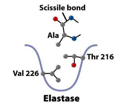
Elastase specificity
Cleaves Ala, Gly, and Val-rich elastin, preferring small neutral residues, especially Ala
The pocket is largely occluded by Val 226 and Thr 216, which replace the Gly residues of chymotrypsin.

Divergent evolution in serine proteases
Chymotrypsin, trypsin, and elastase evolved from a common ancestor
They have very similar 3D structures, but their specificity pockets differ.

Convergent evolution in serine proteases
Subtilisin and serine carboxypeptidase II have very different 3D structures from chymotrypsin, but independently evolved the same active-site residues (Ser, His, Asp)
The order in the linear sequence differs, but the 3D arrangement is the same

Burst phase kinetics (chymotrypsin)
When p-nitrophenylacetate is added, p-nitrophenolate is released rapidly at first, in an amount stoichiometric with the enzyme
A slower steady-state phase follows.

Steady-state phase in burst kinetics
The slower, linear phase after the burst
It is independent of substrate concentration and is limited by deacylation of the acyl-enzyme.
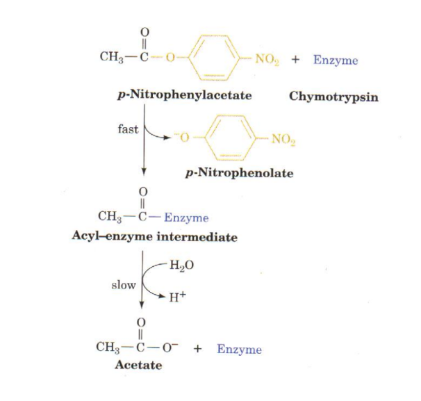
What burst kinetics reveal
A two-step mechanism: fast acylation releases p-nitrophenolate and forms the acyl-enzyme, then slow deacylation (hydrolysis) releases acetate
The burst size is proportional to enzyme concentration (compare the 0.8 vs. 0.4 mg/mL curves).

Active serine
A serine (Ser 195 in chymotrypsin) that forms a covalent bond with the substrate (the acyl-enzyme intermediate)
It is unusually reactive and can be specifically labeled by inhibitors.

Diisopropyl phosphofluoridate (DIPF)
An organophosphate that reacts specifically with the active Ser 195:
Active Ser-CH2OH + DIPF → DIP-enzyme + HF
This irreversibly inactivates ("kills") the enzyme.

Why DIPF is extremely toxic
It also labels the active serine of other enzymes, such as acetylcholinesterase, blocking nerve signal termination

Nerve gases and insecticides
Nerve gases (sarin) and many insecticides (parathion, malathion) are organophosphates that work the same way as DIPF, inactivating the active serine of acetylcholinesterase
Affinity labeling
A substrate analog with a reactive group binds the enzyme's active site and then reacts covalently with a nearby reactive amino acid, inactivating the enzyme
Called a "Trojan horse" effect
It identifies active-site residues.

TPCK
Tosyl-L-phenylalanine chloromethyl ketone, an affinity label for chymotrypsin
Its Phe portion mimics a chymotrypsin substrate, and its chloromethyl ketone (-CO-CH2Cl) reacts with His 57.

Reaction of TPCK with chymotrypsin
His 57's imidazole N attacks the CH2Cl carbon of TPCK and displaces Cl- (HCl released)
This alkylates His 57 and inactivates the enzyme, showing His 57 is a second important catalytic residue.

Catalytic triad
Asp 102, His 57, and Ser 195 in chymotrypsin, linked by hydrogen bonds:
Asp 102 to His 57 to Ser 195
Role of each catalytic triad residue
Ser 195 = nucleophile
His 57 = general base (accepts the Ser proton), then general acid (donates the proton to the leaving group)
Asp 102 = hydrogen bonds to His 57, orienting it and stabilizing its positive charge.
Effect of losing Asp 102
Without Asp 102, the rate of catalysis is only 0.05% of wild-type
Other residues near the chymotrypsin catalytic triad
Gly 193 and Ser 195 backbone NHs form the oxyanion hole
Asp 194 forms a salt bridge with Ile 16.

Serine protease mechanism step 1
After the substrate binds (Michaelis complex), Ser 195 nucleophilically attacks the carbonyl carbon of the scissile peptide bond
This forms the tetrahedral intermediate (covalent catalysis), and His 57 takes up the Ser proton
Asp 102 is hydrogen bonded to His 57.

Serine protease mechanism step 2
The tetrahedral intermediate decomposes to the acyl-enzyme intermediate
His 57 acts as an acid, donating a proton to the leaving amine (R'NH2), which is released as the new N-terminus of the cleaved chain.

Serine protease mechanism step 3
The enzyme is deacylated by the reverse of step 1
Water is the attacking nucleophile (His 57 takes its proton), forming a second tetrahedral intermediate
Ser 195 is the leaving group, releasing the new C-terminus of the cleaved chain and regenerating the active enzyme.

Tetrahedral intermediate
A transition-state-like covalent intermediate in which the former carbonyl carbon is sp3 with four groups and bears a negatively charged oxygen (oxyanion)

Acyl-enzyme intermediate
A covalent ester between Ser 195 and the acyl (N-terminal) portion of the substrate, formed after the first product leaves

Oxyanion hole
A site formed by the backbone NHs of Gly 193 and Ser 195 that hydrogen bonds to the negatively charged oxygen of the tetrahedral intermediate

Transition state stabilization by the oxyanion hole
(1) Conformational distortion forms the tetrahedral intermediate and moves the carbonyl O into the oxyanion hole
(2) There it forms two hydrogen bonds with the enzyme that cannot form when the carbonyl is in its normal (trigonal) conformation
(3) Distortion from enzyme binding makes these hydrogen bonds maximal. This is preferential transition state binding.

Charge relay system
In the catalytic triad, two protons move together
Ser 195's OH proton moves to His 57, and His 57's other N-H proton moves to Asp 102
This leaves Ser 195 as a strongly nucleophilic alkoxide (O-) and Asp 102 neutral.

Triad charge transfer complex stabilization
The Asp 102 to His 57 hydrogen bond stabilizes the charge that develops on His 57 during catalysis, making His 57 a better base and Ser 195 a better nucleophile
Catalytic strategies used by serine proteases
Covalent catalysis (Ser 195 to acyl-enzyme)
general acid-base catalysis (His 57)
transition state stabilization (oxyanion hole)
electrostatic stabilization (Asp 102)
and proximity and orientation (substrate binding in the specificity pocket)