Bones of pectoral girdle and upper extremity
 Call Kai
 Learn
Learn Practice Test
 Spaced Repetition
 Match
Match Flashcards
 Knowt Play
1/53

There's no tags or description
Looks like no tags are added yet.
Name | Mastery | Learn | Test | Matching | Spaced | Call with Kai | Chat |
|---|
No analytics yet
Send a link to your students to track their progress
54 Terms
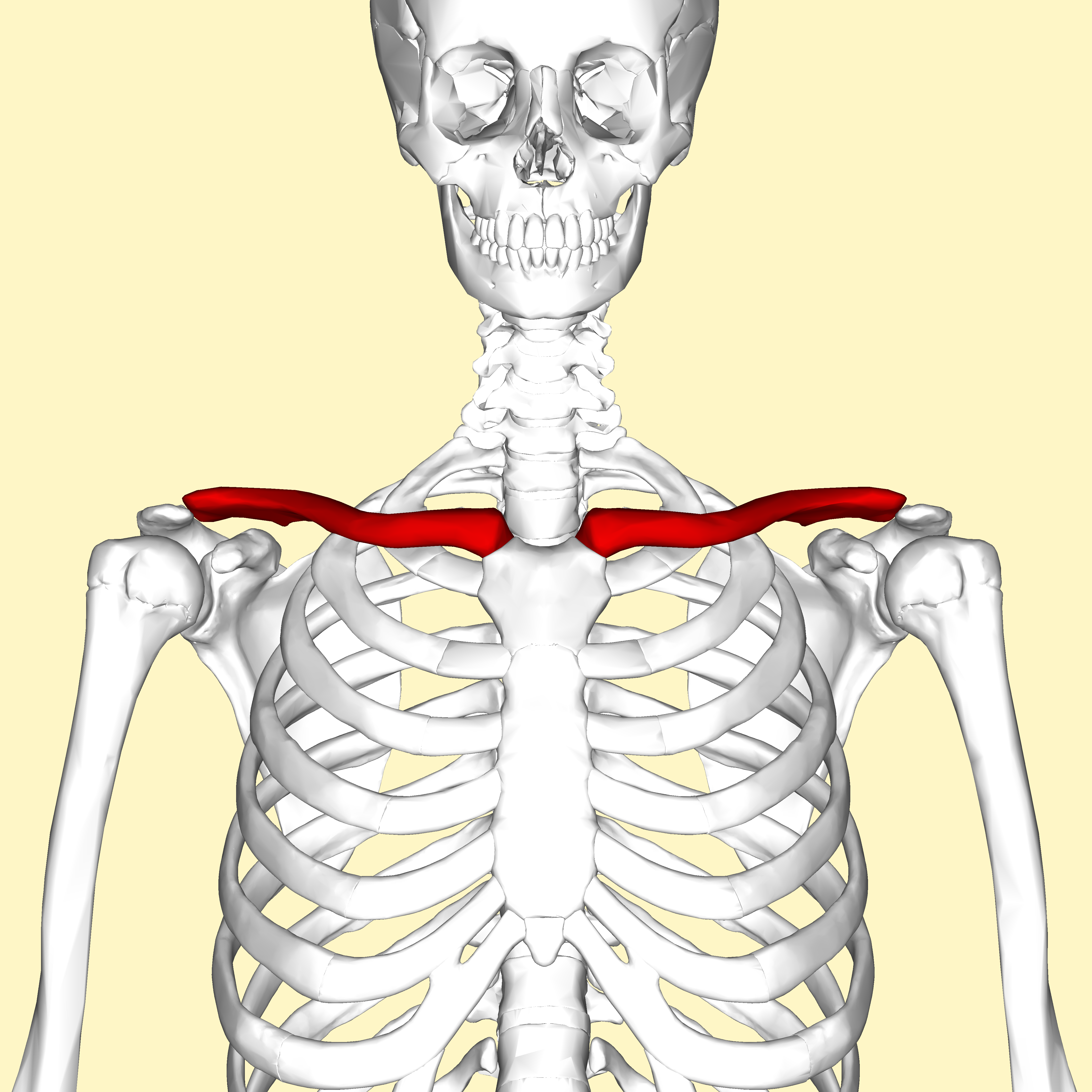
Clavicle
2 bones. the collarbone. elongated, s-shaped bone that extends b/w manubrium of the sternum and the acromion of the scapula


Acromial end, sternal end
Markings of the clavicle

Sternal end of the clavicle
(medial end). roughly pyramidal shaped and articulates w/ the manubrium of the sternum forming the sternoclavicular joint.
Acromial end of the clavicle
(lateral end). broad and flattened and articulates with the acromion of the scapula, forming the acromioclavicular joint

Scapula
2 bones. broad, flat, triangular bone that forms the shoulder blade


spine, acromion, coracoid process, glenoid cavity/fossa, superior angle, inferior angle, medial border, lateral border
markings of the scapula


Spine
ridge of bone on the posterior aspect of the scapula

Acromion
continuous with the spine. larger, flattened, posterior process. articulates w/ the acromial end of the clavicle.

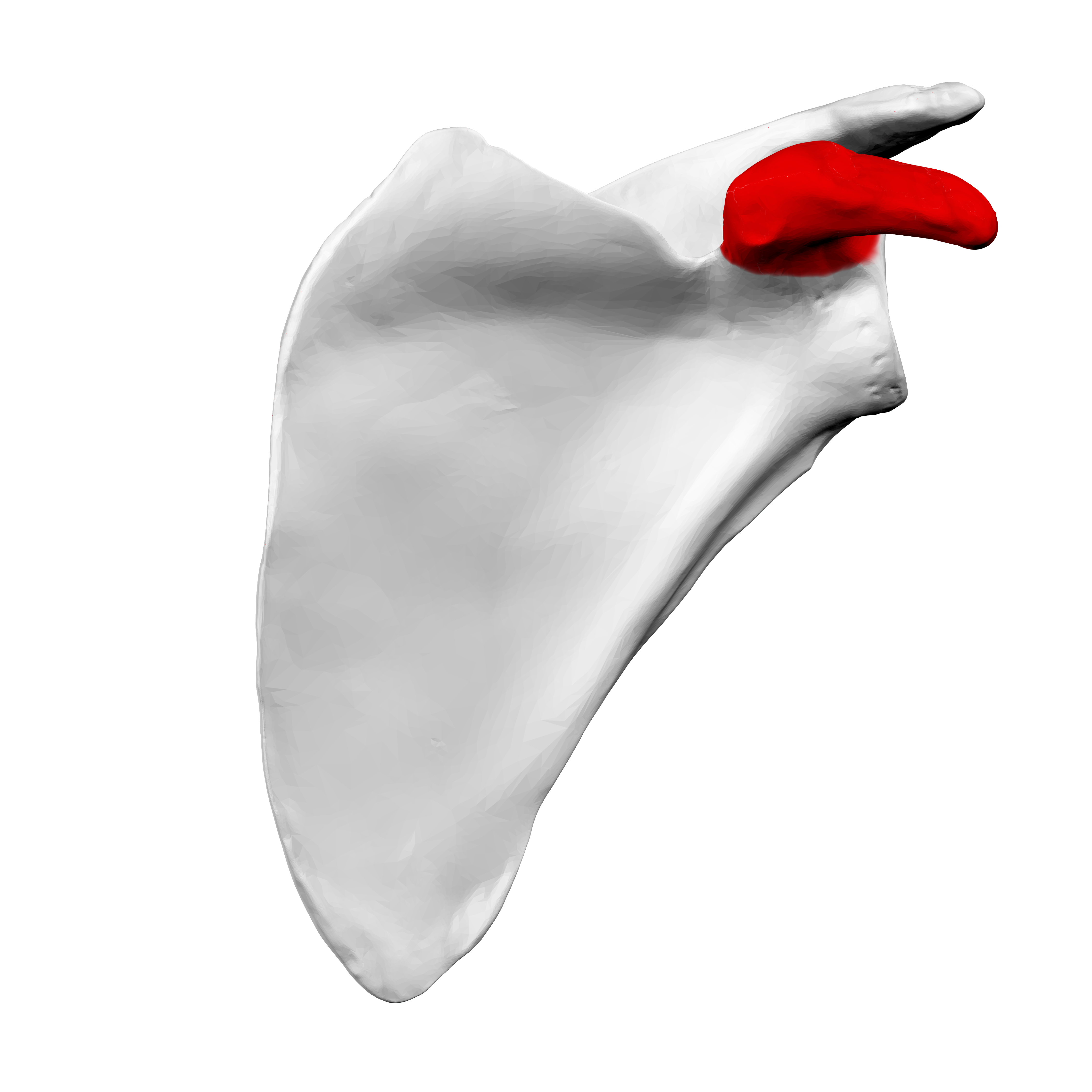
Coracoid process
smaller, more anterior, hook shaped projection that’s a site for muscle attachment


Glenoid cavity/fossa
cup shaped, shallow cavity that articulates w/ the humerus. makes up most of the lateral angle.


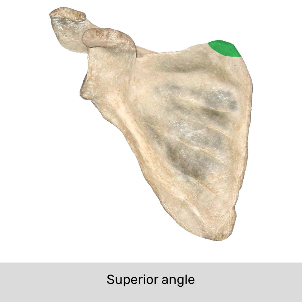
Superior angle
located b/w the superior and medial borders
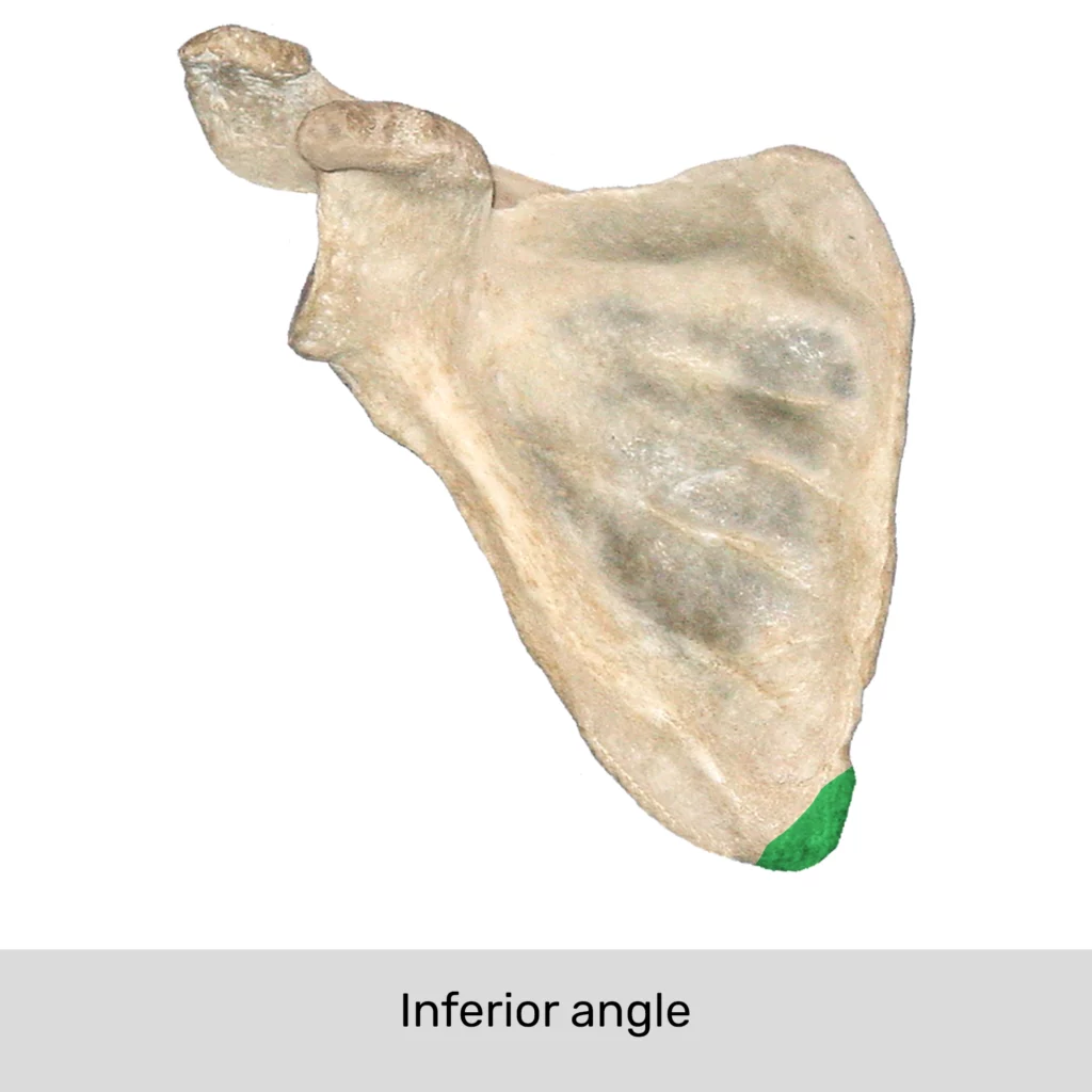
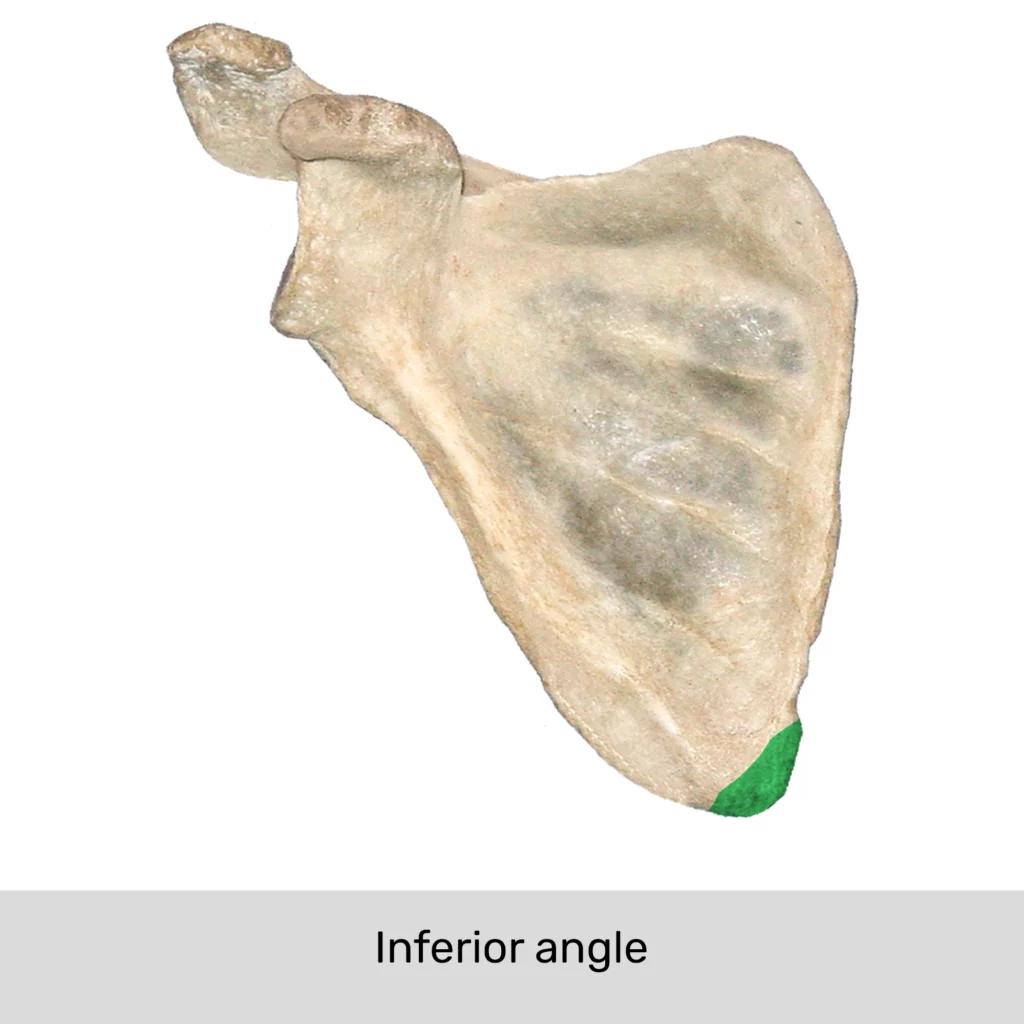
Inferior angle
positioned b/w medial and lateral borders

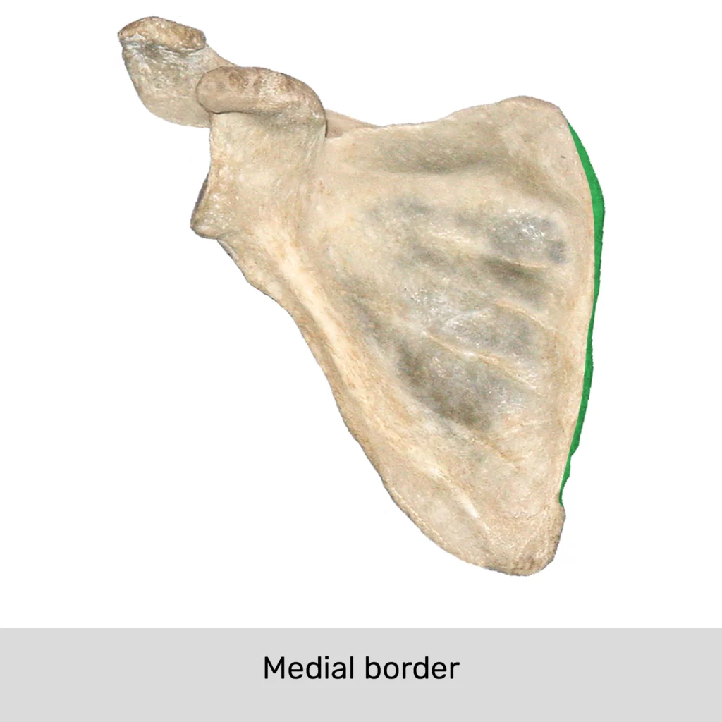
medial border
the edge of the scapula closest to the vertebrae
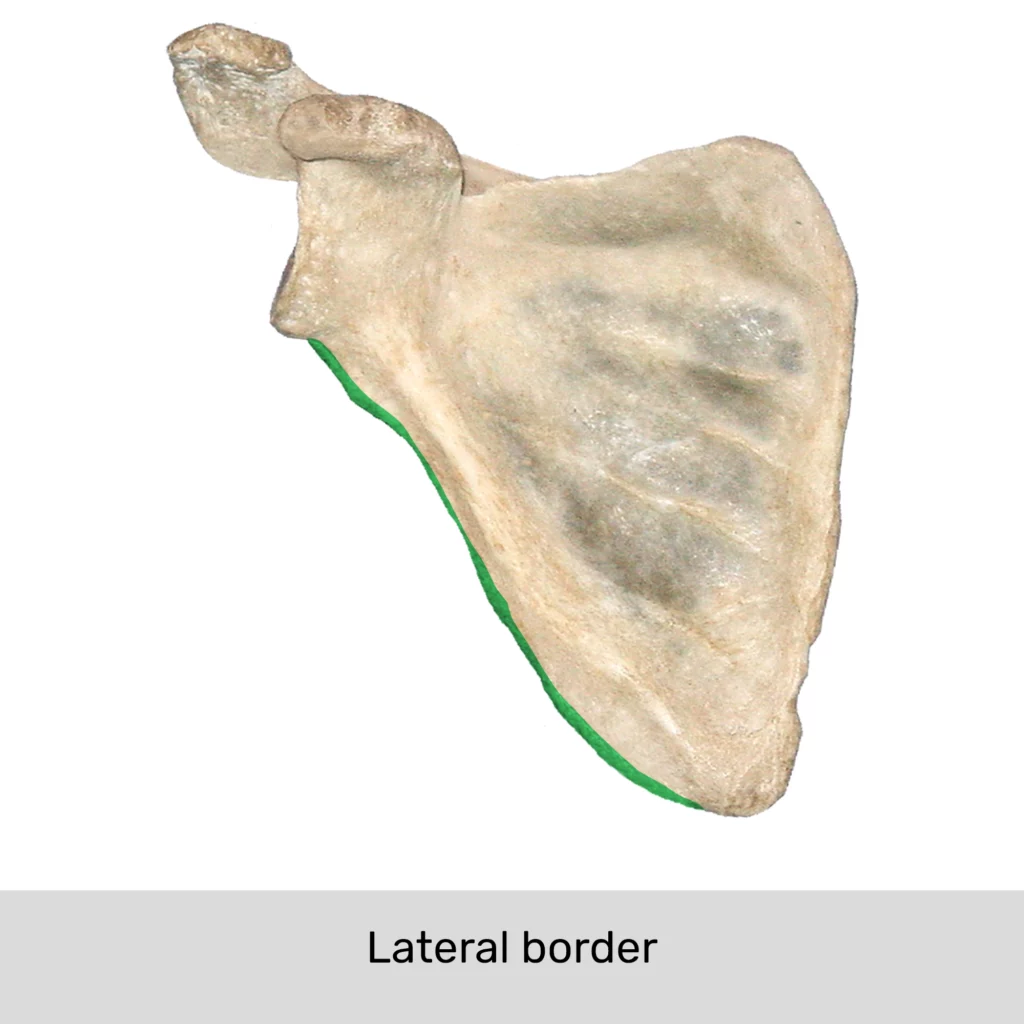
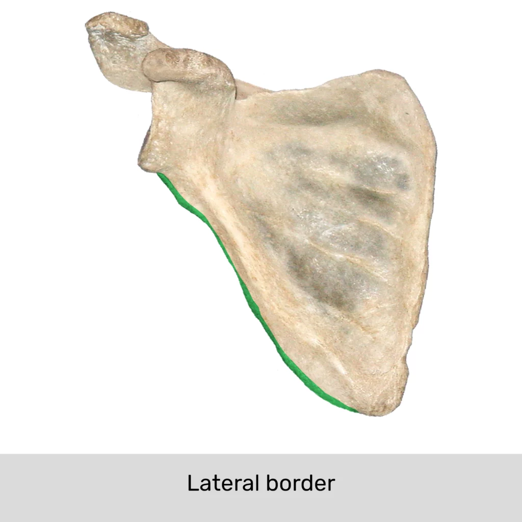
Lateral border
closest to the axilla
clavicle, scapula
Bones of the pectoral girdle

Humerus
2 bones. longest and largest upper limb bone


head, shaft, anatomical neck, surgical neck, greater tubercle, lesser tubercle, intertubercular groove, deltoid tuberosity, trochlea, capitulum, coronoid fossa, radial fossa, olecranon fossa, medial epicondyle, lateral epicondyle
Markings of the humerus
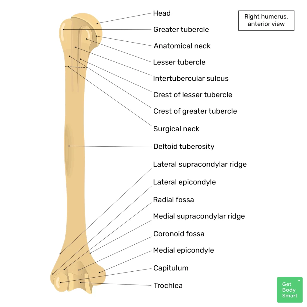
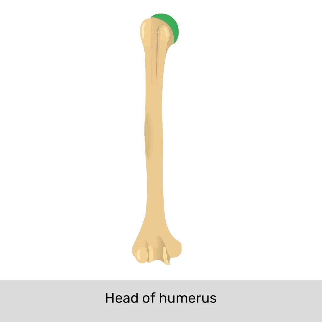
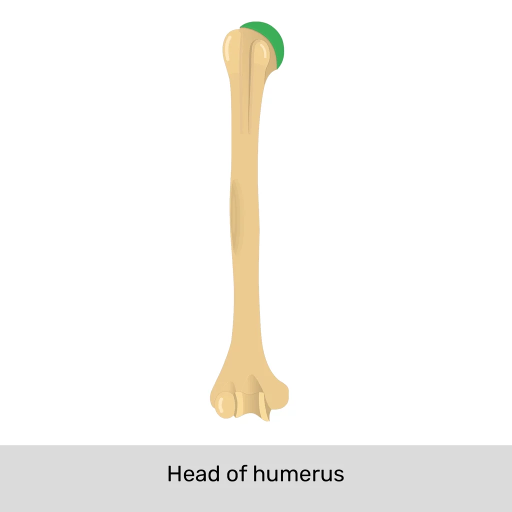
Head of the humerus
on the proximal end. has hemispherical head that articulates w/ the glenoid cavity of the scapula

Shaft of the humerus
long middle portion of the humerus


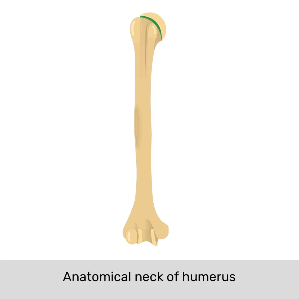
Anatomical neck
b/w the tubercles and the head of the humerus. almost indistinct groove that marks the location of the former epiphyseal plate

Surgical neck
narrowing of the bone immediately distal to the tubercles, at the transition from the head to the shaft. called this bc it’s a common fracture site

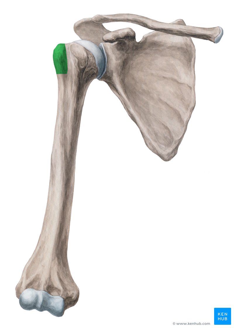
Greater tubercle
positioned lateral to the head and helps form the rounded contour of the shoulder
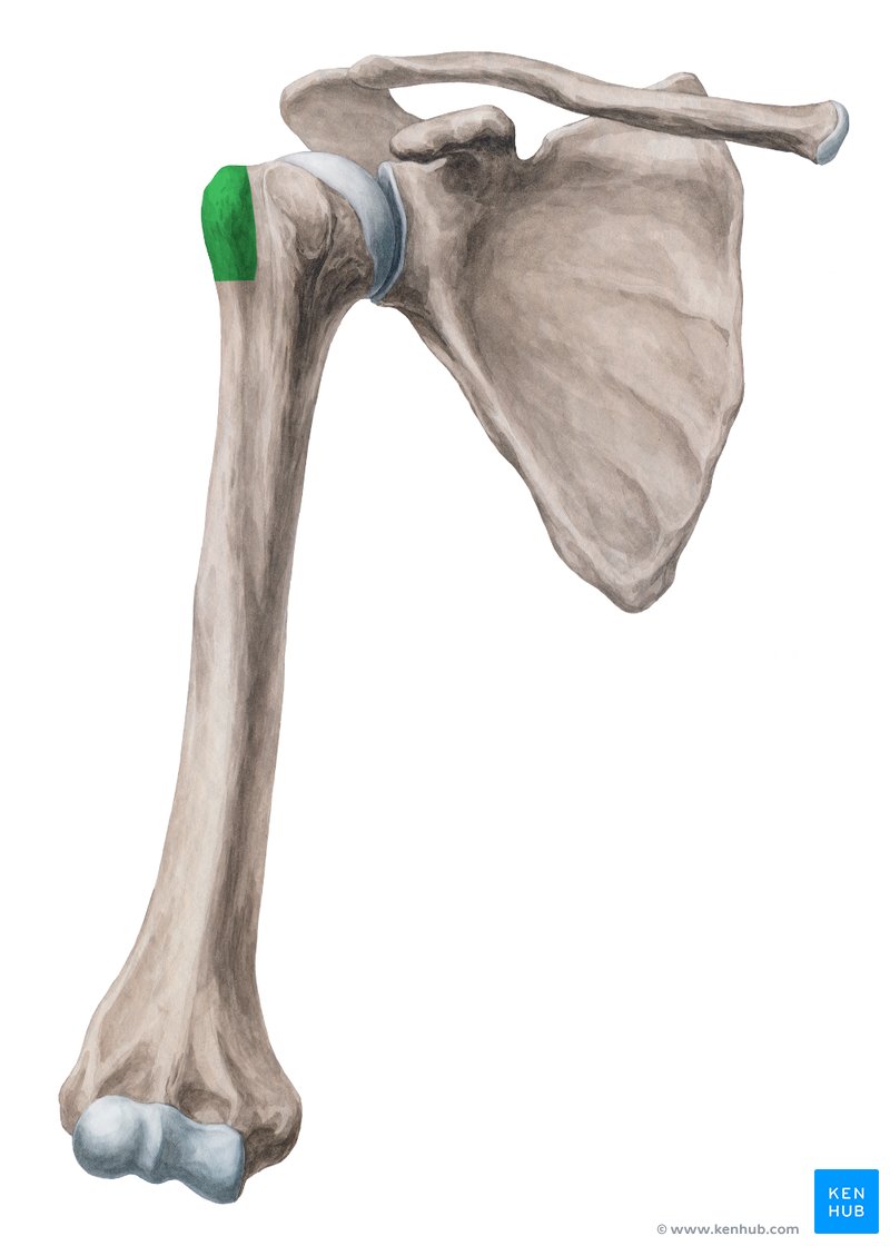

Lesser tubercle
smaller and located more medial to the head
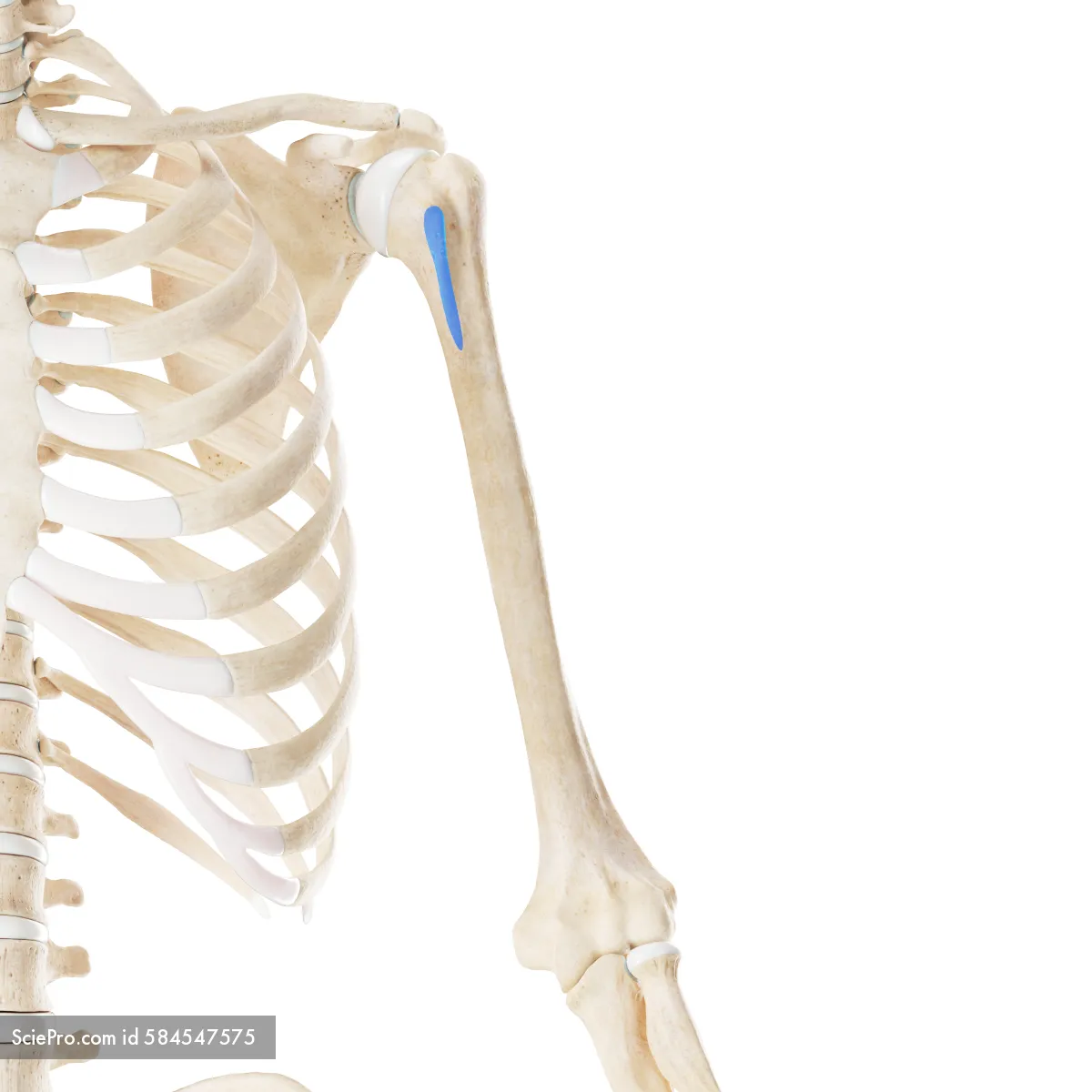

Intertubercular groove
depression that contains the tendon of the long head of the biceps brachii muscle. b/w the 2 tubercles


Deltoid tuberosity
roughened area along the shaft of the humerus. extends along the lateral surface for abt half the length of the humerus. deltoid muscle of the shoulder attaches here


Trochlea
pulley shaped. located medially and articulates w/ the trochlear notch of the ulna


Capitulum
rounded. located laterally and articulates w/ the head of the radius

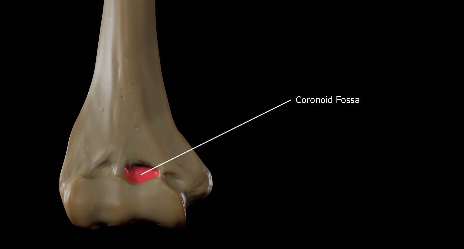
Coronoid fossa
accommodates the coronoid process of the ulna. depression on the anterior surface of distal end of humerus.
Radial fossa
a depression on the anterolateral surface of the distal end of the humerus; articulates w/ the head of the radius
Olecranon fossa
a large depression on the posterior surface at the distal end of the humerus; articulates w/ olecranon of the ulna

Medial epicondyle
small projection medial and proximal to the trochlea

Lateral epicondyle
small projection lateral and proximal to the capitulum

Radius
2 bones. forms the forearm with the ulna. located more laterally
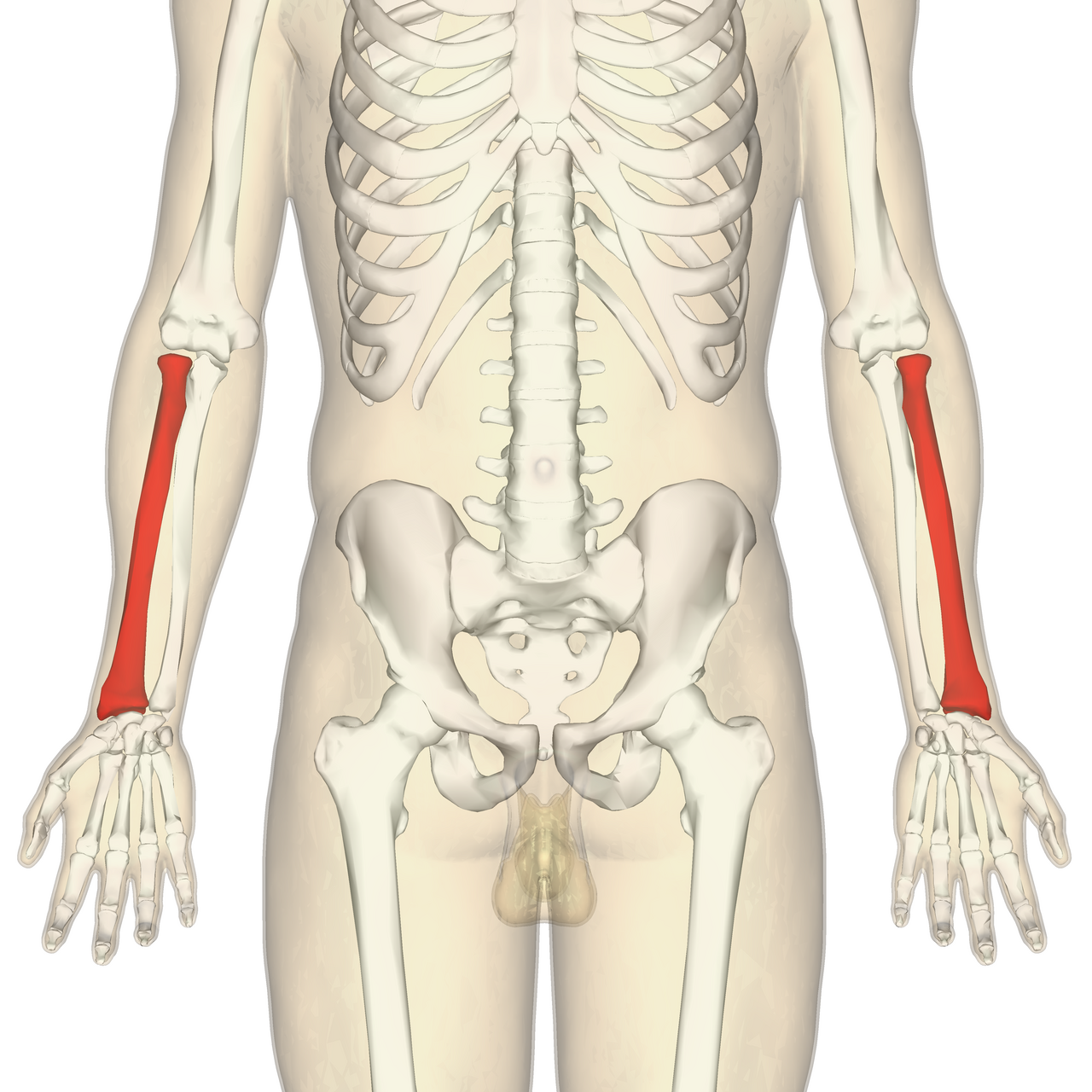

head, neck, radial tuberosity, styloid process of the radius
Markings of the radius
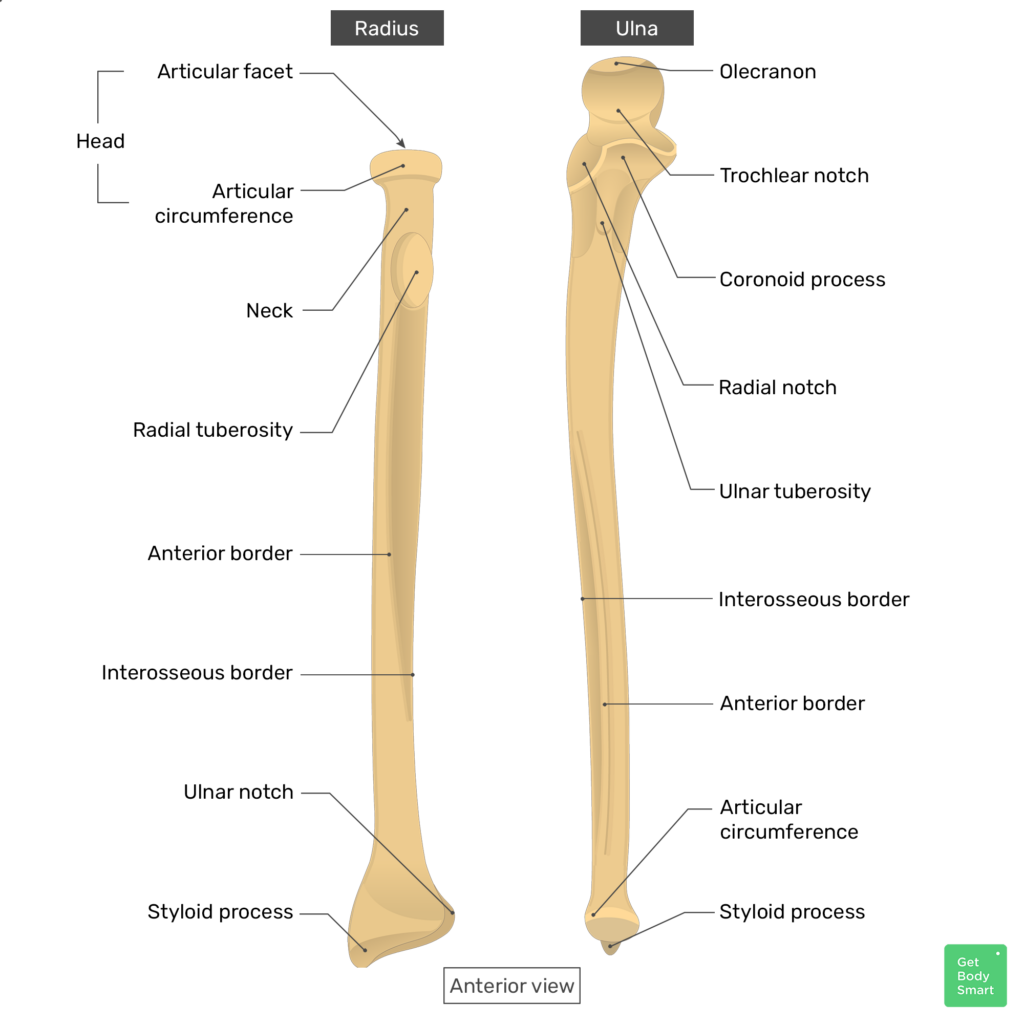
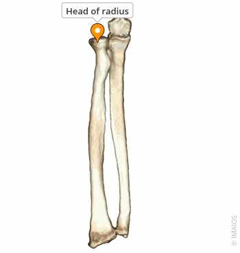
Head of the radius
disc-shaped proximal end of the radius; articulates w/ capitulum of the humerus


Neck of the radius
narrow region just distal to the head of the radius


Radial tuberosity
large projection on the medial surface distal to the neck

Styloid process of the radius
small, pointed projection on the distal radius; forms the lateral aspect of the wrist

Ulna
2 bones. longer, medially placed placed of the forearm


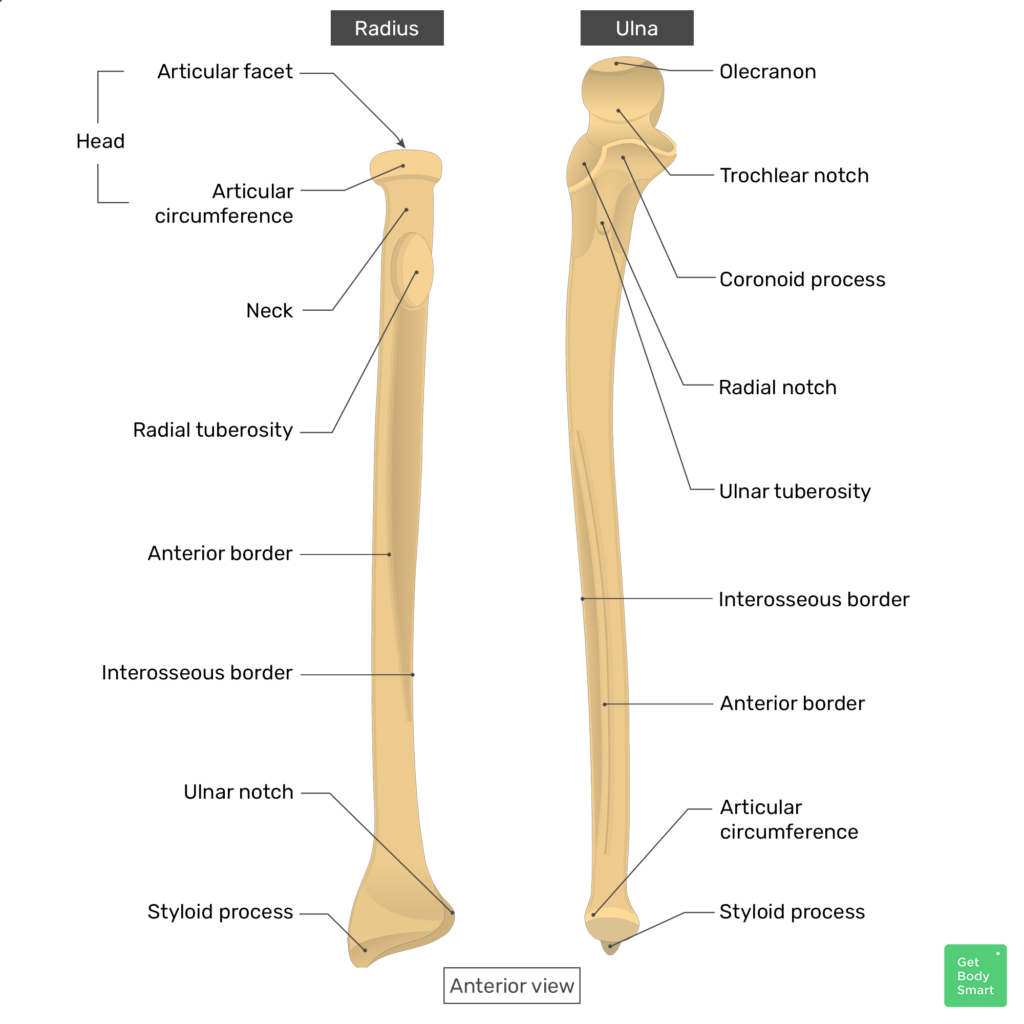
trochlear notch, olecranon, coronoid process, radial notch, styloid process of the ulna
Markings of the ulna

Trochlear notch
a C-shaped groove on the proximal end of the ulna; articulates w/ the trochlea of the humerus


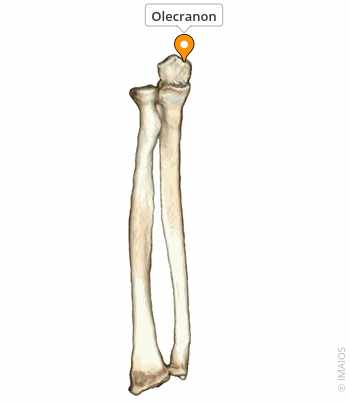
Olecranon
large projection on the proximal, posterior surface of the ulna; forms the point of the elbow and is an attachment for the triceps brachii muscle

Coronoid process
projection on the proximal, anterior surface of the ulna just inferior to the trochlear notch articulates w/ the coronoid fossa of the humerus

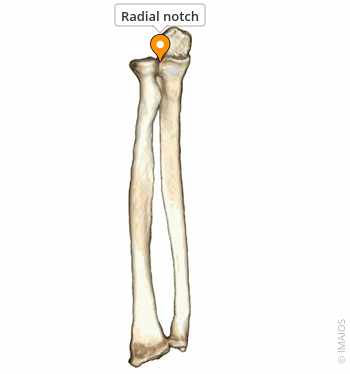
Radial notch
depression on the lateral, proximal surface of the ulna; articulates w/ the head of the radius


Styloid process of the ulna
pointed process on the distal ulna; forms the medial aspect of the wrist

Carpal bones
8 bones. small, short bones that form the wrist
scaphoid, lunate, triquetrum, pisiform, trapezium, trapezoid, capitate, hamate
Names of carpal bones
Metacarpals
10 bones. 5 bones in the palm of the hand; each bone consists of a head, body, and base. Denoted w/ roman numerals I-V w/ metacarpal I located at the base of the thumb and metacarpal V at the base of the pinky.
Phalanges
28 bones. 3 of these found in each of the 2nd-5th fingers, but only 2 found in the thumb
proximal phalanges, intermediate phalanges, distal phalanges
markings of the phalanges

Proximal phalanges
the phalanx closest to the palm of the hand


Intermediate phalanges
the middle phalanx


Distal phalanges
the small, cone shaped phalanx forming the tips of the fingers

Pollex
the 1st phalanx. thumb