biochem p3
 Call Kai
 Learn
 Practice Test
 Spaced Repetition
 Match
 Flashcards
 Knowt Play
1/59

There's no tags or description
Looks like no tags are added yet.
60 Terms
Glycogen
importance of glucose (GLC or Glc)
when to degrade it - glycolysis
when to synthesize it - gluconeogenesis
when to store it - glycogen
glycogen synthesis
glycogen synthase and five other enzymes
glycogen degradation
glycogen phosphorylase and three other enzymes
glycogen storage and disease
Importance of glucose
carbohydrate metabolism is more than just glycolysis. it’s part of an array of anabolic and catabolic pathways
anabolism: pathways that create biomolecules
catabolism: pathways that degrade biomolecules
synthesis: glycogen synthase
don’t typically want to get rid of glucose

Gluconeogenesis and Glycolysis are reciprocally regulated
glycogen synthesis (glucose → glycogen)
glycogen storage OFF when cell needs NADH + H+ and ATP
glycogen degradation (glycogen → glucose)
glycogen degradation ON when cell needs NADH + H+ and ATP
glycolysis (glucose → pyruvate)
On when cell needs: NADH + H+ and ATP
gluconeogenesis (pyruvate → glucose)
OFF when cell has ample: NADH + H+ and ATP
What happens when a cell has ample NADH and ATP?
glycogen synthesis
glycogen storage ON when cell has NADH + H+ and ATP (save the Glc for another day)
glycogen degradation
glycogen degradation OFF when cell has ample NADH + H+ and ATP (save the Glc for another day)
glycolysis
OFF when cell has ample: NADH + H+ and ATP (why make more? save for later)
gluconeogenesis
OFF when cell has ample: NADH + H+ and ATP
What triggers Gluconeogenesis
ON under a very specific set of circumstances:
no influx of glucose (out of glucose)
glycogen stores are depleted in muscle and liver
glucose is badly needed by neuronal tissues (only source of energy is glucose)
think: starvation conditions in higher vertebrates
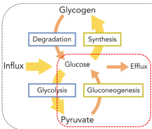
Digging deeper into the bypass reactions, another name for the gluconeogenesis reactions that replace glycolysis steps 10, 3 and 1 :
Dr. Hogan has been out in the woods without food for 72 hours. All his short term energy sources are depleted (no glycogen left and no glucose influx). His thinking is getting ”foggy” because his blood sugar is low and there is no glucose left to fuel his neuronal tissue. What to do? Dr. Hogan realizes if he can gather firewood and start a friction fire, he can signal for help. Where will he get the energy to think through this solution and assemble the kindling?
Gluconeogenesis. Short term synthesis of Glc from 2 Pyr in liver. Glc then exported to blood-->brain + muscles for LIMITED energy
Muscle, Brain and nervous tissue use glucose as primary energy source
Fats in the form of Triacylglycerols (beta oxidation of fatty acids) would also begin to breakdown along with some protein catabolism, but these are slow relative to carbohydrate metabolism
The bypass reactions that replace step 10 of glycolysis: Interconversion of Pyr and PEP
Notice how much energy is being consumed in this step !!! This can’t go on forever

Remaining bypass reactions: Phosphatases

Now Glc exported from Liver
• Transported via bloodstream
• Provides short term energy for neuronal tissue
• Energy expensive
• Requires Pyr
• NOT a long term process for fueling cellular metabolism

Glycogen Synthesis
Insulin and Glucagon: Systemic Control
Glucagon: secreted from the pancreas when GLC is scarce, stimulating glycogen degradation
Insulin: secreted from the pancreas when GLC is abundant, stimulating glycogen synthesis
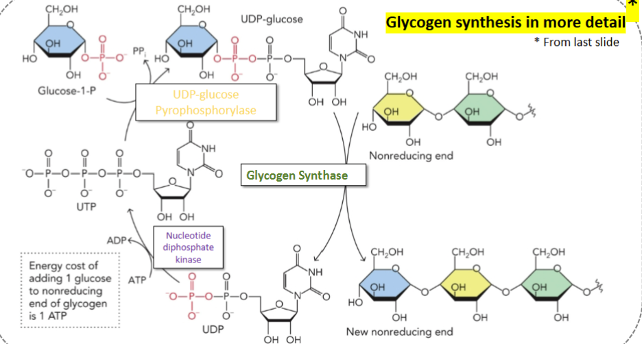
Main enzymes associated with glycogen synthesis
Hexokinase
Phosphoglucosemutase (aka phosphoglucomutase)
UDP-glucose phyrophosphorylase
Glycogen Synthase
Branching enzyme
(Nucleoside diphosphate kinase-indirect) - recreates UDP
degradation only happens at one end
branching enzyme pulls glucoses off chain and sticks then at 6 end; creates more free ends (greater surface area and enzyme cleavage)
inorganic pyrophosphatase degrades PPi
1 position = reducing end
4 position = nonreducing end (receives new glucose)
glycogen alpha 1,4 linked glucose polymer
alpha: at 1 position oxygen preferred to be down
primary linkage between glucose monomers in glycogen alpha-1,4 and to be able to draw two monomer linked that way, for example.

branching enzyme

glycogen degradation
1→6:
glycogen phosphorylase
phosphoglucosemutase (aka phosphoglucomutase) - same as glycogen synthesis
glucose 6 phosphatase (from gluconeogenesis)
glycogen debranching enzyme
Glycogen granules/deposits are large and represent approximately 24 hours worth of energy in higher vertebrates
glycogen phosphorylase w/ PLP coenzyme round 1
Activity of glycogen phosphorylase
(Glcn) → Glcn-1 + G1P

glycogen phosphorylase round 2
cleavage continues until enzyme gets too close to branch point
glycogen phosphorylase stops within 8 glucoses of branch point
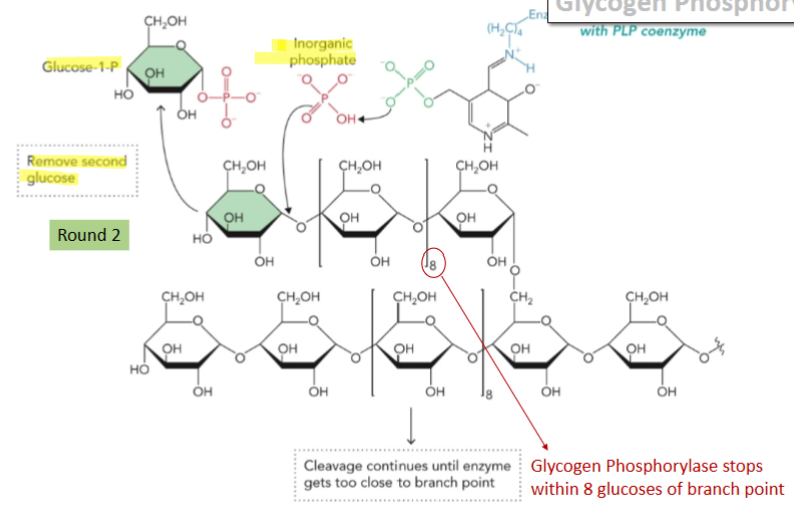
glycogen debranching enzyme: activity #1 transferase
(glycogen phosphorylase) hits a roadblock near a branch point. A transferase activity steps in to pick up a block of three glucose molecules from the branch and moves them to the end of a straight chain, making them accessible

glycogen debranching enzyme: activity #2 glucosidase
After the transferase moves the bulk of the branch, a single remaining glucose molecule is left attached at an α-1,6 branch point. An α-1,6 glucosidase clips off that last stubborn glucose molecule, releasing it as free glucose so breakdown can continue.

Glycolysis: Inputs and Outputs
Net:
glucose → 2 pyruvate
2 ADP → 2 ATP
2 NAD+ → 2 NADH + H+
The TCA cycle is a Central Area of Metabolism at the intersection of several anabolic and catabolic pathways

Pyruvate from Glycolysis can be further oxidized in the presence of O2
The conversion of pyruvate into CO2, reduced cofactors and, ultimately, a lot of ATP occurs in the mitochondria
Net Reaction TCA:
Acetyl-CoA + 3 NAD++ FAD + GDP + Pi + 2 H2O → CoA + 2 CO2 + 3 NADH + 3 H+ + FADH2 + GTP
reduced coenzymes represent potential energy for the cell
CO2 is the most oxidized any carbon can be

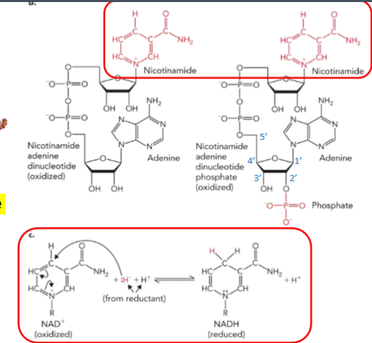
Let’s review the important coenzymes in this process: NAD+ and FAD
NAD+ is derived from Niacin: the nicotinamide portion does the redox chemistry
FAD is derived from Riboflavin (B2): the Flavin portion does the redox chemistry

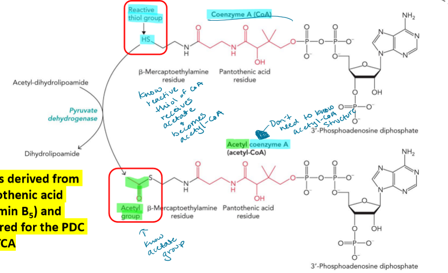
CoA derivation
CoA is derived from pantothenic acid (Vitamin B5) and required for the PDC and TCA
reactive thiol of CoA receives acetate and becomes acetyl-CoA
TPP and Lipoamide
TPP is derived from thiamine (vitamin B1) and required for the PDC and TCA
lipoamide is derived from plant biomass and can be covalently attached to Lys residues
bound to lysine; has 2 sulfurs one of which can be substituted w/ acetyl group

The PDC is an example of a multienzyme complex. What is the advantage of having multienzyme complexes catalyze sequential reactions?
increased frequency of collisions
multiple reactions in one place in cell
coordinate control
reduce side rxns
R = nat end of lysine side chain
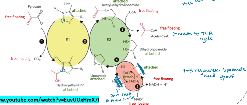
PDC
1. E1: Transfer acetyl group from Pyruvate to TPP→ Hydroxyethyl-TPP
2. E2: Transfer of acetyl group from Hydroxyethel-TPP on E1 to Lipoamide of E2→ Acetyl-dihydrolipoamide
3. E2: Acetyl group transferred to CoA→Acetyl CoA
4. E3: Dihydrolipoamide oxidized, 2 e- to FAD→FADH2
5. FADH2 oxidized by NAD+→NADH +H+
4 and 5 regenerate lipoamide head group
don’t need to know S and SH
Acetyl-CoA heads to TCA cycle
FAD is integrated into 3-D structure of E3
NAD+ is soluble (not part of E3 structure)
TCA cycle is 8 reactions. 5 near eq rxns and 3 far from eq rxns
all FFE and PDC highly regulated enzymes

TCA
dehydrogenase = redox rxn
Can I Keep Selling Skittles For Money Officer
Step 1: oxaloacetate used from step 8 TCA cyclical; Acetyl-CoA has “high-energy” thioester which is hydrolyzed and releases energy
acetyl-CoA binding triggers a conformational change (induced fit)
Step 2: cis-aconitate is an intermediate in the aconitase reaction
step 3: first of 2 oxidative decarboxylations
step 4: second of 2 oxidative decarboxylations; mechanistically identical to PDC: Multienzyme complex, 5 cofactors, 5 reactions
Step 5 (succinyl-CoA synthetase): name for ‘reverse’ reaction; free energy of thioester hydrolysis conserved in the synthesis of GTP via a substrate level phosphorylation
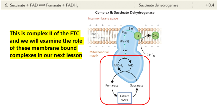
Step 6 (succinate dehydrogenase): this is complex II of the ETC
step 7: fumarase binds ‘trans’ isomer
step 8: energetically couples to reaction 1 which is large and exergonic; last reaction of the TCA generates OAA for reaction 1: citrate synthase
*reduced coenzymes fed into ETC and used to fuel ATP synthesis

ETC
ETC components “ordered” in the IM from low to High Standard Reduction potential
TCA = mitochondrial matrix
ETC = IM

Structure of ATP Synthase
Protons move through a subunit of F0
fixed
Rotation of γ drives conformational changes in β subunits of F1
fixed
Movement of H+ results in rotation of C ring and γ subunit
mobile
H+ flux translates potential energy into rotational energy
F0 in IM space, F1 in matrix/cytoplasm

ETC Proton and Electron Flow
complex I
NADH oxidized and 4 protons pumped into IM space
complex II
FADH2 oxidized and no protons pumped into IM space
complex III
CoQ oxidized and 4 protons pumped into IM space
complex IV
Cytochrome C oxidized, .5 O2 reduced, H2O formed and 2 protons pumped into IM
O2 final electron acceptor formed H2O

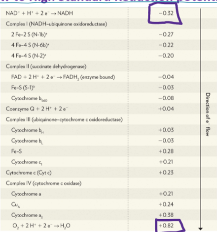
Under standard conditions, will the following reactions proceed spontaneously as written? (You’ll need the table from previous slide)
a) 2 Cytochrome c (Fe3+) + NADH → 2 Cytochrome c (Fe2+) + NAD+
b) Cyto a (Fe2+) + cyto b 560 (Fe3+) → Cyto a (Fe3+) + cyto b 560 (Fe2+)
reaction a

What is the purpose of the ETC and moving H+ into the intermembrane space?
The chemiosmotic theory: The free energy of electron transport is harnessed in the form of a proton gradient that provides the electrochemical potential energy to synthesize ATP
oligomycin: a potent anti-fungal Blocks H+ returning to matrix and therefore producing ATP
Return to this diagram from earlier…Now draw in the structure of ATP synthase and label the F0 and F1 portions. Then, indicate the movement of H+ in relation to ATP synthase.
protons move from IM space into mitochondrial matrix


structure of ATP Synthase is evolutionarily conserved
As H+ flow, potential energy is converted into rotational energy
As H+ flow, the gamma subunit rotates counterclockwise (when viewed from the matrix)
The rotation of gamma induces the conformational changes in the beta subunits that drive ATP synthesis
What is bound to the β subunit in each of the conformational states? (3 subunits w 3 conformational states)
O (Open)= low affinity state for substrate or product
L (Loose) = ADP and Pi binding state
T (Tight) = catalytic state ADP + Pi → ATP
Can multiple β subunits be in the same conformational state at the same time?
no each is in a different conformational state each rotation

ATP Synthase run in reverse (ATP→ADP + Pi) to view the movement of gamma. The is the OPPOSITE of how things work in vivo!!! In cells, rotation of gamma is counterclockwise when F1 is viewed from the matrix
normal everything the same except O state arrows reversed and CCW
O → L → T
In this in vitro experiment, researchers used electromagnets to force rotation of the magnetic bead to drive ATP synthesis in the absence of a proton gradient !

How many ATP do each NADH and FADH2 represent?
NADH: 2.5 or 3 ATP
FADH: 1.5 or 2 ATP

Acid Base Properties of Nucleic Acids
the phosphate backbone is a strong acid
the net charge of the phosphate at pH 7 will be negative
the phosphate group of the nucleotide is polyprotic
both pKa values are < 7; negative charge at physiological pH
purine and pyrimidine
amino and keto tautomers lower energy and more stable at pH 7 for purines and pyrimidines
adenine: 6 amino purine
guanine: 2 amino 6 oxy purine
thymine: 2,4 dioxy 5 methyl pyrimidine
uracil: 2,4 dioxy pyrimidine
cytosine: 2 oxy 4 amino pyrimidine

base pairs

More nomenclature: bases, nucleotides, and nucleotides have unique names

Make a list of the physical and chemical properties of the N-bases What do they share in common?
1. Heterocyclic rings (C, N, H, O)
2. Absorb light at 260 nm (cyclic/aromatic)
3. Delocalized pi electrons
4. Roughly planar
5. Weakly basic
The aromaticity of the bases leads to electron delocalization in the rings, leading to partial double bond character and planarity
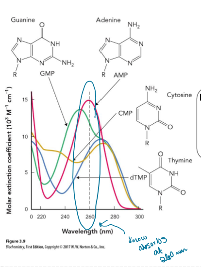
Rank the molecules below from most soluble in aqueous buffer to least soluble in aqueous buffer
Adenosine
Adenine
Deoxyadenylate
Deoxyadenosine 5’ triphosphate
Deoxyadenosine 5’ triphosphate
deoxyadenylate (AMP)
adenosine (nucleoside)
adenine (base)

Ribose and Deoxyribose Chemistry
The furanose ring (5 membered) of nucleosides is puckered. The C-2’ and C-3’ carbons are most often outside of the furanose ring in the endo position, leaving the other four carbons in a conformation that is close to planar.
RNA bases more flexible
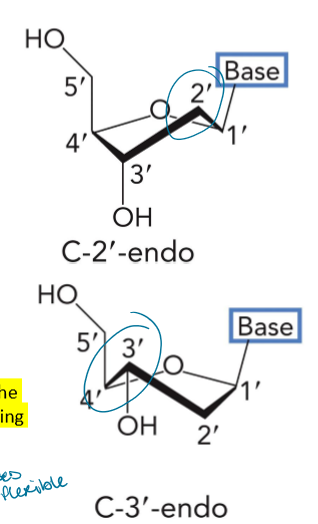
What other factors contribute to the flexibility of Nucleic Acids?
Nucleic acids are highly flexible due to:
1. Conformations of ribose
2. Rotation around contiguous phosphodiester linkages
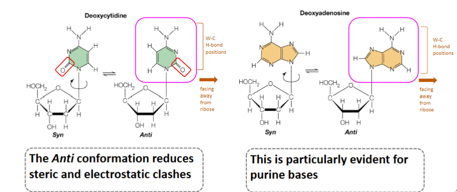
3. Rotation around glycosidic linkage Free rotation around 6 bonds and limited dynamics in the 7th
the anti conformaiton reduces steric and electrostatic clashes; this is particularly evident for purine bases
The 3’→5’ phosphodiester linkage also allows for free rotation
Successive nucleotides are linked covalently through 5’ phosphate of one nucleotide and 3’ -OH of the next
All phosphodiester bonds in DNA & RNA have same orientation along the chain
This gives each linear strand a specific polarity with a distinct 5’ and 3’ end
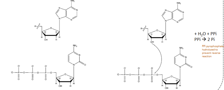
How to make a phosphodiester bond
not energetically favorable but kinetically favorable in cell
PPi pyrophosphate hydrolyzed to prevent reverse reaction


Why flexibility matters
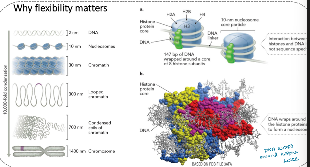
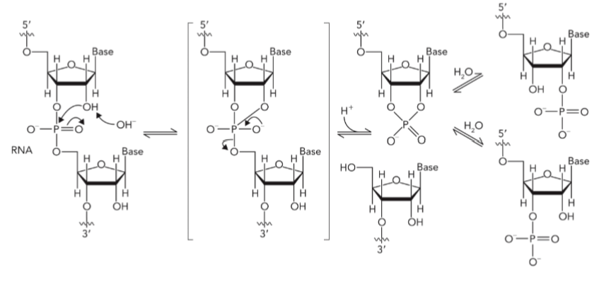
RNA is less stable than DNA due to alkaline hydrolysis of 2’-OH
B/c DNA is 2’ deoxy it doesn’t do this
Single stranded DNA and double stranded DNA are much more stable than RNA in vitro
Structure of double stranded DNA B-DNA
right handed
antiparallel
10.5 bp/turn (36 Angstroms)
1bp 3.4 Angstroms
know major and minor groove
width = 20 Angstroms

Base pairing stability
stacked bases provide stability
sugar phosphate backbone confers great solubility in aq soln

A-form DNA

Thermal denaturation is used to determine stability
The increase in A260 as dsDNA→ssDNA
Is called a “hyperchromic shift”
Thermal denaturation, or melting, can be used to determine stability
melting temp point at with ½ way to denaturation/unzipper

What physical and or chemical factors contribute to the stability of nucleic acids?
G-C content
G-C have 3 hydrogen bonds compared to 2 for A-T, More G-C is more stable
Temperature
As you increase temperature, nucleic acid duplexes (double stranded structures) will un-pair to become single-stranded
Salt
Higher salt concentration screens P-P negative charge repulsion DNA/RNA is more stable in high salt concentrations

Consider the 3 lines (A, B, and C) in the thermal denaturation plot below. Each line represents the thermal denaturation of a 100 nucleotide B-DNA molecule that is 35% G-C under different ionic strengths (salt concentrations). Which line (A, B, or C) would represent the highest ionic strength?
Line C
b/c more salt = higher Tm; less - repulsion
Ionic strength affects TM
Ion pairing with backbone increases TM
Low ionic strength leads to increased electrostatic repulsion and decreased TM

Consider the 3 lines (A, B, and C) in the thermal denaturation plot below. Each line represents the thermal denaturation of B-DNA molecule that is 35% G-C. If A, B, and C all represent dsDNAs of different lengths, which line (A, B, or C) would represent the longest dsDNA?
Line C
b/c Tm increases with chain length
More base stacking and H-bonding
TM increases as Duplex length increases at constant G-C and [NaCl]

Chargaff's rules state that in typical DNA:
a)A = G
b)A = C
c)A = U
d)A + T = G + C
e)A + G = T + C
E

Based on Chargaff's rules, which of the following are possible base compositions for double-stranded DNA?
a)5 %A 45 %G 45 %C 5 %T 0 %U
b)20 %A 20 %G 20 %C 20 %T 20 %U
c) 35 %A 15 %G 35 %C 15 %T 0 %U
d)All of the above
e)None of the above
A
drawing base pairs

Hershey Chase Experiment
Question: is nucleic acid or protein responsible for directing the life cycle of phage?
What gets into the cell?
Conclusion: DNA enters the infected cells and remains in the cells when they are centrifuged; thus, DNA is responsible for directing the life cycle of phage
Phage labelled with 32P or 35S
Isolate labeled phage and infected bacteria
35S in sup; therefore protein does not enter host cell
32P in pellet; therefore DNA entered host cell

Transcription is the first step in gene expression. Every gene needs a promoter to which RNA polymerase binds. The enzyme melts out promoter DNA to access information in the template strand. Why are promoters generally A-T rich?
Promoters of 9 different genes. Note the A-T rich nature of these DNA elements (TATA box)
A-T only have 2 H-bonds so more easily pulled apart and copied
RNAP binds to promoter
closed promoter complex
note how promoter DNA melted out by RNAP providing access to template strand
