CH09 Sensation, Perception, Seeing the World
 Call Kai
 Learn
 Practice Test
 Spaced Repetition
 Match
 Flashcards
 Knowt Play
1/70

There's no tags or description
Looks like no tags are added yet.
71 Terms
CF-1: migraines and a case
migraines
blindsight
a condition in which a person can respond to a visual stimulus without consciously experiencing it
sensory receptors
specialized cells that convert sensory energy into neural activity
sensory system's receptors differing how
designed to respond to narrow energy band
• Vision: Light energy produces chemical energy
• Auditory: Air pressure produces mechanical energy
• Somatosensory: Mechanical energy
• Taste and olfaction: Chemical molecules
receptive fields
region of sensory space in which a stimulus modifies a receptor's activity
receptor density and sensitivty
more tactile receptors onfingers than on arm ==> fingers sensitivity > arm
neural relays
All receptors connect to the cortex through a sequence of 3 or 4 intervening neurons
=> allows sensory systems interact

encoding of sensory information
AP from peripheral nerves to CNS
increase/decrease in discharge rate (frequency?)
Neocortex in the Sensory Fields
The neocortex represents the sensory field of each modality as a spatially organized neural representation of the external world.
each sensory system ---> 1 primary cortical area (maybe secondary areas)
topographic map
neural spatial representation of the body or areas of the sensory world perceived by a sensory organ
reflected by homunculus

sensation
registration of physical stimuli from the environment by the sensory organs
perception
subjective interpretation of sensations by the brain
not accurate, subjective
perceptual illusions

what sensory system is our primary sensory experience
vision
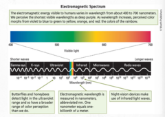
visible light
electromagnetic energy that we see
range of electromagnetic energy visible to humans
400nm violet
to 700nm red
cornea, iris, lens, retina
cornea = clear outer covering
iris = opens and closes to allow in more or less light ; pupil is hole in iris
lens = focuses light, bends to accommodate near and far objects
retina = where light energy initiates neural activity

how the eye works & refractive errors

retina def and functions
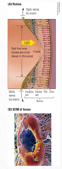
light sensitive surface at the back of the eye consists of neurons & photoreceptors cells,
- translates light into AP
- discriminates wavelengths (colors)
- works in a wide range of light intensities
structure of retina: fovea
- region at center of retina that is specialized for high acuity
- receptive field at center of eye's visual field
- depression at the fovea (light focused)
- where photoreceptors are packed most densely
acuity across visual field?
vision better in center than at margins (hence why we need big letters on our peripheral vision)

structure of retina: blindpsot
- includes region of retina (optic disc) where axons forming the optic nerve leave the eye and where blood vessels enter and leave
- has no photoreceptors
structure of retina: papilledema
- swollen optic disc
- may be due to high intracranial pressure (tumor or brain infection) or inflammation of optic nerve (optic neuritis)
- can cause loss of vision
photoreceptors: rods
More numerous than cones
Sensitive to low levels of light (dim light)
Used mainly for night vision
One type of pigment only

photoreceptors: cones
-Highly responsive to bright light
-Specialized for color and high visual acuity
-In the fovea only
-Three types of pigment

three types of cone pigments & amounts/distribution?
absorb light over range of frequencies but their maximal absorptions are:
419nm (blue or short wavelength)
531nm (green or middle wavelength)
559 (red or long wavelength
equal amount of red/green but FEWER BLUE

CF-2: visual illuminance
- need sufficient light for eye to work
- too little light entering eye produces pb of visual illuminance, in which objects are too dim => hard to see anything
with age, less light allowed through lens / cornea ==> worse vision with age
retinal receptors (mosaic)
retinal mosaic of rods and 3 cone types
near fovea: cones outnumber rods => blue < red (carried on X chromosome)/green

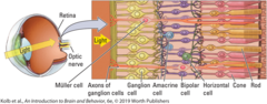
types of retinal neurons
bipolar cell: recieves input from photoreceptors
horizontal cell: links photoreceptors and bipolar cells
amacrine cell: links bipoalr cells and ganglion cells
retinal ganglion cell (RGC): gives rise to the optic nerve
CF-3 Glaucoma
glaucoma: damages optic nerves; most common cause of irreversible blindness
treatment approaches and research (mouse models)

2 types of ganglion cells
Magnocellular cell (M-cell)
• Magno-, large
• Receives input primarily from rods
• Sensitive to light and moving stimuli
Parvocellular cell (P-cell)
• Parvo-, small
• Receives input primarily from cones
• Sensitive to color
optic chiasm
- junction of optic nerves from each eye
- axons from nasal half of each retina cross over to opposite side of brain
- axons from temporal half of each retina remains on same side of brain
=> info from left visual field -> right side of brain and vice versa

3 routes to visual brain
geniculostriate system: projections from retina to lateral geniculate nucleus to visual cortex
tectopulvinar sustem: projections from retina to superior colliculus to pulvinar (thalamus) to parietal and temporal visual areas
retinohypothalamic tract: synapses in tiny suprachiasmatic nucleus in hypothalamus ; roles in regulating circadian rhythms and in pupillary reflex
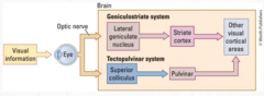
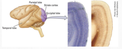
striate cortex
the primary visual cortex
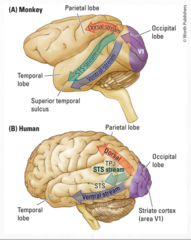
2 visual paths emerge from striate cortex
> one to parietal visual regions
> one to temporal visual regions
dorsal and ventral visual streams
dorsal visual stream: pathway that originates in occipital cortex and projects to parietal cortex
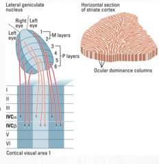
geniculostriate pathway
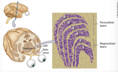
lateral geniuculate nucleus (thalamus)
- right LGN: input from the right half of each retina
- left LGN input fromt he left half of each retina
- 6 layers:
> layer 1, 4, and 6: input from contralateral retina
> layers 2,3 and 5: input from ipsilateral retina
> layes 1 and 2: input from magnocellular cells
> layers 3 to 6: input from parvocellular cells
- P & M retinal ganglion cells send separate pathways to thalamus --> to striate cortex
- same thing for left and right eyes (separate pathways)

tectopulvinar pathway
- magnocellular cells from retina project to superior colliculus, which sends information to pulvinar region of thalamus:
> medial pulvinar => connections to parietal lobe
> lateral pulvinar = sends conections to temporal lobe
occipital cortex
composed of atleast six visual regions
- primary visual cortex (V1; striate cortex) = recieves input from lateral geniculate nucleus
- seondary visual cortex (V2-V5; extrastriate
visual regions of the occipital lobe

heterogeneous layering
Blob (V1) Region in the visual cortex that contains color-sensitive neurons Revealed by staining for cytochrome oxidase
Interblob (V1) Region that separates blobs Participates in the perception of form and motion

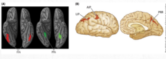
charting the visual streams

Vision Beyond the Occipital Cortex
FFA → faces
PPA → places
what is the "what" and "how" stream?
Ventral = WHAT (object recognition)
Dorsal = HOW (action, movement)
location in the visual world: visual field
- reigon of visual world that is seen by eyes
- divided into left and right fields
- information in the left visaul goes to right hemisphere and vice versa

coding location in the retina
Each retinal ganglion cell (RGC) responds to stimulation on just a small circular patch of the retina—the cell's receptive field.
coding location: light falling on ONE place on retina--> activate ONE ganglion cell
location in the LGN and region V1
• Cells in lateral geniculate nucleus (LGN) also have visual fields.
• Each LGN cell represents a particular place.
• Projects to V1, forming a topographic map
> More cortical tissue is devoted to cells in the fovea than in the periphery
> receptive field in cortex > than retinal ganglion cells

Topographic Organization of Region V1

receptive-field hierarchy

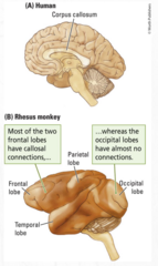
callosal connections: corpus callosum
- corpus callosum connects 2 hemispheres but only specific brain structures
> most frontal lobes connected
> occipital lobes almost no connections: (except cells that lie along midline of visual field connected so that receptive fields overlap)
neuronal activity
As visual information moves through the brain, neurons do different jobs at each stage.
baseline: 12 AP / second
firing rate greater => excitation
firing rate less => inhibition

processing in RGCs: RGCs, on-center cells and off-center cells
RGCs:
- respond only to presence/absence of light, not shape
- concentric circle arrangement: center and surround (periphery)
On-center cells:
- excited when light falls on center portion, inhibited when. falls on surround
- light across whole receptive field => weak excitation
off-center cells:
- OPPOSITE
- => weak inhibition
==> ABOUT CONTRASTS, edge here.... dark there....

overlapping receptive fields
- Neighboring retinal ganglion cells receive their inputs from an overlapping set of photoreceptors
> Receptive fields overlap
- a small spot of light shining --probs-> activity in both on-center and off-center cells

processing in RGCs: luminance contrast
amount of light reflected by object relative to its surroundings ==> we can understand shape

processing shape in primary visual cortex V1
- V1 neurons receive input from multiple RGCs.
- V1 neurons have a much larger receptive field than RGCs.
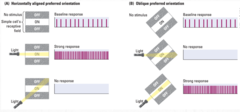
- Cells behave like orientation detectors.
> Excited by bars of light oriented in particular directions
- Simple cells
> Receptive field with a rectangular on-off arrangement
processing shape in V1: complex cells and hypercomplex cells
complex cells: maximally excited by bars of light moving in particular direction through receptive field
hypercomplex:
like complex cells, maximally responsvie to moving bars
have strong inhibitory area at one end of receptive field

V1 receptivity

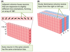
neural circuit in column in visual cortex
sensory input --> cortical column at layer VI
reach stellate c/ in layer IV that synapse with pyramidal c/ in layers III & V
axons of pyramidal cells leave column to join other columns or structures
information flow vertical

organizaiton of functional columns in V1
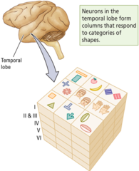
processing shape in TE: complex ft.
- Cells are maximally excited by complex visual stimuli (faces...hands).
> particularly faces seen head-on, viewed in profile, posture of head, specific facial expressions
- complex ft NECESSARY to activate most neurons in TE (orientation, size, color, texture..)
- neurons cluster with similaritt of repsonsiveness to ft.
stimulus equivalence
Recognizing that an object is the same across different viewing orientations
seeing color: subtractive vs additive color mixing
substractive:
obtains entire range of colours in visual world by mixing red/blue/green
additive:
- increases light to make color
- lighter the color => more light it contains

trichromatic theory + limitations
explanation of color vision based on red/green/blue
color perceived
RF-4: color-deficient vision
protanopia (lacking red)
deuteranopia (lacking green)
trichomatic = no lacking

seeing color: opponent-process
opponent-process:
explanation of color vision that emphasizes importance of opposiition of colors: red vs green, blue vs yellow
- occurs in RGCs (on-off, center-surround receptive fields)
opponent-color contrast response at RGCs
center of receptive field = excitatory in some and inhibitory in other
stimulation to periphery has opposite effect
just like on/off light thing in RGCs same thing but with color

seeing color: opponent processes, neurons in V4
do not respond to particular wavelengths but are responsive to various perceived colors
> receptive field center excited by specific color and surround isinhibited
important for color constancy: perceived color is constant relative to other colors regardless of changes in illumination
neuronal activity in dorsal stream: the how-to
Posterior parietal cortex: the how-to stream (for action)
> Neurons in this area are silent to visual stimulation when a person is under anesthesia.
eg. process visual appearance of obj to be grasped
visual brain in action: pathway leading to cortex
injury to pathway leading to cortex
- monocular blindness = destruction of retina or optic nerve of one eye => loss of sight in that eye
- homonymous hemianopia: blindenss of entire left/right visual field
- quadrantanopia: blindness of one quadrant of visual field
- scotoma: small blind spot in visual field caused by small lesion of migraines of visual cortex
IN THE IMAGE ITS V1 BUT FOR MONOCULAR ITS NOT AND FOR THE OTHERS ITS NOT EXCLUSIVELY
injury to what pathway
visual-form agnosia = inability to recognize objects or drawings of objects
color agnosia (achromatopsia) = inability to recognize colors
face agnosia (prosopagnosia) = inability to recognize faces
injury to how pathway
optic ataxia:
> deficit in visual control of reacing & other mvm
> damage to parietal cortex
> retention of ability to recognzie objects normally

plasticity in visual pathways
- manipulation of visual xp during prenatal/early devmt & beyond => profoundly affect visual perception
- people can show remarkably plasticity after injury/loss of visaul pathways
eg. blind individuals and echolocation