Quiz #2 Biochemistry: Chapters 5, 6, and 10
 Call Kai
 Learn
Learn Practice Test
 Spaced Repetition
 Match
Match Flashcards
 Knowt Play
1/100

There's no tags or description
Looks like no tags are added yet.
Name | Mastery | Learn | Test | Matching | Spaced | Call with Kai | Chat |
|---|
No analytics yet
Send a link to your students to track their progress
101 Terms
give some examples of roles of proteins in biological systems
structure, catalysts (enzymes), signal transduction, regulatory, mobility, transport
what determines a proteins specific role
their distinctive structures derived from their amino acid sequence (primary structure)
how do amino acids join?
Join with a peptide bond (covalent bond) between the carboxyl group and the amino group
water is lost within the process

peptide
short polymer of amino acids
dipeptide, tripeptide, 12-20: oligopeptide, many: polypeptide
proteins
a molecule composed of one or more polypeptide chains
homomultimer: one kind of chain, heteromultimer: two or more diff types of chains
simple proteins
proteins that consist of normal amino acids only for their biological function
conjugated proteins
proteins that utilize accessory molecules or chemical groups to carry out their function
prosthetic group
non protein part that is essential to the proteins function
orthologous
proteins from different species with similar sequence and function (common ancestor, for example, hemoglobin in human and cow)
paralogous
proteins from the same species with similar sequence (gene duplication, example: alpha and beta subunits of hemoglobin)
cytochrome c
the electron transport protein found in the mitochondria of all eukaryotic organisms
varies between 103 and 112 amino acids in different species
variation in amino acids between species increases as the phylogenetic difference increases
how do you calculate the net charge and pI of peptides/proteins
consider the n terminus (1st amino acid), and the c terminus (last amino acid) and the side chains of all amino acids between n and c termini
name some common purification techniques
salting in and out, affinity chromatography, ion exchange chromatography, hydrophobic interaction chromatography, gel filtration, etc.
explain salting in and out
salting in: a low concentration of metal salt generally increases protein solubility
salting out: high metal salt concentrations reduce protein solubility
proteins are most insoluble at their isoelectric point
different proteins have different solubilities and therefore require different concentrations of salt to precipitate
when it is no longer soluble in the high salt solution, to precipitates (salting out)

explain dialysis of proteins
allows small molecules and ions to pass through a semipermeable membrane
the macromolecular solution is placed in a semipermeable membrane bag and immersed in a bathing solution
diffusible solutes in the dialysis bag equilibrate across the membrane

explain the chromatographic techniques of protein purification
takes advantage of relative differences in the physical and chemical characteristics of an amino acid, peptide, or protein
the molecule of interest flows through a medium with two phases (solid-liquid, liquid-liquid, or gas-liquid) and partitions between them
explain ion-exchange chromatography
depends on the net charge of the protein
the proteins net charge will depend on the pI of the protein and the pH of the solution in which the protein is (pI of a protein is the pH at which its net charge is 0)
remember: pH> pI overall charge is neg. , pH< pI overall charge is positive
explain the types of protein chromatography
ion-exchange: postivie or negative charged ions
hydrophobic interaction: polarity or hydrophobicity of molecules
affinity: differential affinity of one molecule for others
gel filtration, permeation or size exclusion: molecular size of the molecules
explain gel filtration or size exclusion chromatography
proteins are separated based on their size
column media are composed of beads on varying pore sizes
larger molecules are excluded from the gel beads and emerge from the column sooner than smaller molecules, whose migration is retarded because they can enter the beads

explain affinity chromatography
based on the ability of the protein to bind to a ligand
a small molecule (ligand) is immobilized on a matrix
the protein of interest is then passed through the column, which binds to the target ligand, while other proteins pass through without binding
explain proteins analysis by SDS- PAGE, specifically SDS
SDS molecules disrupt non-covalent interactions that stabilize the tertiary ( or quaternary) structure and denature the protein
SDS wraps itself around the protein backbone and proteins become rod-like
SDS binds at a constant weight ratio of 1.4 g SDS/g of polypeptide through hydrophobic interactions
this way, proteins will have a uniform charge
larger proteins bind more SDS than smaller molecules

explain protein analysis by SDS- PAGE, specifically PAGE
the electrophoretic mobility of shift assay (EMSA) is performed on a polyacrylamide gel (PAGE)
an electric field is applied to the gel to separate proteins
anions = anode (+), cations= cathode (-)
migration is a function of size (smaller go farther than bigger)

how do you measure peptide and protein concentration
proteins and peptides absorb at 215 nm (peptide backbone) amd at 280 nm (specific aromatic side chains)
calculate measuring absorbance at 280 nm and using Beer- Lambert law
A= ecl
explain analysis of peptides and proteins using mass spectrometry (MS)
exploit differences in the mass-to-charge ratio (m/z) of ionized atoms or molecules to separate them from each other
evaporate and ionize the molecules in a vacuum
separate the ions in space and/ or time based on their m/z ratio
measure the number of ions with specific m/z ratios

explain how we analyze peptides and proteins using sequencing
2 ways:
direct amino acid sequencing
sequencing the corresponding DNA in the gene
what are the 6 steps for determining the amino acid sequence of a protein
separation of the polypeptide bonds (if a heteromultimer)
cleavage of the disulfide bridges
analysis of N and C terminals
fragmentation of the polypeptide chain
reconstruction of the amino acid sequence
explain step 1 : separation of polypeptide chains
polypeptide separation is achieved with 8 M Urea, 6 M guanidinium HCl, extreme pH or high salt concentration
if the protein is a heteromultimer, chromatographic methods that can then separate the different chains for individual sequencing
explain step 2: breaking of the disulfide bonds
2 options:
performic acid oxidation
reduction with mercaptoethanol (BME) or dithiothreitol (DTT)
followed by treatment with alkylating agents that modify the -SH groups to prevent recombination of disulfides
explain step 3: analysis of the N- terminus
Edman degradation allows sequential identification of a series of residues starting at the N- terminus
chromatographic techniques are used to identify the PTH- derivative
explain step 4: analysis of the C- terminus
carboxypeptidases cleave the amino acid residues from the C- termini of polypeptides
different carboxypeptidases cleave different amino acid residues
carboxypeptidase A : all residues except Pro, Arg, Asp, Glu, and Lys
carboxypeptidase B : Arg and Lys residues only
carboxypeptidase Y: any residues
c terminal residue is removed enzymatically with the carboxypeptidase first and then reacted with phenyl isothiocyanate to produce the PTH derivative
explain step 5: fragemnataion of the polypeptide
enzymatic fragmentation: trypsin, chymotrypsin, clostripain, staphylococcal proteases
chemical fragmentation: cyanogen bromide
trypsin class on the carboy side of Arg and Lys
generates a set of peptide fragments having either Arg or Lys at their C- termini
chymotrypsin cleaves on the carboxy side of the Phe, Try, and Trp

explain step 5 : fragmentation of the polypeptide
cyanogen bromide (CNBr) reacts with methionine residues and cleaves the peptide bond between the Met and the next amino acid
explain step 6: construction of the complete sequence
compare the sequences of fragments obtained from two or more cleavage procedures
sequence all the peptides produced ( usually by Edman degradation)
the goal here is to find overlapping sequences
align peptides from different fragmentations to reveal the overall amino acid sequence
name some other biomolecules that proteins interact with:
other proteins
nucleic acids (DNA and RNA) to form nucleoprotein complexes
small molecules (metabolites)
metal ions
explain the strength of protein-ligand interactions: dissociation constant (Kd)
Kd=[P][L]/[PL] —> v= [L]/ (Kd+ [L]) where v is the fractional saturation of P with L, [PL]/([P]+[PL])
when v= 0.5, Kd = [L]
i.e., Kd is equal to the concentration of L when half the protein is bound to L
smaller kd indicates better affinity of ligand for protein (typical range - 10^-3 M to 10^-12 M)
name some of the overarching principles link the 3 dimensional structures of proteins
function depends on structure
structure depends on the sequence and non covalent forces
number of protein folding patterns is significant
what bonds form the primary structure of a protein?
covalent bonds
explain hydrogen bonding in proteins
both between atoms in the peptide backbone and between side chains
usually on the surface of proteins, but can also exist on the interior
12-30 kj/mol
explain ionic interactions in proteins
generally located on the surface of proteins
20 kj/mol
explain hydrophobic interactions in proteins
primarily found in the interior of proteins (drives protein folding)
<40 kj/mol
explain van der waals interaction
van der Waals interaction are ubiquitous (appearing everywhere at the same time)
<0.4 - 4 kj/mol
primary structure
the amino acid sequence
secondary structure
organization of amino acids into structures through hydrogen bonds
amide H of one peptide group and the carbonyl O of another
allows the protein to form regular structures
tertiary structure
3- dimensional organization of secondary structures
quantenary structure
organization of several proteins, subunits
explain the amide plane for higher-level protein structures
the peptide bond is planar
rotation is allowed around 2 bonds
the bond linking the alpha carbon with its carbonyl carbon and the bond linking the alpha carbon with the amide nitrogen
the angle for the alpha carbon and the carbonyl carbon is denoted by psi, and the alpha carbon and amide is phi

what are some unfavorable combinations of phi and psi?
0 and 180 for either
0 and 0
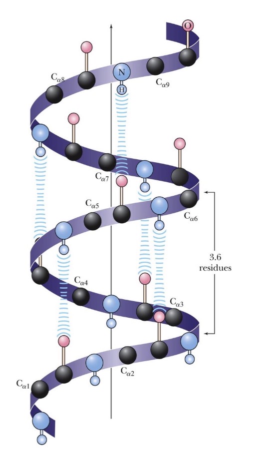
explain alpha helices in secondary structures
alpha helices:
upiquituous component of proteins
stabilized by H- bonds
phi: -60, psi: -45
residue n forms h-bond with the n+4 residue
residues per turn= 3.6
each amino acid extends 1.5 Å along the helix axis
therefore rise per turn= 3.6 * 1.5 Å = 5.4 Å
each peptides bond possess a dipole, meaning the alpha helix itself has a significant dipole moment
In a typical 𝛼-helix of n residues, there are n-4 hydrogen bonds
explain beta strands and sheets in secondary structures
beta strands form when amino acids adopt phi of -120 and psi of 120
beta strands do not exist standalone because the can’t be stand alone
to stabilize, another stretch amino acids should form a beta strand with which the first strand can interact
R groups of consecutive amino acids are on opposite sides of the sheet
beta strands are usually represented by arrows with the arrowhead indicating the direction from N- terminus to C- terminus
at least 5 strands in parallel are needed to be stable whereas antiparallel sheets are stable with 2
parallel rise per residue= 3.25 Å, antiparallel rise per residue= 3.47 Å

what connects alpha helices and beta sheets?
loops
can be divided into structured and unstructured
explain beta turn (or bend) formation
the peptide chain forms a loop with the carbonyl oxygen of one amino acid forming a hydrogen bond with the amide hydrogen of the residue 3 residues down the chain
4 resides are required for a beta turn

explain the tertiary structure of proteins
the arrangement of a single polypeptide chain in 3-dimensional space
proteins fold to form the most stable structure possible, that arises from: the formation of many intramolecular hydrogen bonds and the reduction in the surface area accessible to the solvent that occurs upon folding
proteins are usually a mixture of hydrophilic and hydrophobic elements
the hydrophobic elements usually cluster together in the folded interior of the protein

what are the 3 main classes of biological proteins?
fibrous proteins: simple, linear structure, insoluble in water
globular proteins: spherical in structure, soluble in water
membrane proteins: hydrophobic exterior, insoluble in water
describe fibrous proteins
consist of polypeptide chains organized along a single axis, producing long fibers
tend to be mechanically strong and play a structural role in nature
ex. alpha keratin, fibroin, collagen
explain key characteristics of alpha keratin
predominant constituents of claws, fingernails, hair, etc. in mammals
alpha helical segments dominate their structure
the sequence consists of 311-314- residue- long alpha helical rod segments capped with non-helical N and C termini
the primary structure of helical rods consists of 7 residue repeats (a-b-c-d-e-f-g)n, where a and d are non polar
this structure promotes helix association to form coiled coils
explain key characteristics of fibroin
found in silk fibers and bird feathers
form extensive beta sheets with an alternating sequence Gly-Ala/Ser-Gly-Ala/Ser...
explain key characteristics of collagen
principle constituent of connective tissues
very high proline content
1 out of 3 residues is glycine, forming long stretches of the polypeptide chain consisting of Gly-Pro-Pro repeats
the unusual amino acid composition of collagen is not suitable for alpha helices and beta sheets
it is ideally suited for the collage triple helix: three intertwined helical strands
explain globular proteins
exist in an enormous variety of 3D structures
contain large amounts of alpha helices and beta sheets folded into a compact structure
both polar and non polar interactions stabilize the tertiary structure
include enzymes and the proteins involved in immune and signaling responses
describe bovine ribonuclease A
example of a globular protein
The space between the helices and sheets in the interior of the protein is tightly filled with residues that have mostly hydrophobic side chains
most polar side chains face the outside on the structure and interact with water
how does water play a critical role in globular proteins structures?
there are usually several water molecules per amino acid residue
the polar backbone and side chain groups on the protein surface make h- bonds with solvent water
relatively few water molecules are found inside the protein
what are the protein groups based on secondary structure arrangements?
All alpha groups, in which alpha helices predominate
all beta groups, in which beta sheets predominate
alpha/beta proteins, in which alpha helices and beta sheets are intermingled
alpha + beta proteins, which contain separate alpha helical and beta sheet domains
explain the quantenary structure of proteins
used in cases where proteins are composed of two or more subunits
stabilized by hydrophobic interactions, hydrogen bonding, electrostatic interactions, and, in certain cases, disulfide covalent bonds and interactions with metal ions
how does a protein fold into its functional form: randomly or with some pattern?
the amino acid sequence contains all the information required to fold a polypeptide into its native structure
study of denaturation and renaturation of proteins in the 50’s confirmed this
solutions of ribonuclease were treated with: urea (unfolded the protein) or BME (reduced the disulfide bridges)
explain levinthals paradox
a typical protein can adopt so many conformations that it does not have enough time to reach its most stable state by sampling all possible configurations
therefore proteins must fold by specific folding pathways
explain folding tendencies and patterns in globular proteins
globular proteins adopt the most stable tertiary structure possible by:
satisfying the constraints inherent in their own structure
folding to bury the hydrophobic side chains
polypeptide chains tend to twist slightly in a right-handed direction
explain natural proteins being found multimeric
proteins with 2 or 4 subunits predominate in nature (dimeric or tetrameric forms)
The typical Kd for two subunits: 10−8 to 10−16 M
These values correspond to energies of 50 to 100 kJ/mol at 37° C
entropy loss due to association: unfavorable
entropy gain due to burying of hydrophobic groups: very favorable!
describe intrinsically unstructured proteins (IUP’s)
many proteins exist and function normally in a partially unfolded state
adopt well defined structures in complexes with their target proteins
are characterized by an abundance of polar residues and a lack of hydrophobic residues
what is an example of an IUP?
p53 is a tumor supressor gene
consists of: an N terminal disordered domain of 93 residues, a central DNA binding domain of 200 residues, and a C terminal disordered domain of 100 residues
n terminal binds to over 40 proteins, c terminal binds to over 50 proteins
denaturation
the loss of protein structure and function
weak, non covalent forces maintain the secondary, tertiary, and quantenary structures
a variety of environmental stressors can disrupt these forces
explain denaturation of proteins by heat
as a protein is heated, it maintains its native state until it approaches its unique melting temperature (Tm)
above this temp., the structure unfold and function is lost
explain denaturation of proteins by chemicals
proteins can be denatured by: acids, bases, detergents
denaturation involves disruption of the weak forces that stabilize proteins
the covalent bonds are not affected
explain the central dogma
the flow of genetic information: DNA to RNA to protein
unidirectional and irreversible
replicate, transcribe, translate
nucleotides
the building blocks for nucleic acids
nucleic acids are polymers of nucleotides, the nitrogenous base can vary from nucleotide to nucleotide

pyrimidines
six-membered, heterolytic aromatic rings
planar
cytosine, thymine, and uracil
purines
consists of two heterolytic rings (six and five membered)
slight pucker between the pyrimidines and imidazole portions
adenine and guanine
tautomers
constitutional isomers (two molecules with the same molecular formula) but with different connectivity
can interconvert in a rapid equilibrium

explain what tautomers are dominant at physiological pH
keto tautomers of thymine and guanine predominate
amino tautomers of cytosine and adenine predominate

explain nucleobases UV light absorption
purines and pyrimidines strongly absorb UV light because of their aromaticity
at or near 260nm
describe the structure of a nucleoside
nucleobase plus a pentose sugar
nucleosides are compounds formed when a base is linked to a sugar via a glycosidic bond (C-N)
can have a ribose or deoxyribose sugar
OH vs. H
2 OH found in DNA

name some of the most common naturally occurring ribonucleosides
pyrimidines: cytidine, thymidine, uridine
purines: adenosine, guanosine
describe the structure of a nucleotide
a nucleoside plus a phosphate group
most nucleotides have 5’ phosphate groups
the nucleotides can have multiple phosphate groups
what role do nucleoside 5’ triphosphates play in cells?
indispensable agents in metabolism because their phosphoric anhydride bonds are a source of chemical energy
the cyclic nucleotides are signaling molecules and regulators of cellular metabolism and reproduction
examples
ATP is central to energy metabolism
GTP drives proteins synthesis
CTP drives lipid synthesis
explain what nucleic acids are composed of
linear polymers of nucleotides linked 3’ to 5’ by phosphodiester bridges
they are formed as 5’ nucleoside monophosphate are added to the 3’ OH group of the preceding nucleotide
you read the chains from the 5’ to the 3’ end
explain the structure of DNA
DNA consists of two polynucleotide strands (helices)
the strands are antiparallel
they are held together via the hydrogen bonds of the base pairs
A=T two h-bonds, g-c three h-bonds
the polar sugar-phosphate backbones of the chains are on the outside
who found the structure of DNA?
Watson, Crick, Wilkins, Franklin
explain the path to discovery of DNA
Chargaff examined the base composition of various DNAs but didn’t explain the implications
the four bases did not occur in equimolar amounts
number of purines is always equal to the number of pyrimidines
rosaland franklins x ray fiber diffraction data were crucial
crick showed that it was a helix
Watson found that the base bair h-bonding was specific
explain what determines base paring
large bases pair with small bases
the A-T base pair and the G-C base pairings have virtually identical dimensions
explain Hoogsteen base pairs
bases do not always follow Watson-crick base pairnigs
the purine n7 is a h-bond acceptor and can thus form h-bonds with other donors
the functional groups of A and G that participate in wanton-crick base paring remain accessible in hoogsteen base pairs

explain non-canonical paring
bases can form triples such as TAT and CGC
each purine interacts with one of its pyrimidine partners via a hoogsteen base pairing and the other through a Watson-crick base pairing

what structures can guanine and cytosine nucleobases form?
four guanine bases can form a quadraplex structure due to hoogsteen h-bonds
cytosines can form c+:c pairs at lower pH

explain the difference between DNA and RNA building blocks
DNA contains 2-deoxyribose instead of ribose in RNA
DNA contains thymine instead of uracil in RNA
ribo and deoxyribonucleotides form different conformations
explain the structure of naturally occurring RNA
usually single stranded
more degrees of freedom per nucleotide meaning they have many more conformational possibilities than DNA
however, RNA is rich in double stranded regions that form when complementary sequences within the same chain for intrastrand base pairs
explain why RNA is less chemically stable than DNA
RNA is susceptible to alkaline hydrolysis
OH- acts as a nucleophile and removes a proton from the 2’ OH group
then the 2’O- attacks the phosphodiester bond
leads to the cleavage of the sugar phosphate backbone

explain nucleases and their role
enzymes that hydrolyze nucleic acids
these enzymes are technically phosphodiesterases because they catalyze the cleavage of phosphodiester bonds
because each phosphate in a nucleotide backbone is involved in two phosphodiester linkages, cleavage can technically occur on either side of the phosphorus
name some of the types of nucleases that cleave DNA and RNA
endonuclease: cleave within an olionculeeotide chain
exonucleases: cleave at the end of a polynucleotide chain, can cleave at either the 5’ or 3’ end
Rnase: nuclease specific for RNA
DNase: nuclease specific for DNA
explain how restriction enzyme (DNase) cleaves dsDNA
restriction enzymes are enzymes purified from bacteria that can cleave dsDNA
type 1: require ATP to hydrolyze DNA, catalyze chemical modification of DNA, cleave DNA randomly
type 2: do not require ATP but require Mg2+, do not modify the DNA, cleave at specific sites depending on the DNA sequence
type 3: require ATP, recognize specific nucleotides and cleave at or near the site, catalyze chemical modification of DNA
explain why type 2 restrictive enzymes are very handy in biology
widespread use as a tool in cloning
recognizes a specific DNA sequence and cuts the phosphate backbone at specific sites at or near DNA sequence
recognition site is typically 4-6 bp in length
about 3000 restriction enzymes have been characterized

how do we name restriction enzymes
first is a capital letter denoting the genus of the organism of origin
the next two letters are an abbreviation of the species
if there is more than one strain, then the next will be a capital letter to denote the strain
last there is a roman numeral to indicate the order of purification from a given species (strain)
example: E means genus Escherchia, co means species is coli, R means the strain is RY13, I means first identified from this strain
EcoRI
explain agarose gel electrophoresis
very common DNA analysis method
the gel is submerged in the electrophoresis buffer
DNA is loaded into the wells of the agarose gel
agarose gel separates DNA as a function of size
larger DNA fragments move more slowly through the gel than the smaller fragments
how do we analyze agarose gel electrophoresis
markers: commercially available solutions contain DNA of different lengths and amounts to be used as standards
DNA bands can be visualized by: labeling the DNA ends with 32P (radioactive) and exposing it to photographic film or a phosphor imaging plate, which is highly sensitive, ethidium bromide: intercalates in DNA, and fluoresces when exposed to UV light, DNA is not affected by running on a gel: it can be extracted and used (purification)
