Anatomy Lab Practical 1
 Call Kai
 Learn
 Practice Test
 Spaced Repetition
 Match
 Flashcards
 Knowt Play
1/743

There's no tags or description
Looks like no tags are added yet.
Name | Mastery | Learn | Test | Matching | Spaced | Call with Kai | Chat |
|---|
No analytics yet
Send a link to your students to track their progress
744 Terms

Skin
Skin Information
Epidermis over dermis; subcutis lies deep to the skin.
Skin Function
Barrier, sensation, protection, and temperature regulation.

Hair coat
Hair coat Info
Keratinized hair shafts arise from follicles in the dermis.
Hair coat Function
Insulation and protection.

Compound hair coat
Compound hair coat Info
Several hairs emerge through a common follicular opening; coarse primary hairs accompany finer secondary hairs.
Compound hair coat Function
Combines protective guard hairs with insulating undercoat.

Tactile hairs (vibrissae)
Tactile hairs (vibrissae) Info
Specialized large hairs with richly innervated follicular sinus complexes, especially on face.
Tactile hairs (vibrissae) Function
Detect contact and nearby objects.

Superficial fascia (subcutaneous tissue; hypodermis)
Superficial fascia (subcutaneous tissue; hypodermis) Info
Loose connective tissue and variable fat beneath dermis; contains superficial vessels, nerves, and cutaneous muscles.
Superficial fascia (subcutaneous tissue; hypodermis) Function
Permits skin mobility, cushions, and stores energy.

Deep fascia
Deep fascia Info
Dense connective tissue investing muscles and forming compartments and septa.
Deep fascia Function
Supports muscle groups and transmits force.

Cutaneous trunci muscle
Cutaneous trunci muscle Origin
Superficial fascia of the trunk; broad sheet rather than a single focal bony origin.
Cutaneous trunci muscle Insertion
Axillary and proximal brachial fascia.
Cutaneous trunci muscle Action
Twitches the skin in the cutaneous trunci reflex.
Cutaneous trunci muscle Innervation
Lateral thoracic nerve.
Cutaneous omobrachial muscle Origin
Superficial fascia over the shoulder and brachium.
Cutaneous omobrachial muscle Insertion
Adjacent skin and superficial fascia.
Cutaneous omobrachial muscle Action
Moves the skin over the shoulder and arm.
Cutaneous omobrachial muscle Innervation
Axillary nerve.

Interdigital skin
Interdigital skin Info
Skin between digits, including the web spaces.
Interdigital skin Function
Flexible barrier; inspect for foreign bodies and inflammation.

Claw wall (unguis)
Claw wall (unguis) Info
Hard keratin shell over distal phalanx and claw bed.
Claw wall (unguis) Function
Protects the distal digit and provides traction.

Claw tip (distal free edge/apex)
Claw tip (distal free edge/apex) Info
Unattached distal end of the claw wall beyond its supporting bed.
Claw tip (distal free edge/apex) Function
Traction and wear surface; trimming must avoid vascular living tissues.

Claw fold (periungual fold)
Claw fold (periungual fold) Info
Skin fold surrounding proximal and lateral base of claw.
Claw fold (periungual fold) Function
Protects the emerging claw and its growth region.
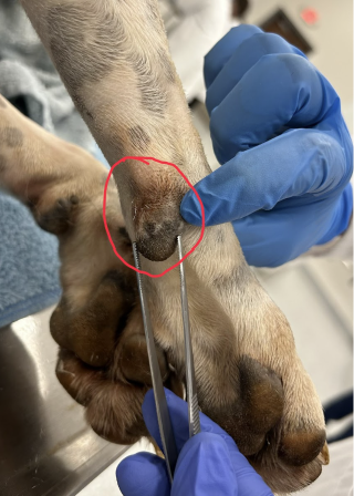
Carpal pad
Carpal pad Info
Hairless keratinized pad on caudal distal forelimb near carpus.
Carpal pad Function
Additional traction during braking; usually not weight bearing in normal stance.

Metacarpal pad
Metacarpal pad Info
Large central palmar forepaw pad; thick epidermis over fibroelastic fatty tissue.
Metacarpal pad Function
Weight bearing, shock absorption, and traction.
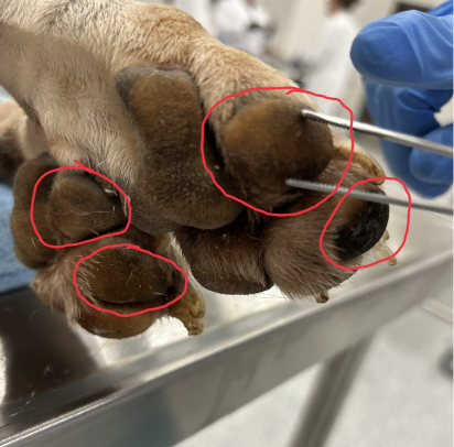
Digital pads
Digital pads Info
Palmar or plantar cushions associated with individual digits.
Digital pads Function
Protect digits and distribute loading.

Nasal planum
Nasal planum Info
Hairless modified skin of external nose.
Nasal planum Function
Protection and tactile sensation; receives moisture from nearby secretions.

Lips and oral commissure
Lips and oral commissure Info
Lips combine external skin, muscle, and oral mucosa; commissure is the junction at the mouth corner.
Lips and oral commissure Function
Retain food and protect oral opening; facial nerve controls labial muscles and trigeminal branches provide sensation.
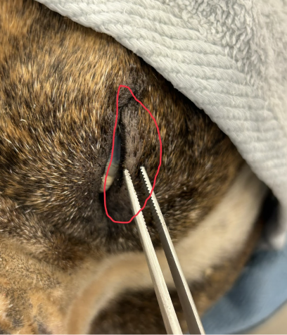
Eyelids
Eyelids Info
Mobile skin folds with conjunctiva internally and tarsal glands in their margins.
Eyelids Function
Protect cornea and spread tear film; facial nerve closes lids, oculomotor nerve elevates upper lid, trigeminal branches carry sensation.
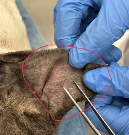
External ear (pinna)
External ear (pinna) Info
Skin-covered elastic auricular cartilage directing sound toward the external acoustic meatus.
External ear (pinna) Function
Sound collection and communication; auricular muscles are supplied by facial nerve.
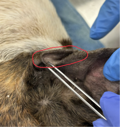
Cutaneous marginal pouch of external ear (Henry's pocket)
Cutaneous marginal pouch of external ear (Henry's pocket) Info
Fold or pouch along the caudolateral margin of pinna.
Cutaneous marginal pouch of external ear (Henry's pocket) Function
Normal identification landmark; specific function is not established.

Superficial Pectoral

Superficial Pectoral
Superficial Pectoral Origin
First two sternebrae and median fibrous raphe; lab manual also allows part of the third sternebra.
Superficial Pectoral Insertion
Entire crest of the greater tubercle of the humerus.
Superficial Pectoral Action
Adducts the free limb; prevents abduction during weight bearing.
Superficial Pectoral Innervation
Cranial pectoral nerves, ventral rami C7–C8.
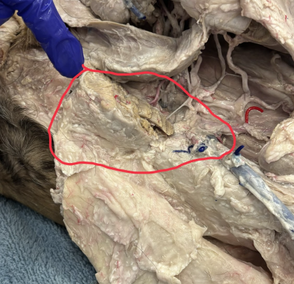
Deep Pectoral

Deep Pectoral
Deep Pectoral Origin
Ventral sternum, median raphe, and associated abdominal fascia.
Deep Pectoral Insertion
Lesser and greater tubercles of the humerus.
Deep Pectoral Action
Adducts and retracts the free limb; draws the trunk forward and extends the shoulder when the advanced limb is fixed.
Deep Pectoral Innervation
Caudal pectoral nerves, principally C8–T1.

Brachiocephalicus

Clavicular intersection of brachiocephalicus
Brachiocephalicus and clavicular intersection Origin
Clavicular intersection is the fibrous junction between cleidobrachialis and cleidocephalicus; it contains a clavicular vestige in the dog.
Brachiocephalicus and clavicular intersection Insertion
Cleidobrachialis attaches to the humeral crest; cleidocephalicus attaches to the mastoid process and middorsal cervical raphe.
Brachiocephalicus and clavicular intersection Action
Advances the limb and extends the shoulder; with the limb fixed, bends the head and neck to the side.
Brachiocephalicus and clavicular intersection Innervation
Brachial part: brachiocephalic nerve from cervical ventral rami; cephalic part: accessory nerve and cervical branches.
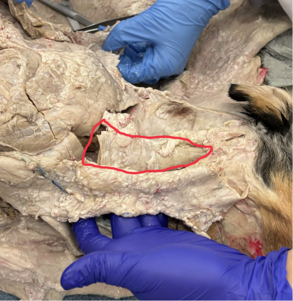
Omotransversarius
Omotransversarius Origin
Wing of the atlas.
Omotransversarius Insertion
Distal scapular spine and acromion.
Omotransversarius Action
Advances the limb; bends the neck laterally with the limb fixed.
Omotransversarius Innervation
Accessory nerve. Dyce lists scapula as origin and atlas as insertion; attachments are the same.
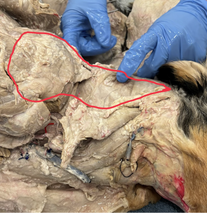
Trapezius
Trapezius Origin
Cervical part: median neck raphe; thoracic part: supraspinous ligament over cranial thoracic vertebrae, approximately T1–T9.
Trapezius Insertion
Scapular spine.
Trapezius Action
Elevates and abducts the limb; cervical and thoracic portions move the scapula cranially and caudally, respectively.
Trapezius Innervation
Accessory nerve.
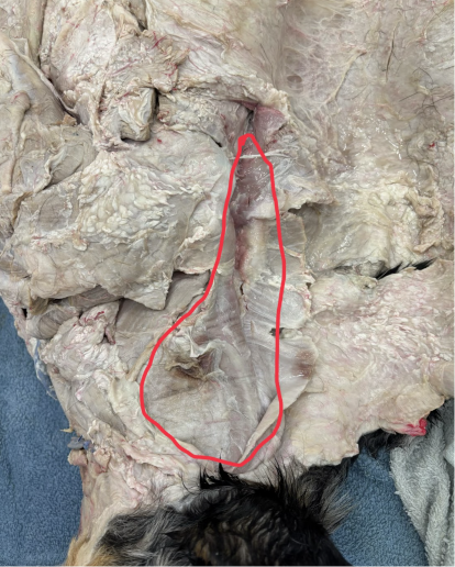
Rhomboideus

Rhomboideus capitis (yellow), cervicis (blue), thoracis (red)
Rhomboideus Origin
Capitis: nuchal crest; Cervicis: neck raphe; Thoracis: cranial thoracic spinous processes and supraspinous ligament.
Rhomboideus Insertion
Dorsal border and adjacent medial surface of the scapula.
Rhomboideus Action
Elevates the limb and holds the scapula against the thorax.
Rhomboideus Innervation
Cervical and thoracic spinal nerve branches; Dyce canine Table 16.2 specifies ventral branches.
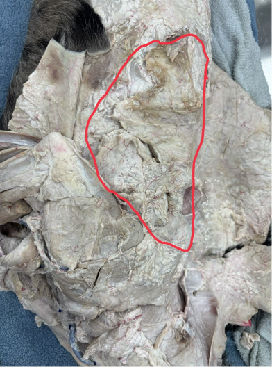
Latissimus dorsi
Latissimus dorsi Origin
Thoracolumbar fascia over caudal thoracic and lumbar vertebrae; attachments to caudal ribs.