BIO 112: Unit 2 Macromolecules the building blocks of the cell
 Call Kai
 Learn
 Practice Test
 Spaced Repetition
 Match
 Flashcards
 Knowt Play
1/49

There's no tags or description
Looks like no tags are added yet.
50 Terms
Covalent bonds
Molecules are formed when atoms share electrons
Strong and permanent
Two types: Polar and non-polar
Polar Covalent bonds
Electrons are not evenly shared: creates a partial charge
Large difference in electronegativity
The molecule has a Permanent Dipole (PD)
O, N, S (high electronegativity) bonded with C, H, P (low electronegativity)
Non-Polar Covalent bonds
Electrons are shared evenly
Very little difference in electronegativity
Common examples: C-H, C-C
A partial charge can be induced: Induced Dipole (ID)
Aside:
Hydrophobic molecules are non-polar because non-polar species lack partial charges and they can’t form hydrogen bonds
Non-covalent interactions
Molecules can interact with each other through electrostatic interactions: no shared electrons
Weaker than covalent bonds
Strongest to weakest of the non-covalent interactions

What is a hydrogen bond?
Permanent Dipole- Permanent Dipole but the partial positive charge is on the Hydrogen atom
Hydrogen can be bonded to O or N
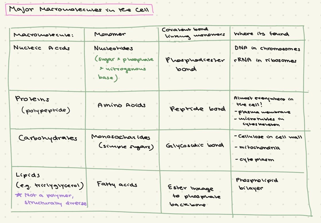
What are the 4 main catagories of macromolecules in the cell?
List their:
Monomer
The name of the covalent bond that links monomers
Where its found
Describe the difference between Hydrophobic, Hydrophilic, and Amphipathic molecules

Why doesn’t directionality and polarity apply to Lipids?

What is polarity and directionality in macromolecules?
Polarity:
A structural property
comes from the asymmetric structure of the repeating monomer units and how they are linked together
Each end of the monomers and polymer chain are chemically distinct from the other (causes polarity)
Directionality:
as a result of having polarity
the chain can only be built and read in one direction
It allows cells, enzymes, and replication machinery to navigate, read, or synethize the polymer in a specific sequential orientation
How do Nucleic Acids exhibit directionality

How do polypeptides exhibit directionality?

Describe how the cell membrane is both a container and a semipermeable barrier
Container:
The hydrophobic (non-polar) interior blocks large, charged, and polar molecules keeping the cell’s contents in and the outside out
Semipermeable:
The core lets small, non-polar molecules pass freely
Large, charged, and polar molecules need membrane proteins to cross
What formations do phospholipids in water spontaneously form?

What is the hydrophobic effect?
The observation that non-polar molecules aggregate (cluster/group together) in aqueous solutions
It is energetically favourable and therefore spontaneous because the water movement is less constrained
Explain the thermodynamic process (hydrophobic effect) driving the assembly of lipids into membranes
Lipids form into membranes because it increases the entropy of the surrounding water molecules, not because the tails strongly attract each other
Individually each lipid molecule forces surrounding water molecules to become highly ordered (unfavourable decrease in entropy)
When lipids aggregate together fewer molecules are associated with lipids (favourable increase in entropy)

Why do phospholipids form bilayers?
Hydrophobic effect
It is energetically favourable
▵G < 0 (its spontaneous)
Lower energy = more stable
Describe the amphipathic nature of phospholipids
The hydrophilic head is made up of polar and charged molecules (phosphate charged, glycerol is polar)
The hydrophobic tail is made up of non-polar covalent bonds (C-C and C-H bonds)
Explain how proteins are embedded in or interact with membrane bilayers.
Integral membrane proteins have non-polar functional groups which stick out into the hydrophobic membrane and interact non-covalently
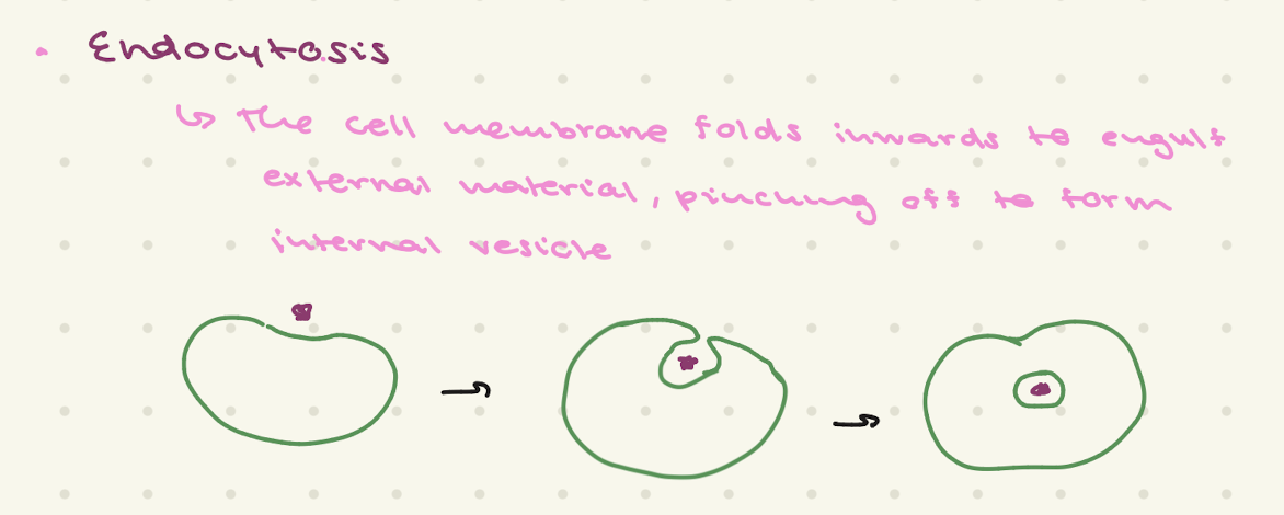
Endocytosis
Exocytosis

Why is it important for the lipid bilayer to be selectively permeable
Required to maintain homeostasis (stable internal environment)
Cells must be able to control the movement of molecules across the membrane: it needs nutrients
What are important molecules cells uptake

What are the types of Membrane Transport?
Passive Transport (doesn’t require ATP): Diffusion, and facilitated diffusion
Active Transport (Requires ATP): Primary and Secondary Active Transport
Simple Diffusion
Net movement of particles directly through the lipid bilayer from high to low concentration
Particles move down their concentration gradients
At equilibrium there is no net movement, but random movement does not stop
It is energetically favourable for molecules to diffuse down their concentration gradient: Results in increased entropy
Facilitated Diffusion and two types of transporters
Proteins transport large, charged, and polar molecules down their concentration gradients
There are two types of proteins: Channels and Carriers
Channel Proteins
Used for facilitated diffusion
Channels are selective for a specific type of molecule
Aids molecules moving down their concentration gradient
Channel is lined with polar amino acids: provides a hydrophilic channel for the movement of molecules
Channels can be leak (slow movement of ions) or gated (open or close due to signal)
Carrier proteins
Used for facilitated diffusion
Carrier proteins are “gated”
Transports a specific type of molecule
Protein undergoes conformational change to open and close the carrier
Active Transport
Movement of molecules across a membrane from a region of lower concentration to higher concentration
Goes against the concentration gradient using cellular energy
Primary Active Transport
Uses a chemical energy source (ATP) to pump molecules across the membrane
Common example: Na/K pump —> Na moves out of cell, K moves in both against their concentration gradients

Secondary Active Transport
Uses an electrochemical gradient previously created by primary transport instead of ATP
One solute goes DOWN it concentration gradient as another solute is transport UP its gradient

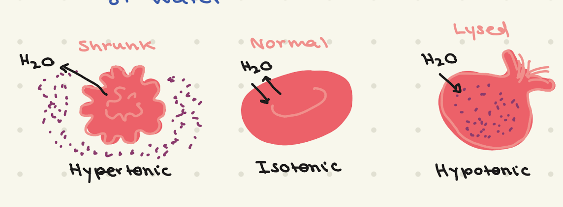
Osmosis
Movement of water from a region of low solute concentration to high solute concentration
Driven by pressure, not the diffusion of water
During the formation of a peptide bond (polymerization), what molecule is a byproduct?
h2o

Peptide
short chain of amino acids < 50
Polypeptide
chain of amino acids >50
Protein
chain of amino acids folded into a tertiary structure
Amino Acid Residue
what remains of the amino acid after peptide bond formation (h2o is a byproduct)
What occurs in the primary stage of protein folding
Linear chain of amino acids form
Covalent peptide bonds form between amino (N) and carboxyl (C) ends

What occurs in the secondary stage of protein folding
Two types of structures form:
Alpha Helices
Beta Sheets
Non-covalent hydrogen bonds between the backbone atoms

What occurs in the tertiary stage of protein folding
3D shape of a single polypeptide protein
Determines the proteins function
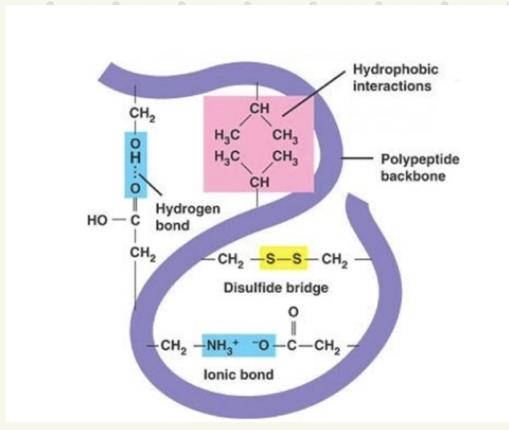
Many types of interactions occur:
Covalent bonds between R groups (Disulphide bridge from cysteine R groups)
Non-covalent bonds between R groups and backbone (ionic interactions, hydrogen bonds)
Non-covalent bonds between R groups (hydrophobic, ionic, hydrogen bonds, ID-ID, PD-ID, PD-PD
What occurs in the quaternary stage of protein folding
interactions between one of more proteins
Types of interactions:
Hydrophobic: nonpolar side chains cluster together away from water—> ID-ID
Hydrogen Bonds
Ionic (salt) bridges
Protein Denaturation
A process that changes a protein’s shape, destroying its function
Its due to the disruption of non-covalent interactions
Causes: changing pH, temperature, chemicals (urea)
Most proteins cannot “re-nature” and do not re-gain function
RNAase A is an exception
Strong interactions (covalent) between side chains are more difficult to denature
What is the driving force of protein folding
Thermodynamics:
(△G= △H - (T△S)
Bond formation releases energy making △H (enthalpy) negative
The favourable release of heat/bond formation is the primary driving force: it overcomes the unfavourable loss of entropy due to ordered protein folding

How is the Hydrophobic effect related to Protein Folding

Explain the Hydrophobic effect in relation to DNA
Charged phosphates: Hydrophilic Backbone stays on the outside
Hydrophobic Nitrogenous bases go inwards
Tucking the bases inwards increases entropy: favourable
Base Stacking Interactions
Bases have planar molecular shape: they stack vertically on top of each other
Provides vertical stability: keeps the double helix rigid and secure
(Hydrogen Bonding between bases)
Chargaff’s Rule
The amount of:
A=T
G=C
Therefore the total number of purines always equals total number of pyrimidines
A+G=T+C
It proves that bases don’t pair randomly, but form specific complimentary pairs
Purines
Adenine and Guanine
Pyrimidines
Thymine and Cytosine
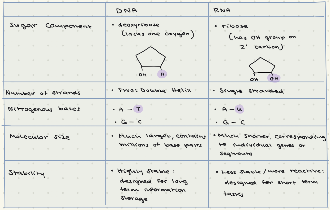
Differences in DNA and RNA