TDP: 1st Practical (Integumentary)
 Call Kai
 Learn
Learn Practice Test
 Spaced Repetition
 Match
Match Flashcards
 Knowt Play
1/65

Earn XP
Description and Tags
Integumentary System
Name | Mastery | Learn | Test | Matching | Spaced | Call with Kai | Chat |
|---|
No analytics yet
Send a link to your students to track their progress
66 Terms
On a skin exam, what are you examining on patient’s skin?
Color changes (make sure to ask patient as well—cyanosis, jaundice)
Moisture (dryness, sweating, oiliness)
Temperature (use back of your fingers)
Texture (rough, smooth, scaly, )
Mobility (lift fold of skin; note ease or not)
Turgor (lift fold of skin; note speed of return)
If there are abnormal findings on a patient’s skin, what are you documenting?
Location
Size (use a ruler to measure in cm or mm)
Number (single or multiple)
Distribution (local, general, symmetrical, sun-exposed)
Configuration (shape)
Texture (smooth, fleshy, warty, scaly)
Color (erythema, hypo/hyperpigmented, skin-colored, pallor)

Describe this lesion
Location
Size
Number
Distribution
Configuration (annular: ring-like)
Texture
Color

Describe this lesion
Location
Size
Number
Distribution
Configuration (Coalescing: early process of merging)
Texture
Color
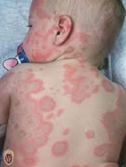
Describe this lesion
Location
Size
Number
Distribution
Configuration (Confluent: have already merged early on)
Texture
Color

Describe this lesion
Location
Size
Number
Distribution
Configuration (Crescentic)
Texture
Color
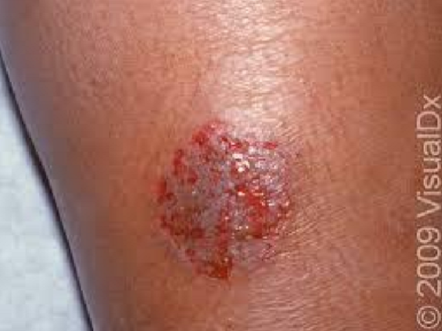
Describe this lesion
Location
Size
Number
Distribution
Configuration (Discoid/Nummular: coin-shaped)
Texture
Color

Describe this lesion
Location
Size
Number
Distribution
Configuration (Discrete)
Texture
Color
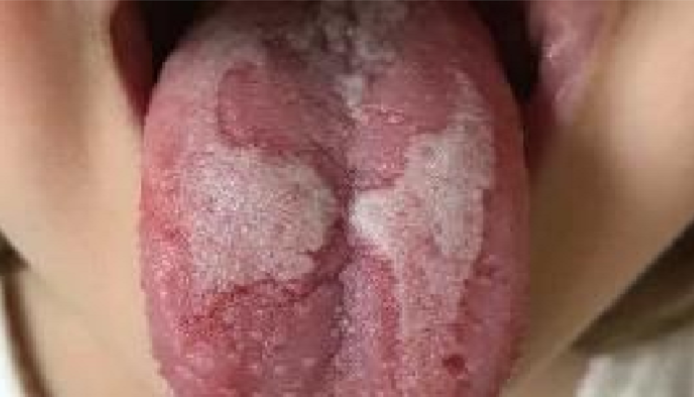
Describe this lesion
Location
Size
Number
Distribution
Configuration (Geographic)
Texture
Color

Describe this lesion
Location
Size
Number
Distribution
Configuration (Grouped)
Texture
Color

Describe this lesion
Location
Size
Number
Distribution
Configuration (Gyrate)
Texture
Color

Describe this lesion
Location
Size
Number
Distribution
Configuration (Herpetiform: grouped vesicles)
Texture
Color

Describe this lesion
Location
Size
Number
Distribution
Configuration (Linear/striate)
Texture
Color

Describe this lesion
Location
Size
Number
Distribution
Configuration (Morbilliform: generalized, small dusky-red macules)
Texture
Color
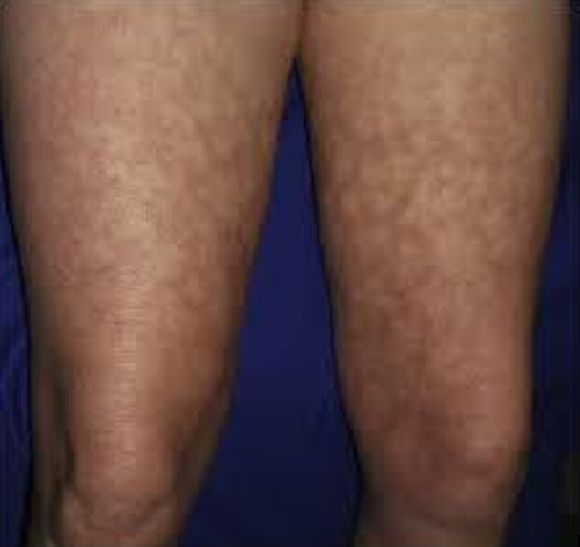
Describe this lesion
Location
Size
Number
Distribution
Configuration (Reticular: netlike)
Texture
Color

Describe this lesion
Location
Size
Number
Distribution
Configuration (Target/Bullseye)
Texture
Color
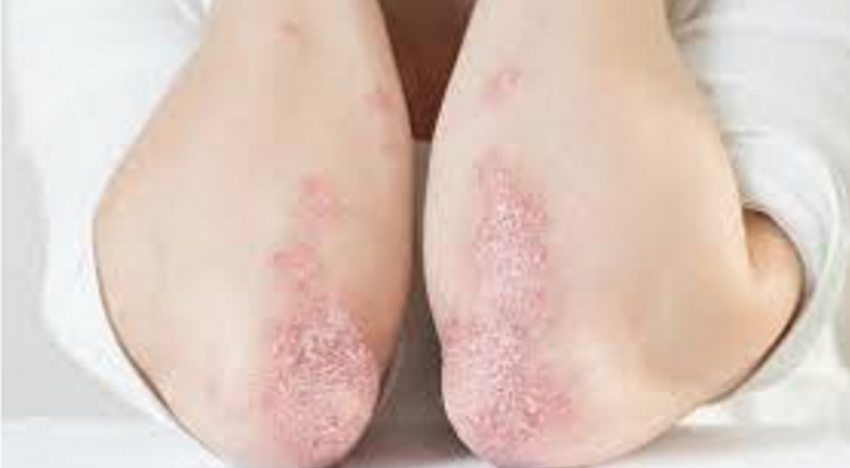
Describe this lesion
Location
Size
Number
Distribution
Configuration
Texture (Scaly)
Color
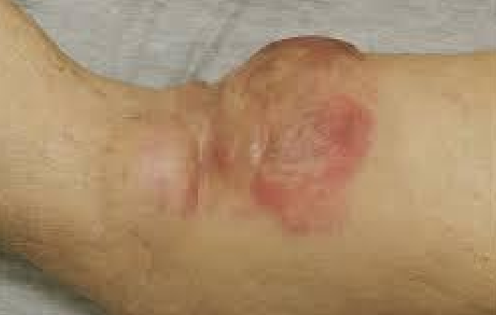
Describe this lesion
Location
Size
Number
Distribution
Configuration
Texture (Indurated: firm or hardened)
Color
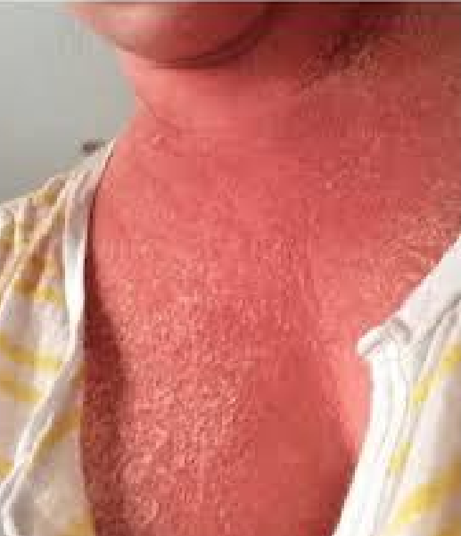
Describe this lesion
Location
Size
Number
Distribution
Configuration
Texture (Friable: easily bleeds or broken)
Color
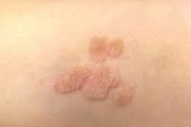
Describe this lesion
Location
Size
Number
Distribution
Configuration
Texture (Verrucous: warty)
Color

Describe this lesion
Location
Size
Number
Distribution
Configuration
Texture (Fluctuant: compressible and fluid-filled)
Color

Describe this lesion
Location
Size
Number
Distribution
Configuration
Texture (Umbilicated: central depression)
Color

Describe this lesion
Location
Size
Number
Distribution
Configuration
Texture (Pedunculated: attached by stalk)
Color

Describe this lesion
Location
Size
Number
Distribution
Configuration
Texture (Sessile: attached w/o stalk)
Color
Inspect patient’s skin sitting down
Hair and scalp (distribution, texture, quantity)
Head and neck (forehead, eyebrows, eyelids, eyelashes, conjunctivae, sclerae, nose, ears, cheeks, lips, oral cavity, chin, beard)
Upper back
Shoulders
Arms
Hands
Fingernails
Chest
Abdomen
Anterior thighs, legs
Feet and toes (including soles, interdigital areas, and toenails)
Palpate patient’s skin sitting down
Fingernails
Chest
Abdomen
Anterior thighs and legs
Feet and toes (including soles, interdigital areas, and toenails)
Inspect patient’s skin standing up
Lower back
Posterior thighs and legs
Breasts*
Axillae*
Genitalia* (including axillary and pubic hair)
*sensitive areas won’t be done in lab
What are the screening criteria for melanoma?
A - Asymmetry (each side of mole)
B - Border irregularity
C - Color variation (more than 2 colors)
D - Diameter (> 6mm)
E - Evolving in size, symptoms, or morphology


What kind of lesion is this?
Classification: Primary
Type: Macule
Definition: Flat, non-palpable change in color of skin
Size: < 1 cm
Example: Freckles, lentigo, pityriasis versicolor

What kind of lesion is this?
Classification: Primary
Type: Patch
Definition: Flat, non-palpable change in color
Size: > 1 cm
Example: Vitiligo, eczema, psoriasis

What kind of lesion is this?
Classification: Primary
Type: Papule
Definition: Small, solid, elevated
Size: < 1 cm
Example: Acne, insect bites, warts
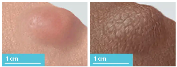
What kind of lesion is this?
Classification: Primary
Type: Nodule
Definition: Solid, elevated (bumpy)
Size: > 1 cm
Example: Cysts, lipomas, dermatofibroma

What kind of lesion is this?
Classification: Primary
Type: Plaque
Definition: Elevated, flat-topped
Size: > 1 cm
Example: Psoriasis, lichen planus

What kind of lesion is this?
Classification: Primary
Type: Tumor
Definition: Large, solid mass
Size: Usually > 2 cm
Example: Skin cancers, benign tumors

What kind of lesion is this?
Classification: Primary
Type: Pustule
Definition: Elevated lesion containing pus
Size: Usually < 1 cm
Example: Acne pustules, folliculitis

What kind of lesion is this?
Classification: Primary
Type: Vesicle
Definition: Small blister containing clear fluid
Size: < 1 cm
Example: Herpes simplex, contact dermatitis

What kind of lesion is this?
Classification: Primary
Type: Bulla
Definition: Large blister containing clear fluid
Size: > 1 cm
Example: Bullous pemphigoid, burns
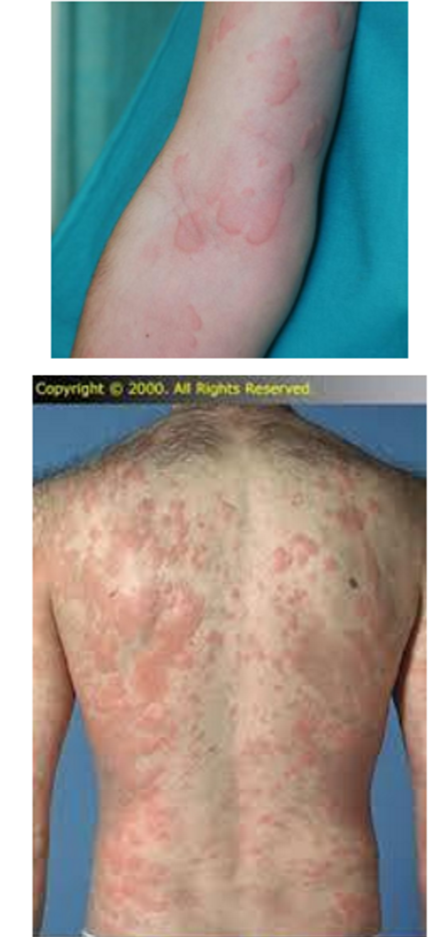
What kind of lesion is this?
Classification: Primary
Type: Wheal
Definition: Elevated, transient, erythematous
Size: Variable
Example: Urticaria (hives), allergic reactions
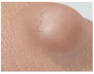
What kind of lesion is this?
Classification: Primary
Type: Cyst
Definition: Encapsulated, fluid, or semisolid
Size: Variable
Example: Epidermoid cysts, piloidal cyst

What kind of lesion is this?
Classification: Secondary
Type: Scale
Definition: Flakes of keratin
Example: Psoriasis, seborrheic dermatitis

What kind of lesion is this?
Classification: Secondary
Type: Crust
Definition: Dried serum, blood, or pus
Example: Impetigo, eczema
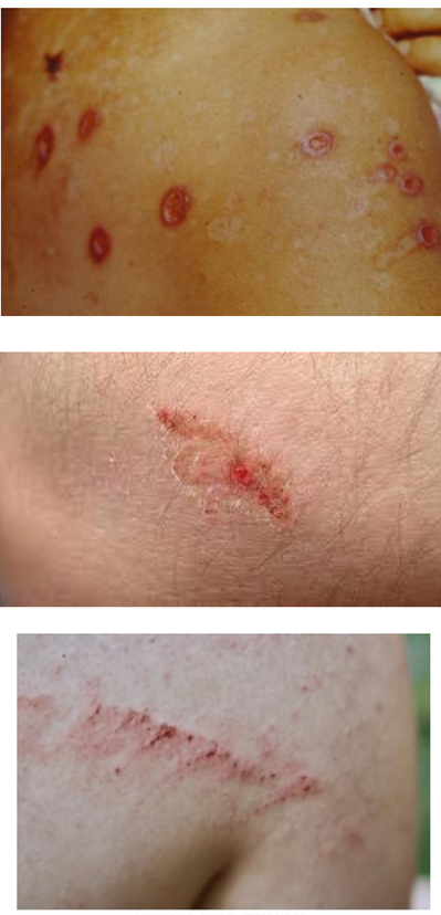
What kind of lesion is this?
Classification: Secondary
Type: Excoriation
Definition: Linear erosion from scratching
Example: Pruritus, scabies

What kind of lesion is this?
Classification: Secondary
Type: Fissure
Definition: Linear crack or split
Example: Eczema, xerosis

What kind of lesion is this?
Classification: Secondary
Type: Erosion
Definition: Loss of epidermis
Example: Abrasion, bullous disorders

What kind of lesion is this?
Classification: Secondary
Type: Ulcer
Definition: Loss of epidermis AND dermis
Example: Venous stasis ulcer, pressure ulcer

What kind of lesion is this?
Classification: Secondary
Type: Lichenification
Definition: Thickened skin with accentuated lines
Example: Chronic eczema
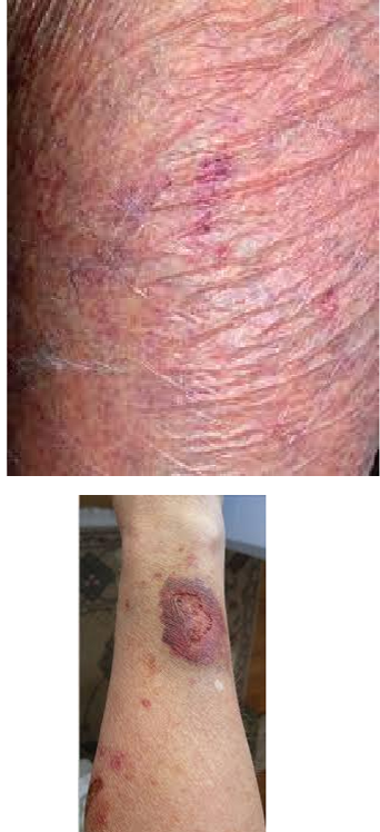
What kind of lesion is this?
Classification: Secondary
Type: Atrophy
Definition: Thinning of the skin
Example: Steroid use, chronic sun damage
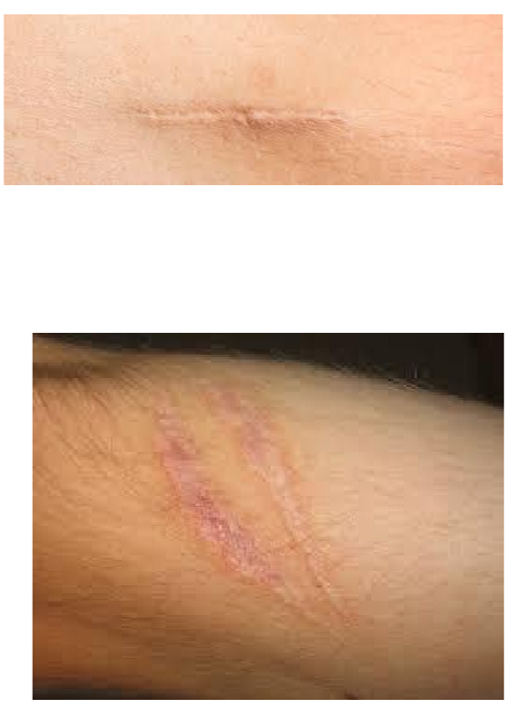
What kind of lesion is this?
Classification: Secondary
Type: Scar
Definition: Fibrous tissue
Example: Post-surgical, post-acne

What kind of lesion is this?
Classification: Secondary
Type: Keloid
Definition: Thick, raised scar (extending beyond original site)
Example: Keloid scars

What kind of lesion is this?
Classification: Secondary
Type: Sclerosis
Definition: Hardening of skin
Example: Scleroderma, morphea

What kind of lesion is this?
Type: Petechaie
Definition: Pinpoint, non-blanching red or purple spots
Size: 1-4 mm

What kind of lesion is this?
Type: Purpura
Definition: Non-blanching red or purple spots
Size: 4 mm - 1 cm

What kind of lesion is this?
Type: Ecchymosis
Definition: Large non-blanching area
Size: > 1 cm

What kind of lesion is this?
Type: Telangiectasia
Definition: Dilated superficial blood vessels
Size: Variable

What kind of lesion is this?
Type: Furnucle
Definition: Infection of hair follicle
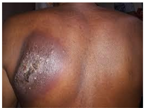
What kind of lesion is this?
Type: Carbuncle
Definition: Group of collected furnucles

What kind of lesion is this?
Type: Abscess
Definition: Collection of pus in tissue
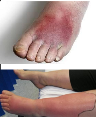
What kind of lesion is this?
Type: Cellulitis
Definition: Infection of dermis and subcutaneous tissue
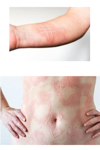
What kind of lesion is this?
Type: Dermatographia
Definition: Raised wheals after scratching skin

What kind of lesion is this?
Type: Abrasion
Definition: Superficial loss of epidermis
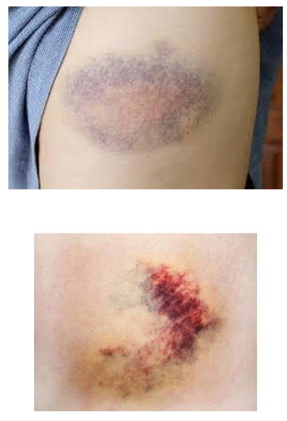
What kind of lesion is this?
Type: Contusion
Definition: Bruising without skin break
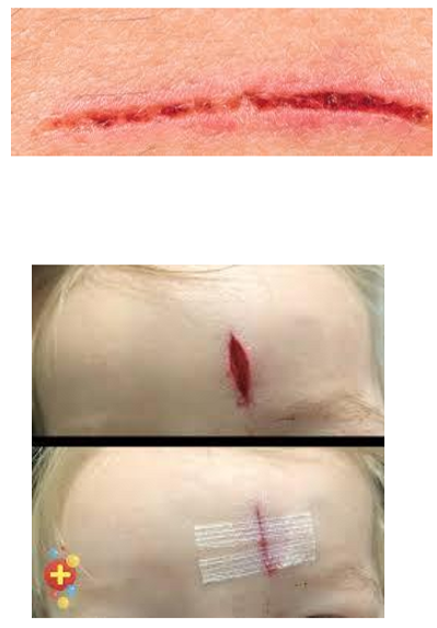
What kind of lesion is this?
Type: Laceration
Definition: Irregular cut or tear in skin
Inspect the patient’s hair
Observe for hair loss or thinning
Inspect scalp for erythema, scaling, bruising, tenderness, bogginess, scarring
Observe hair width part at various sections of scalp
Perform the hair pull test
Instructions
Grasp ~50-60 hairs with thumb and index/middle finger
Pull away from scalp
Results
Positive test if > 10% (>6) hairs pulled out
Telogen Effluvium (hair shedding)
Perform the hair tug test
Instructions
Hold a group of hairs in one hand
Pull along hair shaft with other hand
Results
If hair breaks, then abnormal (hair fragility)
Examine the patient’s nails
Examine all labeled parts, and compare hands.
Note shape
Nail pitting (dents on nail plate) → Psoriasis
Beau lines (lines across nail plate) → High fever/sepsis, stress, malnutrition
Muehrcke lines (paired lines across) → Hypoalbuminemia
Clubbing (enlargement of distal finger + increased nail curvature) → Lung, cystic fibrosis, congenital heart disease
Koilonychia (concave, spoon-shaped) → Iron deficiency anemia
Onychomycosis (fungal infection) → Dermatophytes, diabetes
Note color
Terry nails (white) → Cirrhosis
Subungual hematoma (black spot) → Injury
*red box in photo = epidermal layer under the nail plate
