Cells

0.0(0)
Studied by 0 people
call kaiCall Kai
learnLearn
examPractice Test
spaced repetitionSpaced Repetition
heart puzzleMatch
flashcardsFlashcards
GameKnowt Play
Card Sorting

1/32

encourage image

There's no tags or description

Looks like no tags are added yet.

33 Terms

1
New cards

What does cell theory state?

Cells are a fundamental unit of structure, function & organisation in all living organisms

2
New cards

What is a tissue, organ & organ system?

  • Tissue: a group of similar cells that work together to perform a single function

  • Organ: a group of different tissues that work together to perform a specific function

  • Organ system: a group of organs that work together to perform a specific function


3
New cards

What are the two types of cells?

  • Prokaryotic cells: do not have a nucleus or membrane-bound organelles

  • Eukaryotic cells: contain a nucleus & membrane-bound organelles


4
New cards

What is the structure & function of the cell membrane?

Structure:

  • found in all cells

  • phospholipid bilayer → molecules embedded within & attached on the outside (proteins, carbohydrates, cholesterol)

Function:

  • controls the entrance & exit of materials → acts as a barrier between the inside & outside of the cell
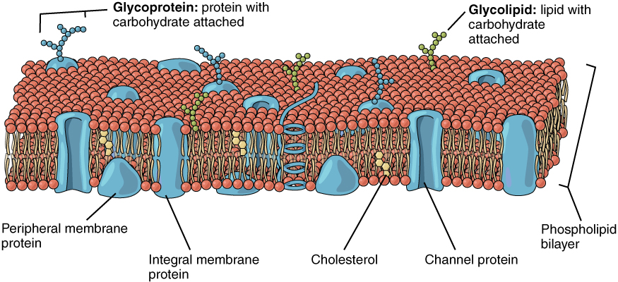

<p><strong><u>Structure:</u></strong></p><ul><li><p>found in all cells</p></li><li><p>phospholipid bilayer → molecules embedded within &amp; attached on the outside (proteins, carbohydrates, cholesterol)</p></li></ul><p><strong><u>Function:</u></strong></p><ul><li><p>controls the entrance &amp; exit of materials → acts as a barrier between the inside &amp; outside of the cell</p></li></ul><p></p>
5
New cards

What is the structure & function of the nucleus?

Structure:

  • nuclear envelope (double membrane)

  • nuclear pores

  • nucleoplasm (granular, jelly-like material)

  • chromosomes (protein-bound, linear DNA)

  • nucleolus (smaller sphere inside - site of rRNA production → makes ribosomes)

Function:

  • site of DNA replication & transcription (making mRNA)

  • contains the genetic material for each cell


<p><strong><u>Structure:</u></strong></p><ul><li><p>nuclear envelope (double membrane)</p></li><li><p>nuclear pores</p></li><li><p>nucleoplasm (granular, jelly-like material)</p></li><li><p>chromosomes (protein-bound, linear DNA)</p></li><li><p>nucleolus (smaller sphere inside - site of rRNA production → makes ribosomes)</p></li></ul><p><strong><u>Function:</u></strong></p><ul><li><p>site of DNA replication &amp; transcription (making mRNA)</p></li><li><p>contains the genetic material for each cell</p></li></ul><p></p>
6
New cards

What is the structure & function of mitochondria?

Structure:

  • double membrane (inner membrane called cisternae)

  • fluid centre called matrix

  • loop of mitochondrial DNA

Function:

  • site of aerobic respiration & ATP production

  • DNA to code for enzymes needed in respiration


<p><strong><u>Structure:</u></strong></p><ul><li><p>double membrane (inner membrane called cisternae)</p></li><li><p>fluid centre called matrix</p></li><li><p>loop of mitochondrial DNA</p></li></ul><p><strong><u>Function:</u></strong></p><ul><li><p>site of aerobic respiration &amp; ATP production</p></li><li><p>DNA to code for enzymes needed in respiration</p></li></ul><p></p>
7
New cards

What is the structure & function of chloroplasts?

Structure:

  • surrounded by a double membrane

  • thylakoids arranged into circular piles called grana (contains chlorophyll)

  • fluid filled stroma contains enzymes for photosynthesis

Function:

  • found in plants → site of photosynthesis


<p><strong><u>Structure:</u></strong></p><ul><li><p>surrounded by a double membrane</p></li><li><p>thylakoids arranged into circular piles called grana (contains chlorophyll)</p></li><li><p>fluid filled stroma contains enzymes for photosynthesis</p></li></ul><p><strong><u>Function:</u></strong></p><ul><li><p>found in plants → site of photosynthesis</p></li></ul><p></p>
8
New cards

What is the structure & function of ribosomes?

Structure:

  • small & made up of two sub-units of protein & rRNA

  • larger ribosomes (80s) found in eukaryotic cells

  • smaller ribosomes (70s) found in prokaryotic cells

Function:

  • site of protein synthesis


<p><strong><u>Structure:</u></strong></p><ul><li><p>small &amp; made up of two sub-units of protein &amp; rRNA</p></li><li><p>larger ribosomes (80s) found in eukaryotic cells</p></li><li><p>smaller ribosomes (70s) found in prokaryotic cells</p></li></ul><p><strong><u>Function:</u></strong></p><ul><li><p>site of protein synthesis</p></li></ul><p></p>
9
New cards

What is the structure & function of the endoplasmic reticulum?

Structure:

  • RER & SER both have cisternae (highly folded membranes)

  • RER has ribosomes on the cisternae

Function:

  • RER → protein synthesis

  • SER → synthesise & store lipids/carbohydrates
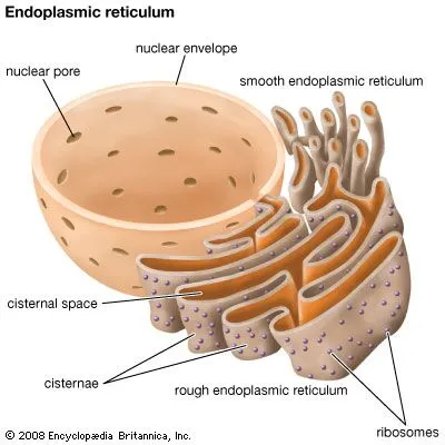

<p><strong><u>Structure:</u></strong></p><ul><li><p>RER &amp; SER both have cisternae (highly folded membranes)</p></li><li><p>RER has ribosomes on the cisternae</p></li></ul><p><strong><u>Function:</u></strong></p><ul><li><p>RER → protein synthesis</p></li><li><p>SER → synthesise &amp; store lipids/carbohydrates</p></li></ul><p></p>
10
New cards

What is the structure & function of the Golgi Body?

Structure:

  • highly folded membranes forming cisternae, which secretary vesicles pinch off from

Function:

  • transports proteins & lipids for extracellular use from the RER to cell membrane for export (exocytosis)


<p><strong><u>Structure:</u></strong></p><ul><li><p>highly folded membranes forming cisternae, which secretary vesicles pinch off from</p></li></ul><p><strong><u>Function:</u></strong></p><ul><li><p>transports proteins &amp; lipids for extracellular use from the RER to cell membrane for export (exocytosis)</p></li></ul><p></p>
11
New cards

What is the structure & function of the vacuole?

Structure:

  • filled with fluid surrounded by a single membrane called the tonoplast

Function:

  • makes cells turgid & provides support

  • temporary store of sugars & amino acids

  • the pigments colour petals to attract pollinators
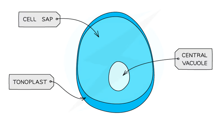

<p><strong><u>Structure:</u></strong></p><ul><li><p>filled with fluid surrounded by a single membrane called the tonoplast</p></li></ul><p><strong><u>Function:</u></strong></p><ul><li><p>makes cells turgid &amp; provides support</p></li><li><p>temporary store of sugars &amp; amino acids</p></li><li><p>the pigments colour petals to attract pollinators</p></li></ul><p></p>
12
New cards

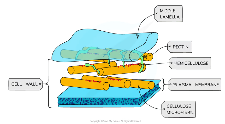
What is the structure & function of the cell wall?

Structure:

  • made of microfibrils of the cellulose polymer in plants

  • made of chitin (nitrogen-containing polysaccharide) in fungi

Function:

  • provides structure & strength to the cell

<p><strong><u>Structure:</u></strong></p><ul><li><p>made of microfibrils of the cellulose polymer in plants</p></li><li><p>made of chitin (nitrogen-containing polysaccharide) in fungi</p></li></ul><p><strong><u>Function:</u></strong></p><ul><li><p>provides structure &amp; strength to the cell</p></li></ul><p></p>
13
New cards

What is the structure & function of lysosomes?

Structure:

  • contains digestive enzymes (lysozymes)

Function:

  • hydrolyse phagocytic cells

  • completely break down dead cells

  • exocytosis (release enzymes to the outside of the cell to destroy material)

  • digest worn out organelles for reuse of material


<p><strong><u>Structure:</u></strong></p><ul><li><p>contains digestive enzymes (lysozymes)</p></li></ul><p><strong><u>Function:</u></strong></p><ul><li><p>hydrolyse phagocytic cells</p></li><li><p>completely break down dead cells</p></li><li><p>exocytosis (release enzymes to the outside of the cell to destroy material)</p></li><li><p>digest worn out organelles for reuse of material</p></li></ul><p></p>
14
New cards

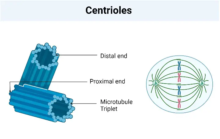
What is the structure & function of the centriole?

Structure:

  • always two found near the nucleus

  • part of the cytoskeleton

Function:

  • used in cell division to make spindle fibres that move chromosomes

<p><strong><u>Structure:</u></strong></p><ul><li><p>always two found near the nucleus</p></li><li><p>part of the cytoskeleton</p></li></ul><p><strong><u>Function:</u></strong></p><ul><li><p>used in cell division to make spindle fibres that move chromosomes</p></li></ul><p></p>
15
New cards

What is the structure & function of the cytoskeleton?

Structure:

  • made up of three types of protein fibres (microfilaments, intermediate filaments & microtubules) that are arranged in a network of interconnected fibres

Function:

  • provides structural support

  • facilitates cell division

  • enables movement & aids in intracellular transport


<p><strong>Structure:</strong></p><ul><li><p>made up of three types of protein fibres (microfilaments, intermediate filaments &amp; microtubules) that are arranged in a network of interconnected fibres</p></li></ul><p><strong>Function:</strong></p><ul><li><p>provides structural support</p></li><li><p>facilitates cell division</p></li><li><p>enables movement &amp; aids in intracellular transport</p></li></ul><p></p>
16
New cards

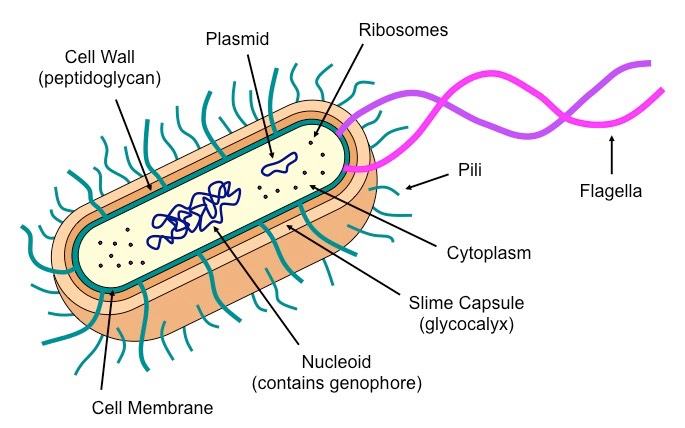
What is the nucleoid?

A region found in prokaryotic cells (e.g. bacteria) of the cytoplasm that contains DNA
<p>A region found in prokaryotic cells (e.g. bacteria) of the cytoplasm that contains DNA</p>
17
New cards

What is the structure & function of the plasmid?

Structure:

  • small, circles of DNA which are separate from the main DNA loop

Function:

  • used to exchange DNA between bacterial cells & in genetic engineering


<p><strong><u>Structure:</u></strong></p><ul><li><p>small, circles of DNA which are separate from the main DNA loop</p></li></ul><p><strong><u>Function:</u></strong></p><ul><li><p>used to exchange DNA between bacterial cells &amp; in genetic engineering</p></li></ul><p></p>
18
New cards

What is the structure & function of the cell wall in prokaryotic cells?

Structure:

  • thick layer outside the cell membrane made of peptidoglycan

Function:

  • used to give the cell strength, rigidity & resist osmotic lysis


19
New cards

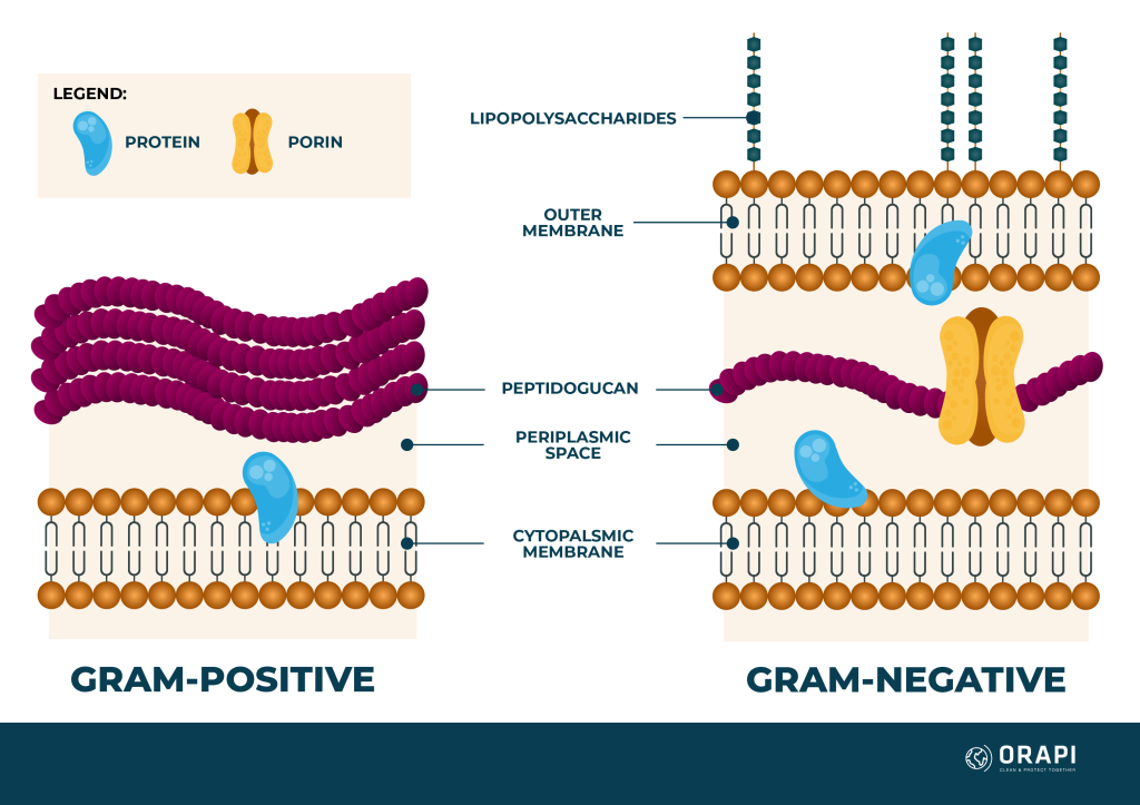
What is a Gram positive cell wall?

  • Stains purple

  • Have a thick peptidoglycan cell wall outside their cell membrane

  • Cell wall is very strong & allows these bacteria to withstand severe physical conditions

<ul><li><p>Stains <strong>purple</strong></p></li><li><p>Have a <strong>thick peptidoglycan cell wall</strong> outside their cell membrane</p></li><li><p>Cell wall is <strong>very strong </strong>&amp; allows these bacteria to withstand severe physical conditions </p></li></ul><p></p>
20
New cards

What is a Gram negative cell wall?

  • Stains pink

  • Have a thick layer of periplasm, a thin peptidoglycan cell wall & a second, outer membrane which contains lipopolysaccharides

  • This layer resists antibiotics & lysozyme enzymes → more difficult to kill
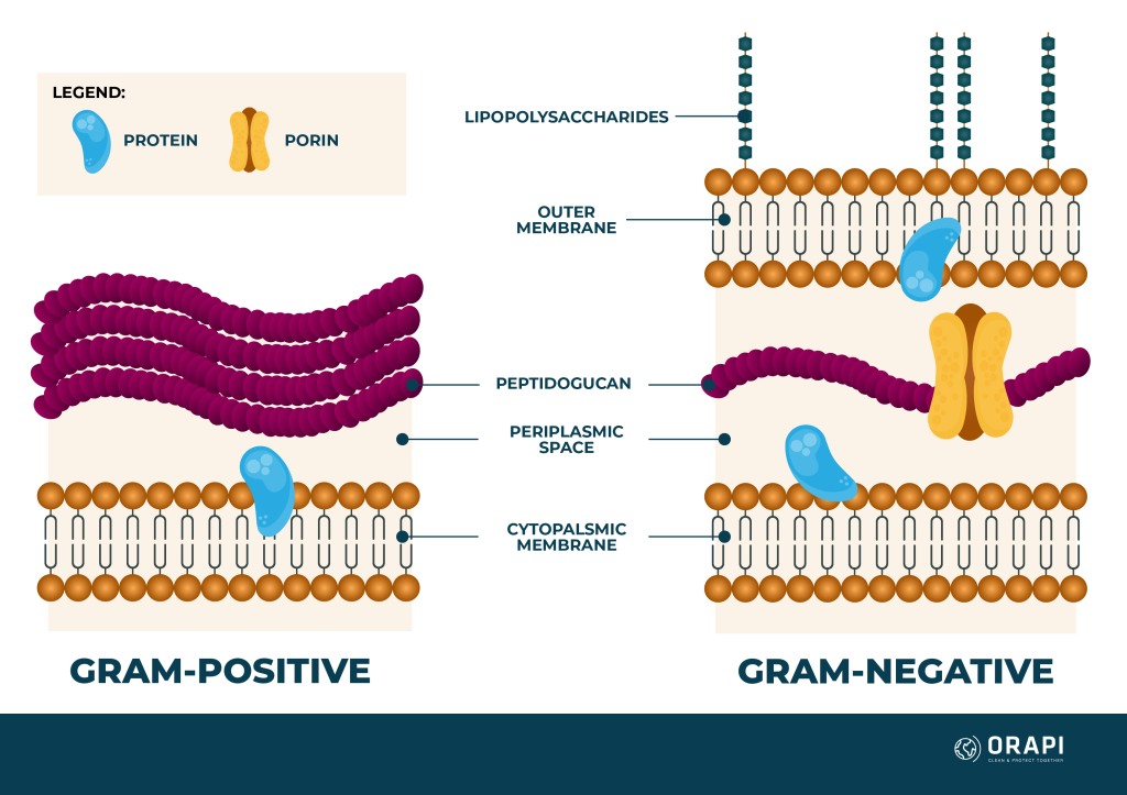

<ul><li><p>Stains<strong> pink</strong></p></li><li><p>Have a<strong> thick layer of periplasm</strong>, a<strong> thin peptidoglycan cell wall</strong> &amp; a second, outer membrane which contains <strong>lipopolysaccharides</strong></p></li><li><p>This layer<strong> resists antibiotics &amp; lysozyme enzymes</strong> → more difficult to kill </p></li></ul><p></p>
21
New cards

Explain why Gram positive & Gram negative bacteria react differently to some antibiotics

  • Gram positive bacteria have a thick layer of peptidoglycan in their cell wall

  • Some antibiotics inhibit formation of peptidoglycan, so are effective against Gram positive bacteria

  • Some antibiotics are not able to cross the peptidoglycan layer, so they are only effective against Gram negative bacteria


22
New cards

What is magnification & resolution?

  • Magnification: how much bigger the image is compared to the original object

  • Resolution: the smallest separation at which two separate objects can be distinguished


23
New cards

Outline the features of a light microscope

  • Specimens are illuminated with visible light, which is focused using glass lenses & viewed using the eye

  • Have 2 lenses:

    • an objective lens → magnifies the image

    • an eyepiece lens → focuses the image on the eye

  • Specimens can be living or dead but need to be thin so enough light can be transmitted through them

  • Most biological specimens usually need to be coloured with a chemical stain (e.g. iodine or methylene blue)
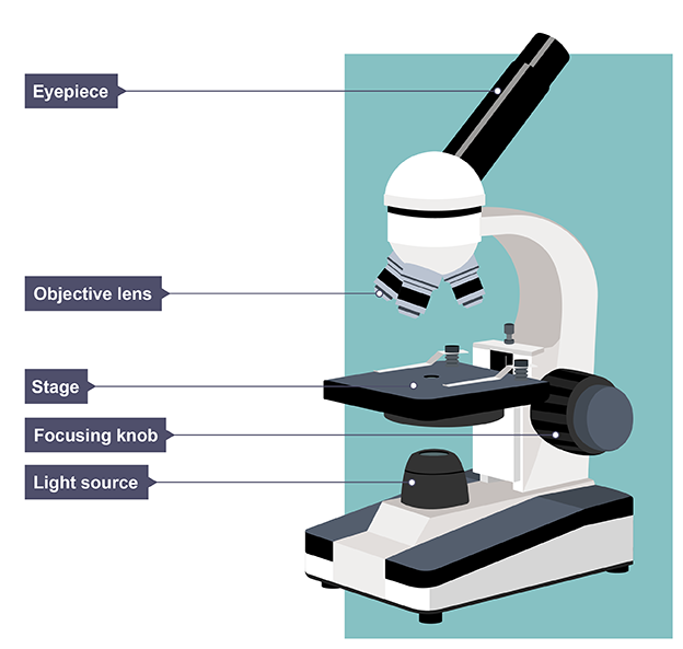

<ul><li><p>Specimens are illuminated with visible light, which is focused using glass lenses &amp; viewed using the eye</p></li><li><p>Have 2 lenses:</p><ul><li><p>an <strong>objective lens</strong> → magnifies the image</p></li><li><p>an<strong> eyepiece lens </strong>→ focuses the image on the eye</p></li></ul></li><li><p>Specimens can be living or dead but need to be thin so enough light can be transmitted through them</p></li><li><p>Most biological specimens usually need to be coloured with a chemical stain (e.g. iodine or methylene blue)</p></li></ul><p></p>
24
New cards

Explain the importance of staining specimens in microscopy

Stains increase contrast as different organelles take up stains to different degrees, allowing organelles to become visible so they can be identified

25
New cards

What is the equation to calculate the total magnification in a microscope?

Total magnification = objective lens x eyepiece lens

26
New cards

Outline the transmission electron microscope (TEM)

Uses a beam of electrons to “illuminate” the specimen → used to resolve small sub-cellular ultrastructure:

  • specimens need to be very thin, & to prevent them from breaking have to be embedded in plastic

  • specimens also have to be stained with electron-dense heavy metal compounds (images are colourless)

  • there must be a vacuum inside the electron microscope, so living organisms cannot be used
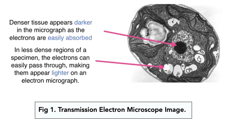

<p>Uses a <strong>beam of electrons </strong>to<strong> “illuminate”</strong> the specimen → used to resolve<strong> small sub-cellular ultrastructure:</strong></p><ul><li><p>specimens need to be <strong>very thin</strong>, &amp; to prevent them from breaking have to be <strong>embedded in plastic</strong></p></li><li><p>specimens also have to be <strong>stained with electron-dense heavy metal compounds</strong> (images are colourless)</p></li><li><p>there must be a vacuum inside the electron microscope, so<strong> living organisms cannot be used</strong></p></li></ul><p></p>
27
New cards

Outline the scanning electron microscope (SEM)

  • Scans a fine beam of electrons onto a specimen & detects the electrons scattered back by the surface

  • The scattered electron signal is then converted into a computer-generated image of the surface of the specimen → images are detailed but do not have very high magnification
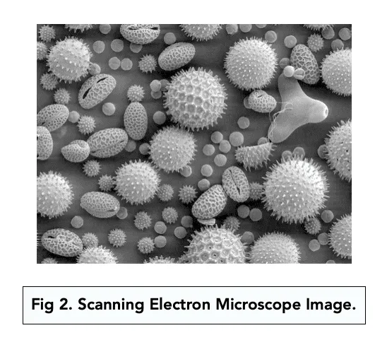

<ul><li><p><strong>Scans a fine beam of electrons</strong> onto a specimen &amp; detects the <strong>electrons scattered back by the surface</strong></p></li><li><p>The scattered electron signal is then converted into a <strong>computer-generated image of the surface </strong>of the specimen → images are <strong>detailed </strong>but <strong>do not have very high magnification</strong></p></li></ul><p></p>
28
New cards

Explain how to calibrate an eyepiece graticule

  1. Line up the stage micrometer & eyepiece graticule whilst looking through the eyepiece

  2. Count up how many divisions on the eyepiece graticule fit into one division on the micrometer scale

  3. Each division on the micrometer is 10 μm, so this can be used to calculate what one division on the eyepiece graticule is at that current magnification
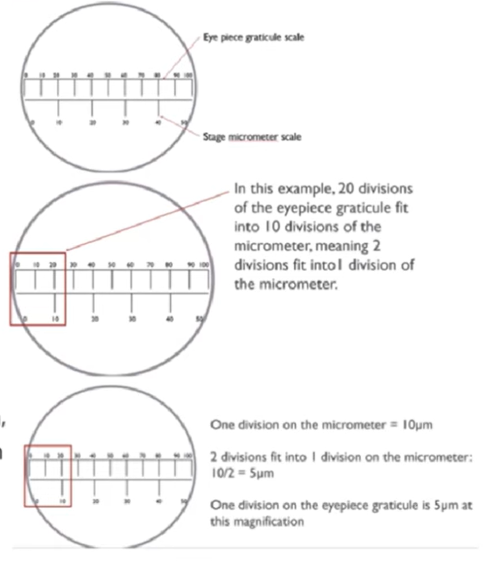

<ol><li><p>Line up the stage micrometer &amp; eyepiece graticule whilst looking through the eyepiece</p></li><li><p>Count up how many divisions on the eyepiece graticule fit into one division on the micrometer scale</p></li><li><p>Each division on the micrometer is 10 μm, so this can be used to calculate what one division on the eyepiece graticule is at that current magnification</p></li></ol><p></p>
29
New cards

Why can you see the detailed structure of a prokaryotic cell with an electron microscope but not light microscope?

Light has a longer wavelength than electrons, so a light microscope has a lower resolution than an electron microscope

30
New cards

What is the equation for magnification?

Magnification = image size / actual size

31
New cards

How to calculate the actual size using a scale bar?

Actual size = (image size/bar size) x bar scale

32
New cards

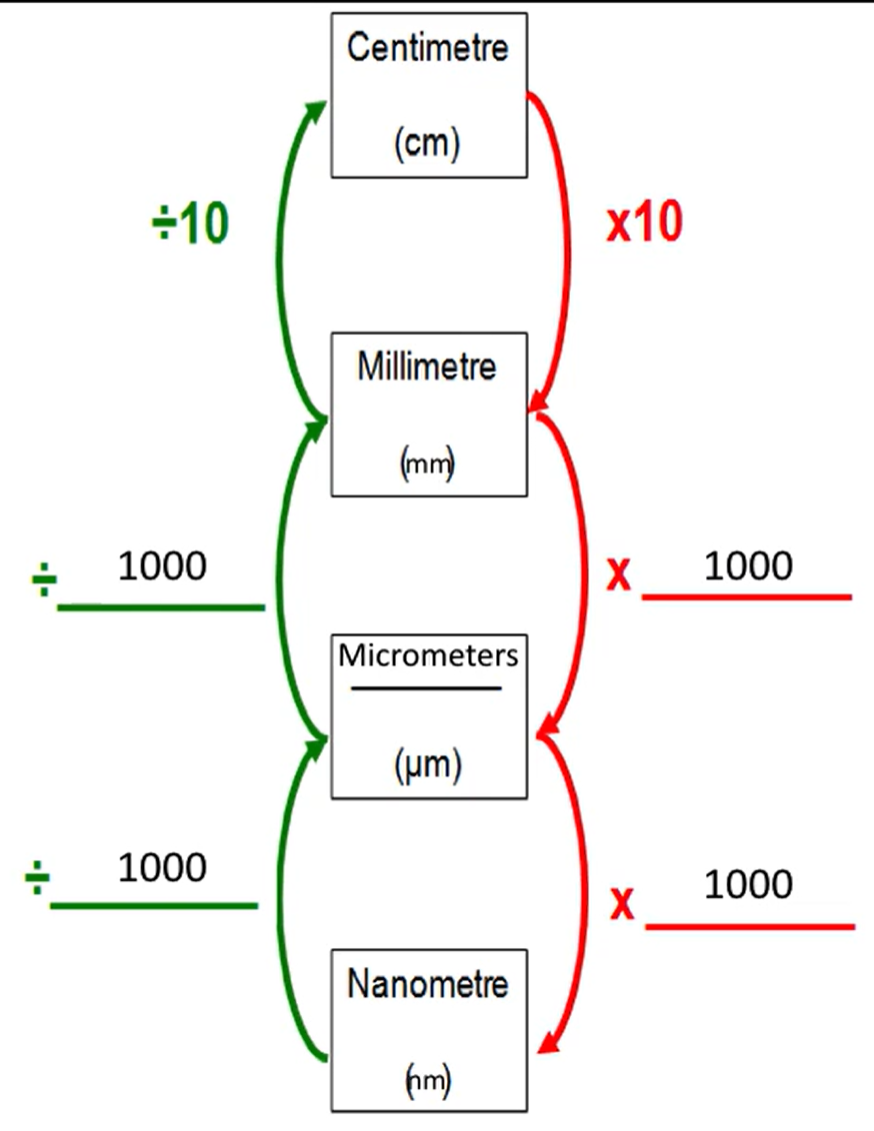
How do you convert between units?

knowt flashcard image
33
New cards

What happens when two numbers in scientific notation are:

  • multiplied

  • divided


  • Multiplied: the numbers from 1 to <10 are multiplied & the powers of ten are added

  • Divided: the numbers between 1 to <10 are divided & the powers of ten are subtracted

    • how to remember → “indices are mad, to multiply you add. Indices are cracked, to divide you subtract”


<ul><li><p><strong>Multiplied</strong>: the numbers from 1 to &lt;10 are multiplied &amp; the powers of ten are added</p></li><li><p><strong>Divided: </strong>the numbers between 1 to &lt;10 are divided &amp; the powers of ten are subtracted</p><ul><li><p>how to remember → “indices are mad, to multiply you add. Indices are cracked, to divide you subtract”</p></li></ul></li></ul><p></p>