Head and Neck Anatomy: Arteries, Nerves, Muscles & Glands
 Call Kai
 Learn
 Practice Test
 Spaced Repetition
 Match
 Flashcards
 Knowt Play
1/57

There's no tags or description
Looks like no tags are added yet.
58 Terms

Identify this structure
Subclavian artery: Neck artery; right side arises from the brachiocephalic trunk, left from the aortic arch. Branches: vertebral, internal thoracic, thyrocervical trunk. Passes between the anterior and middle scalene muscles.

Identify this structure
Common carotid artery: Right: brachiocephalic trunk; left: aortic arch. Bifurcates at about C4 into internal and external carotid arteries. Innervation: sympathetic/vagal fibers in the carotid sheath.

Identify this structure
External carotid artery: Origin: common carotid bifurcation (about C4). Branches (proximal to distal): superior thyroid, ascending pharyngeal, lingual, facial, occipital, posterior auricular, maxillary, superficial temporal. Supplies the face, scalp, neck, and thyroid.

Identify this structure
Superior thyroid artery: Origin: external carotid (first branch). Supplies thyroid gland and larynx. Exam tagged.

Identify this structure
Facial artery: Origin: external carotid. Runs deep to the posterior belly of the digastric and stylohyoid, through the submandibular gland, and over the mandible. Supplies face and submandibular region. Exam tagged.

Identify this structure
Submental artery: Origin: facial artery in the submandibular triangle. Supplies the submental region, mylohyoid, and anterior digastric.

Identify this structure
Lingual artery: Origin: external carotid. Runs deep to the hyoglossus. Supplies the tongue. Exam tagged.

Identify this structure
Internal carotid artery: Origin: common carotid bifurcation (about C4). Enters the carotid canal in the petrous temporal bone. Supplies the brain and eye. Foramen: carotid canal (petrous temporal bone).

Identify this structure
Vertebral artery: Origin: first branch of subclavian artery. Enters transverse foramina of C6 to C1, then ascends through the foramen magnum to supply the brain. Foramina: transverse foramina C6-C1; foramen magnum.
Identify this structure
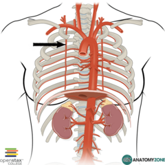
Internal thoracic artery: Origin: subclavian artery. Descends behind the costal cartilages along the inner chest wall.

Identify this structure
Thyrocervical trunk: Origin: subclavian artery. Branches: inferior thyroid, suprascapular, transverse cervical, ascending cervical.

Identify this structure
Transverse cervical artery: Origin: thyrocervical trunk. Supplies the trapezius and levator scapulae.

Identify this structure
Suprascapular artery: Origin: thyrocervical trunk. Passes over the suprascapular notch to supply the supraspinatus and infraspinatus. Foramen: suprascapular notch (under the transverse scapular ligament).

Identify this structure
Internal jugular vein: Origin: sigmoid sinus at the jugular foramen. Descends in the carotid sheath and joins the subclavian vein to form the brachiocephalic vein. Receives the common facial vein. Foramen: jugular foramen.

Identify this structure: 11
Common facial vein: Origin: union of the facial and retromandibular veins. Drains into the internal jugular vein.

Identify this structure
Facial vein: Origin: angular vein at the medial eye. Passes superficial to the submandibular gland and joins the retromandibular vein to form the common facial vein.

Identify this structure
Ansa cervicalis: Nerve Loop formed by C1 (superior root, joins CN XII) and C2-C3 (inferior root). Innervates the sternohyoid, sternothyroid, and omohyoid.

Identify this structure
Vagus nerve (CN X): Origin: medulla (dorsal motor nucleus and nucleus ambiguus). Exits the skull through the jugular foramen. Innervates the pharynx, larynx, thoracic, and abdominal viscera. Foramen: jugular foramen.

Identify this structure. 11
Superior laryngeal nerve: Origin: vagus nerve at the inferior ganglion. Divides into an internal branch (sensory above the vocal folds, passing through the thyrohyoid membrane) and an external branch (motor to the cricothyroid). Foramen: thyrohyoid membrane.

Identify this structure: 2
Internal laryngeal nerve: Origin: superior laryngeal nerve. Sensory to the laryngeal mucosa above the vocal folds. Passes through the thyrohyoid membrane with the superior laryngeal artery. Foramen: thyrohyoid membrane.

Identify this structure: 3
External laryngeal nerve: Origin: superior laryngeal nerve (CN X). Innervates the cricothyroid. Exam tagged in the lecture outline.

Identify this structure: 2
Recurrent laryngeal nerve: Origin: vagus. Right: loops under the subclavian artery; left: loops under the aortic arch. Ascends in the tracheoesophageal groove. Innervates all intrinsic laryngeal muscles except the cricothyroid.

Name this nerve
Nerve to the mylohyoid (CN V3): Origin: mandibular division (V3) via the inferior alveolar branch. Innervates the mylohyoid and anterior belly of the digastric. Foramen: foramen ovale (V3 exits here).

Identify this structure: 4
Hypoglossal nerve (CN XII): Origin: medulla (hypoglossal nucleus). Exits through the hypoglossal canal at the occipital condyle. Innervates all intrinsic and most extrinsic tongue muscles. Foramen: hypoglossal canal.

Identify this structure
Nerve to the thyrohyoid: Origin: C1 fibers traveling with CN XII. Innervates the thyrohyoid and geniohyoid. Foramen: hypoglossal canal (with CN XII).

Identify this structure
Sternocleidomastoid (SCM): Origin: manubrium and medial clavicle. Insertion: mastoid process and superior nuchal line. Innervation: spinal accessory nerve (CN XI), with C2-C3 proprioception. Foramen: jugular foramen (CN XI spinal part exits here).

Identify this structure
Sternohyoid: Origin: posterior manubrium and medial clavicle. Insertion: body of the hyoid. Innervation: ansa cervicalis (C1-C3).

Identify this structure
Sternothyroid: Origin: posterior manubrium and first costal cartilage. Insertion: oblique line of the thyroid lamina. Innervation: ansa cervicalis (C1-C3).

Identify this structure
Cricothyroid: Origin: anterior arch of the cricoid. Insertion: inferior horn and lamina of the thyroid cartilage. Innervation: external laryngeal nerve (CN X). Only laryngeal muscle in the anterior neck.
Identify this structure
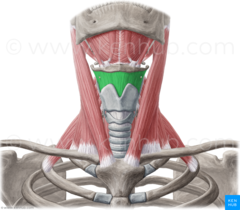
Thyrohyoid: Origin: oblique line of the thyroid lamina. Insertion: greater horn of the hyoid. Innervation: nerve to thyrohyoid (C1 via CN XII).

Identify this structure
Mylohyoid: Origin: mylohyoid line of the mandible. Insertion: median raphe and body of the hyoid. Innervation: nerve to mylohyoid (CN V3). Forms the floor of the submandibular triangle.

Identify this structure: B
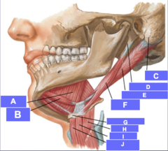
Anterior digastric: Origin: digastric fossa of the mandible. Insertion: intermediate tendon (anchored to the hyoid by a fascial sling). Innervation: nerve to mylohyoid (CN V3).
Identify this structure: E
Posterior digastric: Origin: mastoid notch of the temporal bone. Insertion: intermediate tendon. Innervation: CN VII (posterior belly branch). Foramen: stylomastoid foramen (CN VII exits here). Active during chewing and swallowing.
Identify this structure
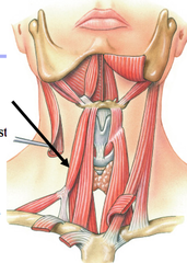
Omohyoid: Origin: inferior belly from the superior border of the scapula; superior belly from the intermediate tendon. Insertion: body of the hyoid. Innervation: ansa cervicalis (C1-C3).

Identify this structure
Stylohyoid: Origin: styloid process of the temporal bone. Insertion: greater horn and body of the hyoid. Innervation: CN VII (stylohyoid branch). Foramen: stylomastoid foramen (CN VII exits here).

Identify this structure
Hyoglossus: Origin: greater horn of the hyoid. Insertion: side of the tongue. Innervation: hypoglossal nerve (CN XII).

Identify this structure
Thyroid gland: (endocrine gland). Arterial supply: superior and inferior thyroid arteries. Innervation: sympathetic and parasympathetic fibers. .

Identify this structure
Parotid gland: (Salivory gland) Duct: Stensen's duct, opening opposite the upper second molar. Innervation: CN IX (parasympathetic via otic ganglion); CN VII (facial nerve passes through it). Foramen: stylomastoid foramen region (CN VII runs through the gland).

Identify this structure
Submandibular gland: (salivary gland). Duct: Wharton's duct, opening at the sublingual papilla. Innervation: CN VII (parasympathetic via submandibular ganglion, chorda tympani).

Identify this structure
Hyoid bone: (bone). Attachments: suprahyoid and infrahyoid muscles. No direct bony articulation.

Identify this structure
Thyroid cartilage: Attachments: thyrohyoid membrane, cricothyroid, sternothyroid, thyrohyoid.

Identify this structure
Laryngeal prominence: (anterior projection of the thyroid lamina). Palpable in the midline of the neck.

Identify this structure
Cricoid cartilage: (cartilage). Attachments: cricothyroid, posterior cricoarytenoid, and the cricotracheal ligament. I

Identify this structure
Cricothyroid membrane/ligament: (membrane). Spans from the cricoid to the thyroid cartilage. Used for emergency airway access (cricothyrotomy).
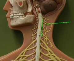
Identify this structure
Cervical plexus: Origin: ventral rami C1-C4. Predominantly cutaneous; also innervates the phrenic nerve and ansa cervicalis contributions. Foramen: intervertebral foramina C1-C4.

Identify this structure
Supraclavicular nerves: Origin: cervical plexus (C3-C4). Innervates skin over the clavicle and shoulder.

Identify this structure
Transverse cervical nerve: Origin: cervical plexus (C2-C3). Innervates the anterior neck skin.

Identify this structure
Great auricular nerve: Origin: cervical plexus (C2-C3). Innervates the skin over the parotid gland and earlobe.

Identify this structure
Lesser occipital nerve: Origin: cervical plexus (C2). Innervates the skin of the posterolateral scalp and neck.

Identify this structure
Phrenic nerve: Origin: cervical plexus (C3-C5). Innervates the diaphragm.

Identify this structure
Brachial plexus (ventral rami): Origin: ventral rami C5-T1. Passes through the posterior triangle and scalene triangle between the anterior and middle scalenes. Foramina: intervertebral foramina C5-T1.

Identify this structure
Spinal accessory nerve (CN XI): Origin: spinal part from the accessory nucleus (C1-C5/6). Cranial part from the medulla. Enters the foramen magnum and exits the jugular foramen. Innervates the SCM and trapezius. Foramen: jugular foramen.

Identify this structure
Trapezius: Origin: occipital bone (superior nuchal line), ligamentum nuchae, spinous processes of C7-T12. Insertion: lateral clavicle, acromion, and spine of the scapula. Innervation: CN XI and C3-C4 proprioception.

Identify this structure
Anterior scalene: Origin: anterior tubercles of the C3-C6 transverse processes. Insertion: scalene tubercle of the first rib. Innervation: ventral rami C4-C6. The phrenic nerve runs directly on the anterior scalene muscle. It forms from C3-C5, descends across the anterior surface of anterior scalene. Subclavian artery passes behind it.

Identify this structure
Middle scalene: Origin: posterior tubercles of C2-C7 transverse processes. Insertion: first rib (posterior to the subclavian groove). Innervation: ventral rami C3-C8.

Identify this structure
Posterior scalene: Origin: posterior tubercles of C5-C7 transverse processes. Insertion: outer surface of the second rib. Innervation: ventral rami C6-C8.

Identify this structure
Levator scapulae: Origin: posterior tubercles of C1-C4 transverse processes. Insertion: superior angle of the scapula. Innervation: dorsal scapular nerve (C5) and C3-C4.

Identify these structures
Splenius (capitis-yellow/cervicis-red): Origin: ligamentum nuchae and spinous processes C7-T3 (capitis); T3-T6 (cervicis). Insertion: mastoid process and superior nuchal line (capitis); posterior tubercles of C1-C3 transverse processes (cervicis). Innervation: dorsal rami of cervical nerves.