Physiological Psychology - Quiz 5
 Call Kai
 Learn
 Practice Test
 Spaced Repetition
 Match
 Flashcards
 Knowt Play
1/45

There's no tags or description
Looks like no tags are added yet.
46 Terms
Guiding principles of sensory systems
Not neurons but cells in our eyes, nose, etc. that transmit information
Specialized sensory receptors in the body detect and convert physical signals into interpretable neural signals (action potentials) by extracting features that carry information relevant to understanding our world around us
The above process of converting sensory signals into action potentials that our brain interprets is known as “sensory transduction” and occurs in peripheral sensory receptors/organs
There is topographic mapping of sensory space at the level of the sensory organ and in sensory brain regions (auditory cortex), with adjacent regions of sensory organs (such as the retina, cochlea, or skin) mapping onto adjacent regions of the brain–this organization allows the brain to represent and interpret sensory input in a systematic and orderly manner
Adjacent regions of sensory organs that map onto adjacent regions of the brain
Nervous system is organized in a certain way
Mail analogy, sorted by area, street, box
Information from all sensory systems (with the exception of olfaction) pass through the thalamus
Information from most of our senses passes through the thalamus, which then relays info to primary sensory cortices
Flow of sensory information
Most of our sensory receptors are on our head (cuz it goes first when we walk)

Spinal cord → Relay brainstem → Thalamus → Primary sensory cortical areas → either back to thalamus or to secondary sensory cortex
Ex. a visual signal the thalamus sends to the visual cortex (thalamus = gateway to the cortex)
Primary sensory cortex
Receives direct sensory input from the thalamus
There are different primary sensory cortices for the different sensory modalities
Each of these cortical regions has a separate map of the same receptive surface, but the different cortical regions process the information differently and make different contributions to perceptual experiences
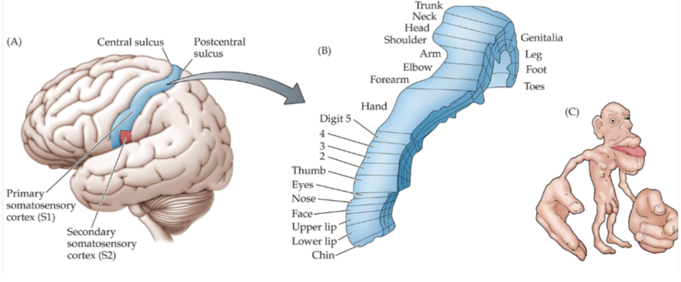
Secondary sensory cortex
Receives direct projections from primary sensory cortex and generally does higher-order sensory computations
Wernike = language understanding
Humans have higher brain regions for language understanding
Sensory information from the body goes to the spinal cord then the brainstem and then thalamus
Sensory information from the head (eyes, ears, nose, tongue) goes through sensory cranial nerves to the brainstem
Sensory information from the body (touch, pain, temperature, itch)
Flow of Auditory sensory information

Non-neuronal sensory cells (in cochlea → hair cells), sensory transduction
Relayed to neuronal sensory cells, makes up auditory nerve, projects to relay nuclei, then thalamus, then primary sensory cortex, then higher order cortices
Sound travels in air in 3D sinusoidal waves
Displacement of air molecules, like waves, pushing molecules in front of it, that’s how sound waves travel, there are areas where the air molecules compress and expand
Sound as frequency of physical compression and rarefaction (expansion)
Compressed air = peak of sound waves graph
In space there's no air molecules to push

Sound frequency vs. Amplitude
Waveform = shape of wave
Phase = peak(compressions) or trough (expansions) of air
Amplitude = height of wave, loudness, intensity, measured as sound pressure level
Larger amplitude = louder
Frequency = pitch, how fast wave repeats (cycles per second) = 100hz, measured in Hz
→ Our perception of frequency is called pitch
Higher pitch = more cycles per second
Elephants make really low frequency sounds
Humans cannot hear this

Human hearing is in the range of 20Hz - 20,000Hz
We can hear 20 to 20,000hz

Components of the human ear *image
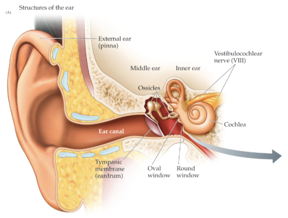
Outer ear
Pinna
Captures, focuses, and filters sound
Funnels sound waves into the ear canal (part of outer/external ear) and vibrates ear drum → all 3 are outer ear, starts shaking middle ear ossicles), middle ear(ossicles, amplifies sound waves, only 3 ossicle bones), inner ear (cochlea = snail thing)
Consists of the pinna, auditory/ear canal, and tympanic membrane (ear drum)
The pinna & auditory canal focus sound energy on the tympanic membrane
The ridges and valleys of the human pinnae evolved to enhance the perception of sounds important to speech (~2000-5000Hz)
We humans turn our heads to locate sounds, while other mammals can direct/swivel their pinnae toward the source of a sound
Ears
Funnel information/sound waves
Sea otters can fold in ears, so they can swim without getting ear infections
Outer & Middle ear
Ossicles (middle ear)
3 bones
Tiny bones: Malleus, incus, and stapes
When the eardrum goes back and forth due to a sound vibration, it amplifies the sound waves, and the last bone, stapes, comes in contact with the cochlea
Stapes beats on the cochlea and causes turbulence in cochlear fluid
Tympanic membrane/”eardrum” (outer ear), boundary structure
Sound waves in the air strike the tympanic membrana and cause the tympanic membrane to vibrate and transmit sound waves to the ossicles
The ossicles vibrate to amplify sound pressure waves by 200% and transfer sound waves from air to liquid via the oval window (cochlear fluid inside the cochlea)
The middle ear is between the tympanic membrane and the cochlea
Inner ear
Converts sound into neural activity
Consists of the cochlea and its constituents (parts)
The complex structures of the inner ear convert sound into neural activity
In mammals the auditory portion of the inner ear is a coiled, fluid-filled structure called the cochlea
Contains three fluid-filled chambers “scala” that can be seen in a cross section (scala vestibuli, scala media, and scala tympani)
Scala media - A tiny organ in it called the organ of corti is responsible for auditory transduction
Contains endolymph (high K+)
Has a high concentration of cochlear fluid with a high concentration of potassium, flows in and depolarizes
Contains the organ of Corti, where sound waves are transduced into neural impulses

Organ of Corti
The organ of Corti is where the “magic” happens (“magic” = sensory transduction)
Contains the auditory sensory cells, called hair cells (inner & outer)
Hair cells are embedded in the flexible basilar membrane, which is wider at the apex than at the base (it’s tapered), as we’ll see, the mechanical properties of the basilar membrane are important for the phenomenon of tonotopy
The auditory nerve terminals that transmit neural signals to and from the brain leave organ of Corti to go to the brainstem
Purple looking things are not neurons, avocado looking with hairs = hair cells
Basilar membrane - hair cells embedded in here, at the very end it is wider and floppy, and at the base it is stiff
Innermost part = apex
Outermost part = base

Tonotopy along the flexible basilar membrane
Tonotopy = systematic representation of sound frequency along the length of the cochlea (high → low)
The basilar membrane vibrates in response to sound waves at specific locations depending on the frequency of the sound wave
It’s about 5 times wider at the apex of the cochlea than at the base, it separates the scala tympani from the scala media and vibrates in response to sound
Because of the physical properties of the membrane, it will maximally deflect/activate the basilar membrane at different locations based on frequency
A soundwave of low frequency maximally deflects the apex (the wider end)
When sound pressure waves reach the basilar membrane, it ripples like shaking out a rug
Because of the changing physical properties of the membrane, each location along the membrane shows the strongest deflection to a specific sound frequency (tonotopy!)
When the stapes moves in and out as a result of sound waves hitting the eardrum, it sets up waves or ripples in the fluid of the scala vestibuli, which in turn cause the basilar membrane to ripple
*High frequencies have their greatest effect near the base, where the basilar membrane is narrow and relatively stiff, and low-frequency sounds produce a larger response near the apex, where the membrane is wider and floppier and promotes low-frequency hearing
Low frequencies on inside, going from high to low outside to inside

Traveling cochlea waves
Turbulence in cochlear fluid, if high freq, turbulence maximally activate/deflect
If low, maximally reflect → like ignore this

Tonotopy: High → Low frequency sound waves in the cochlea
Sound waves of different frequencies vibrate (or maximally deflect) a specific part of the membrane, this frequency information is left intact all the way to the level of auditory cortex
The hair cells arranged along the length of the organ of corti then transduce movements of the basilar membrane into electrical signals
100hz low freq
Turbulence → The part of the membrane that is most
Changes in width and stiffness of the membrane
Base: narrow/stiff = high frequency sound waves
Apex: wide/floppy = low frequency sound waves
Where is the sound coming from on the basilar membrane
On the base vs. on the rest of it
Low frequency on flatter/wider end

Organ of corti contains inner and outer hair cells
Hairs = steriocilla
(looks like eyelashes)
Outer hair cells: fine-tune cochlear responses by expanding and contracting in response to movement of the basilar membrane (they enhance frequency discrimination and amplify sound
Inner hair cells: have the role of auditory transduction, start the process of turning sounds waves into neural impulses
The sensory (auditory) receptors that transduce movement of the basilar membrane into neural code/action potentials, not neurons

3 chambers of cochlea
Middle: solimedia
Organ of corti
3 to 1 ratio of outer to inner hair cells
Basilar membrane
Sound wave jostles hair cells
Key to how auditory transduction works
Hair cell structure, stereocilia are arranged by height and connected by tip links
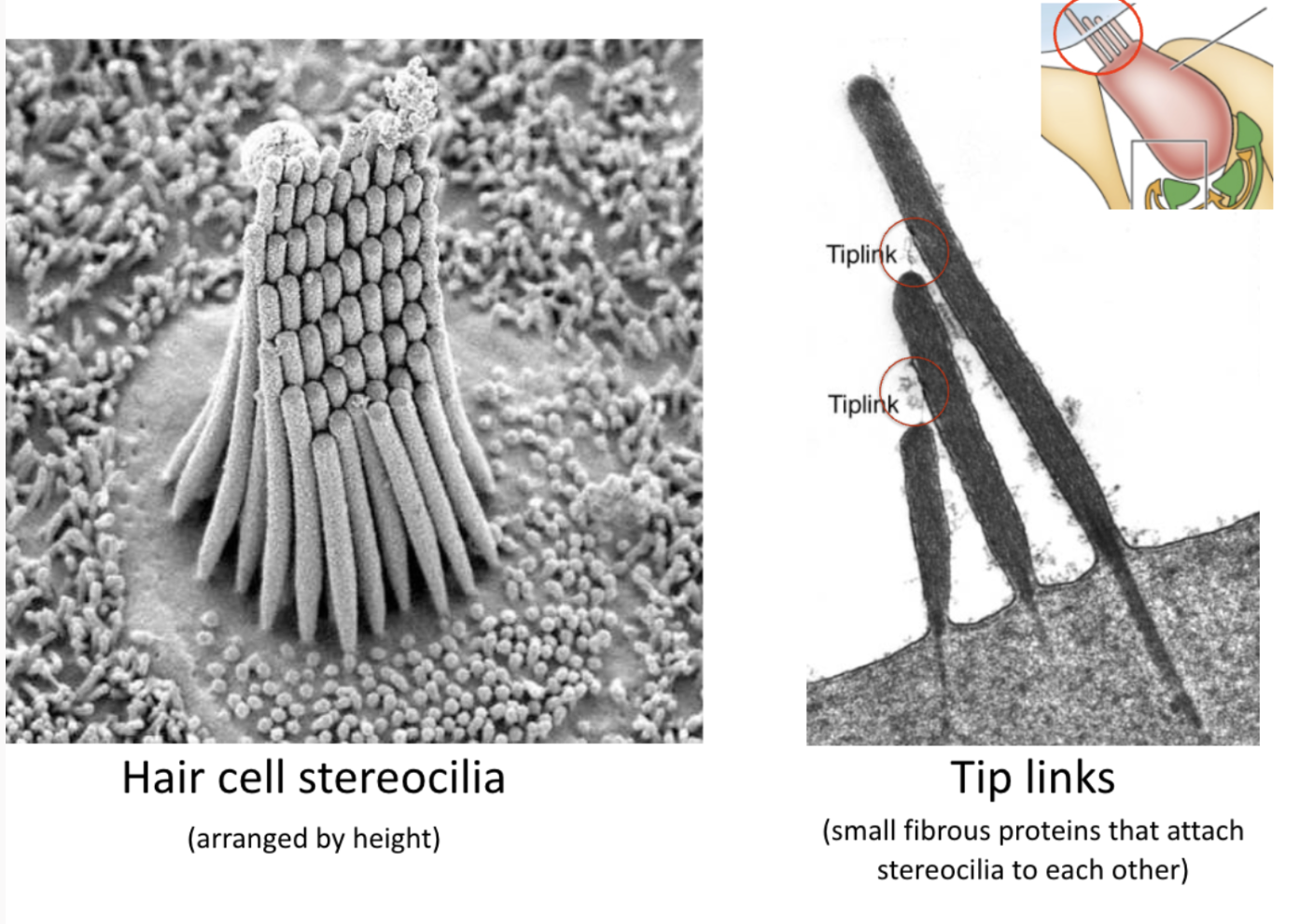
Arranged by height → important for how they transduce sound into an electrical potential
*don’t freely move independently of one another
Attached to one another by proteins called tip links
So if one moves in a certain direction, it pulls the other ones too
When stereocilia are deflected toward the tallest stereocilium, tip links physically pull open cation (K+/Ca2+) channels

How the process starts:
Tip links are like springs to attach the hair cells together, you can put tension on them, if a sound wave comes in and makes the springs move they sway as a unit, what causes the swaying is the basilar membrane going up and down?
Springs are attached to the side of one and the top of another, ion channel
When stereocillia are deflected towards the tallest stereocillia, it puts tension on tip links and yanks open ion channels (have trap doors) when stereocillia move in a direction, they open ion channels
Sounds that cause the stereocilia to sway, even only very slightly, increase the tension on the tip links and pop open the ion channels to which they are attached
The channels snap shut again in a fraction of a millisecond as the hair cell sways back
The ion channels of stereocilia thus resemble trapdoors, with springlike mechanisms in the top links’ anchors providing the force to rapidly shut the channels after they’ve been pulled open
So in a real sense, a stereocilium ion channel is spring-loaded with a hair trigger
Auditory transduction: The bending of Stereocilia opens cation channels
At top of each stereocilia, is an ion channel
Way down low is the basilar membrane that moves up and down in response to a sound wave, then shakes these stereocillum, when they go towards the tallest, tip links stretch open the ion channels and the ions flow in (bring pos charge) and depolarize inner hair cells.
*It goes right because if it goes left there's no tension on the springs for the channels to open, this is why they are arranged by height

Hair cells transduce sound when their stereocilia are bent

The stereocilia project into the endolymph in the scala media
Stereocilia are wedged into tectorial membrane (gel like), physically in contact with the membrane, which is anchored on one edge
If there is turbulence from sound pressure, if basilar membrane is moving up and down, as it moves upward, it pushes inner hair cells up, and as they get deflected upwards, the tectorial membrane produces a shearing force on the stereocilia, deflecting/bending them, stereocilia shear/scrape the tectorial membrane (bend towards right, direction of tallest) up depolarization (open ion channels, action potentials, not fired from hair cells, from auditory nerve fibers, sends info to brain), bending left is hyperpolarization
This is the transduction event that generates an electrical signal and encodes information about sound
Opening tip-link channels allows K+/Ca2+ entry, depolarizing hair cells
Hair cell stereocilia protrude into the endolymph of the scala media, which has a high [K+] and an electrical potential of +80mV relative to perilymph, where the basal portion of the hair cells sit
Resting potential of hair cells is -45mV, creating a 125 mV negative potential (known as the endocochlear potential)
The mechanical opening of the stereocilium ion channels allows an inrush of K+ and Ca2+ ions.
This concentration gradient drives K+ (and Ca2+) into hair cells through cation channels when they are pulled open by sound waves
Scolamedia contains fluid called endolymph, high concentration of potassium ions in it, and in each hair cell there is a low concentration of K+, so when channels open, K+ flows into the cells, depolarizes hair cell, concentration gradient
Potassium + Calcium flow into the hair cell
Membrane becomes more positive, rapid depolarization of the hair cell
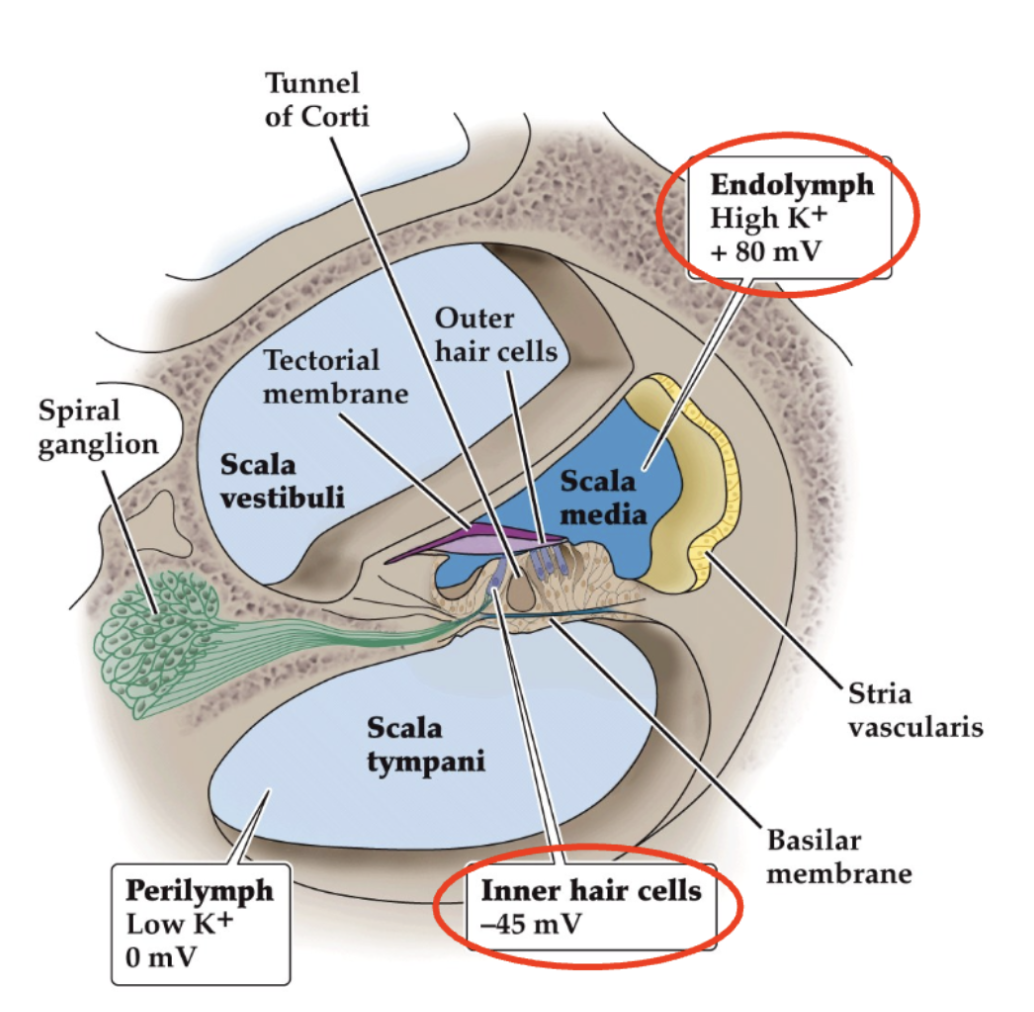
When inner hair cells depolarize via ion channels in the stereocilia they release glutamate onto SGCs

The influx of cations (K+ and Ca2+) depolarizes the hair cell (EPSP), causing the opening of voltage gated calcium channels leading to glutamate release
This initial depolarization leads to a rapid influx of Ca at the base of the IHC(inner hair cell), which causes synaptic vesicles there to fuse with the presynaptic membrane and release their transmitter contents, probably glutamate, from the base of the hair cell, triggering the production of action potentials in the afferent axons
This process excites the spiral ganglion cell or SGC (yellow, bottom; part of the auditory nerve), causing it to fire action potentials that travel to the brain–this is auditory transduction
SGCs fire action potentials, but hair cells are auditory receptors (not neurons) and do not fire action potentials!
Just like neurons have voltage gated calcium channels, these hair cells have them too, when there is enough depolarization. Inner hair cells release glutamate, vesicles in hair cells, but hair cells aren't firing action potentials, don’t have to. Signal doesn’t have to travel down a long axon, the positive charge just has to diffuse a little bit, activating VGCaChannels, vesicle fusion, release glutamate, spiral ganglion cell (makes up nerves) projects all the way into the brain, these cells fire AP’s cuz its a long way to the brain, then the brain interprets the signal
Hair cells are very sensitive to tiny changes!
It’s like this building, it sways 5cm at the top due to wind, which is approximately the same amount of relative deflection of stereocilia that drives a detectable signal in an inner hair cell

Action potentials encoding information about sound frequency are sent to primary auditory cortex (A1) via the thalamus for sound frequency perception
Action potentials ultimately end up in the cortex
Inputs cross over from left to right side (opposite side of the brain controls the other side of the body)
Decosate = cross over
Projections go directly into the brain stem, thalamus, to auditory cortex
Left side is responsible for hearing in your right ear
Cochlea is where auditory transduction and sensation happens
Perception starts in primary auditory cortex
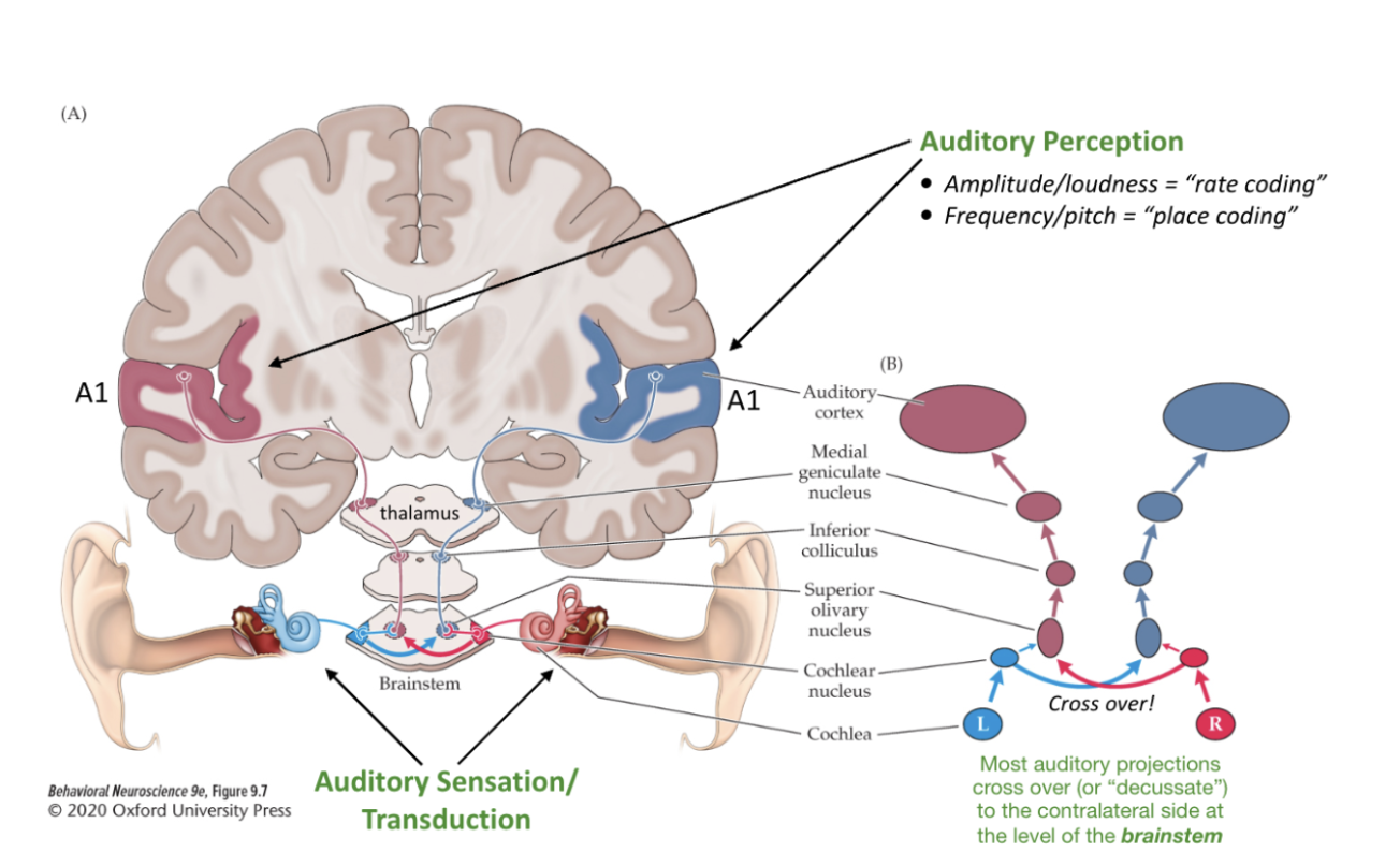
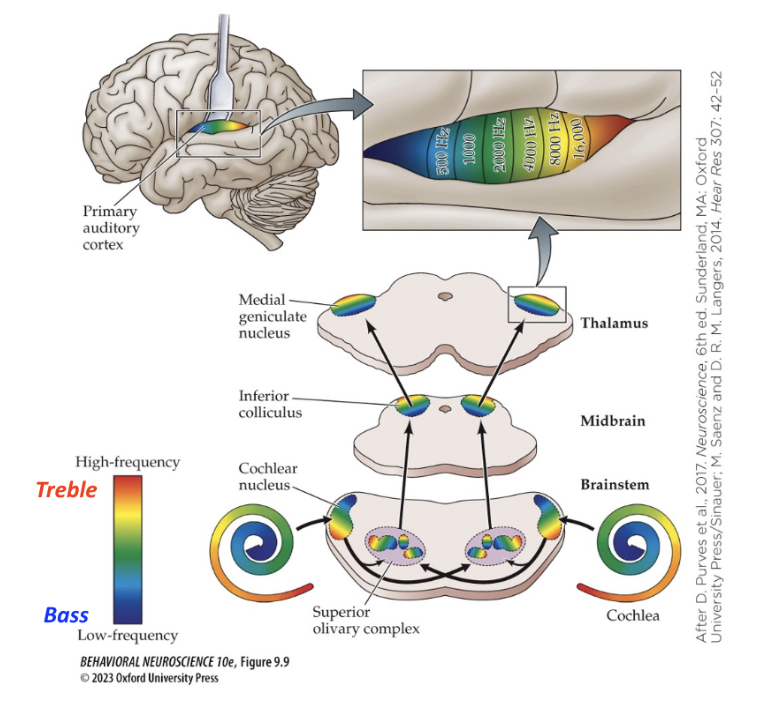
Place coding: Primary auditory cortex (A1) has a “tonotopic map” of sound frequencies preserved from the cochlea
Loudness = rate coding = number of action potentials
Louder, same areas activate but just more action potentials
Frequency/pitch = uses place coding, projections of the auditory nerve that come from a specific place on the basilar membrane project to a specific place on the auditory cortex
Auditory cortex in temporal lobe, there’s different bands
Goes from low to high hz, so neurons in those regions respond specifically to those frequency
So a high pitched/frequency noise would make action potentials in that certain band, stimulates the neurons
The frequency from the cochlea is preserved all the way to the level of A1 (place coding), (frequencies stay separate, rainbow colors don’t mix, frequencies don’t mix), if its high, it jostles the hair cells, then the specific auditory cortex region will be activated
→ This is tonotopy(the arrangement/structure of the cochlea by frequencies)
There is a tonotopic map of different sound frequencies that are preserved
Tonotopy explains how we perceive different frequencies “pitches”, “loudness” (amplitude) is encoded by increased firing rate (known as “rate coding”)

Every level of the auditory system shows orderly, segregated tonotopic mapping of the spectrum of audible frequencies
The neurons within all levels of the auditory pathway display tonotopic organization, they are arranged in the form of an orderly map of the auditory frequencies to which they respond
This organization begins with the cochlea
Because the inner hair cell fibers exiting the cochlea are organized according to their points of origin along the basilar membrane, analysis of the auditory projection reveals an orderly map from low frequencies to high frequencies
This can be seen when the responses of auditory brain regions to tones of different frequencies are mapped
Human auditory cortex responses to random sounds vs. speech
Top: fMRI scans showing that pure tones “noise” activate primary auditory cortex (A1) in the temporal lobe
Auditory cortex lights up (increased activity)
Bottom: Speech sounds also activate A1, but additionally activate specialized secondary or “higher” auditory cortices (ex. Wernicke’s Area) important for interpreting speech sounds
Wernike’s Area: (Posterodorsal temporal lobe) is uniquely human, and critical for speech/language comprehension (a “higher order” auditory computation)
Wernike’s area - important for language comprehension (writing, sign language also) main job is to make sense of language (its activated from speech, puts sounds together to make meaning)
Damage to Wernike’s area → results in loss of the ability to understand speech, called Wernike’s aphasia, in their mind they can make sense(they know what they want to say, but it comes out uninterpretable/random)
Patients have trouble comprehending others, and don’t recognize errors in their own “fluent” speech
World salad - speech is filled with random words
Way more activity for speech
Some frontal lobe activation (for making meaning of speech)

What do outer hair cells do?

3:1 ratio, more outer hair cells than inner hair cells
Not directly related to auditory transduction
But important
Frequency discrimination
Sound amplification
They fine-tune cochlear responses by expanding and contracting in response to movement of the basilar membrane (they enhance frequency discrimination and amplify sound
We can discriminate sounds by 2Hz
Outer hair cells in cochlea amplify sound and fine tune hearing sensitivity
Contract and expand in response to depolarization by sound
Outer hair cells can lengthen and shorten in response to movement of basilar membrane
Arrow shows that when an outer hair cell depolarizes they shrink
Presto = react quickly (think music)
*Outer hair cells have motor proteins called prestins that cause the hair cell to shorten or contract in response to depolarization–this modifies the stiffness of the basilar membrane, helping with frequency discrimination and sound amplification
Depolarization because basilar membrane goes up, being opens channels, K+ influx leads to the activation of prestin proteins and causes the contraction of the outer hair cells
Movement injects mechanical energy into the basilar membrane
When it contracts it brings the membranes together
Cochlear amplifiers, can increase standing soundwaves
Blocking outer hair cell mobility (ex. with drug furosemide) decreases the amplitude of sound evoked standing waves on the cochlea, reducing sound amplification and discrimination
Motility = ability to move
Outer hair cells have this ability to lengthen and shorten because of the prestins
Hyperpolarization = expand
Those mice had their outer hair cells removed and are auditorily impaired

Outer hair cells have contractile proteins called Prestins, which contract in response to depolarization
Put electrode in bottom of hair cell
Basilar membrane is causing the contractions
This example shows movement in response to injected current rather than basilar membrane movement, but note the immediate motility (lengthening/shortening) of this “dancing” outer hair cell–this process amplifies sound and enhances frequency discrimination in the cochlea
How do we localize where sounds are coming from if we can’t see the source of the sound?
Auditory cortex has nothing to do with this
Brainstem is where
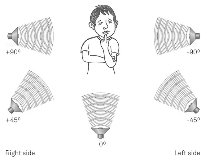
Duplex theory: We localize sounds in horizontal space using interaural intensity and interaural time differences
There are two levels to this if you’re talking about a high pitched or low pitched sound
High pitch/frequency is easier
Low pitch is more difficult
Interoral = between two ears
For high frequency: can detect differences in intensity
If there's a high pitch sound, if it's closer to your left ear than right, on your auditory nerve in left ear there will be a lot of action potentials
But on the auditory nerve of your right ear, it will be less
It will sound louder in the ear its closest to, cuz head is a buffer, will reduce loudness to the ear that has ur head in the way (right ear in this case)
As frequency becomes higher, it becomes easier to differentiate, because of intraoral
Low frequency sounds, no matter where they originate, reach the two ears with comparable intensity
For these frequencies, differences in times of arrival are the principal cues for sound localization (and at very low frequencies, neither cue is much help; this is why you can place the subwoofer of an audio system anywhere you want
Big sound waves (low frequency sounds) → so it hits both ears equally, so the brainstem computer intraoral time difference (of hitting the right vs. left ear)
Onset disparity
Ongoing phase disparity - maybe it hits the left ear first and short delay to the other ear
Interaural intensity differences (IIDs): For high frequency sounds, the head casts a “sound shadow” such that the sound is perceived louder by the ear closer to the source of sound through rate coding (A & B)
Result from comparison of the intensity of the sound–the physical property that we consciously perceive as loudness (in the left versus right ear)
Interaural temporal/time differences (ITDs): For low frequency sound, our auditory brainstem nuclei keep track of the disparity (or latency) for sound to reach one ear vs. the other (C; next three slides)
They arise because one ear is always a little closer to an off-axis sound than is the other ear
Brainstem coincidence detection for low frequency (low pitch) sound localization

For low freq sounds, the brain uses interaural temporal differences
Projections from auditory nerve, action potentials make their way to the brainstem
Axons from the right also go up the neurons
Delay lines

Coincidence detectors = work on spatial summation (multiple incoming neurons creates the strongest summation)
Maximally excited by a specific latency difference between inputs from the two ears, corresponding to particular place in space
Dog + running scenario
Low rumbling noise hits left ear first
Left ear fires action potentials
Action potentials go to neurons
Activates all of the neurons
With a delay, the sound then hits the right ear
Then same thing happens on the right side (goes to neuron 1, then 2, then 3(farthest))
Neuron 3 receives action potentials from both sides, it fires the most, and that perceives the sound that is coming from the dog
Neuron 2 if sound coming from right in front of you
Neuron 3 is a coincidence detector
*textbook images
Sound to the left of the midline…
One neuron is going to be maximally stimulated and that tells you where the sound comes from
When hearing goes wrong: There are three major categories of hearing loss and deafness *image

Conduction deafness
Hearing impairment associated with pathology of outer or middle ear cavities, disorders of the outer or middle ear prevent vibrations produced by auditory stimuli from reaching the cochlea
In one form of conduction deafness, the ossicles become fused and can no longer transmit sound vibrations effectively
Bones start to grow together, they don’t vibrate as much
Can be corrected with hearing aids, amplify what the ossicles/eardrum is now lacking
Anything that affects the outer or smth ear
Sound waves are physically prevented from reaching the cochlea.
Sensorineural deafness
Issue with the cochlea or with its projections that interferes with auditory transduction sound waves → action potentials)
Noise pollution, infections, toxins, trauma, hereditary disorders
Can cause permanent damage to hair cells, irreversible
The basilar membrane is going up and down, shatters hair cells
Some people are born without functional hair cells
Even if vibrations are successfully conducted to the cochlea, the sensory apparatus of the cochlea (the organ of Corti and the hair cells it contains) may fail to encode the ripples created in the basilar membrane into the volleys of action potentials that the brain understands
The middle ear works fine, but the structures that convert physical waves into electrical signals are destroyed.
Central deafness
Hearing impairment caused by a lesion or a stroke in auditory brain centers (ex. A1 → primary auditory cortex)
Strokes also cause deafness
People show various acoustic reflexes mediated by the brainstem, such as a startle response to a sudden, loud noise, and they deny hearing the sounds to which they are reacting
Inability to discriminate high frequency sounds
Affected individuals can often hear a normal range of pure tones but are impaired in the perception of complex, behaviorally relevant sounds
Rare
Deafness to certain sounds, words, or frequencies
Wernike’s area - if a stroke here, that would be central deafness
The entire ear works perfectly, but the brain's auditory processing centers are damaged.
How loud is too loud?
Kills inner hair cells
Auditory transduction can no longer take place
People who listen to music using headphones at 89 decibels or louder for more than 5hrs/week expose themselves to levels known to induce permanent hearing loss
Noise pollution and very loud sounds like industrial noise, loud concerts, engines, and gunfire can severely damage the cochlea, and hair cells bare the brunt of the damage–stereocilia can shatter, looking like a flattened forest

Cochlear damage due to loud noise
Damage in middle band between apex and base
Don't grow back
Permanent hearing loss
Left image: Normal cochlea showing healthy hair cells and stereocilia lining the organ of Corti
Right image:
Top: Hair cells are completely missing from a wide extent of the cochlea, corresponding to the frequency of intense sounds that the person was exposed to
Bottom: Stereocilia have been crushed/shattered by the noise exposure, like a flattened forest
Perception of approximately what sound frequency range would be most affected by the damage shown in “B” above?
Mid frequency

Lecture 17 Learning Objectives:
Appreciate some general guiding principles of sensory systems (Ch. 8)
Understand how sound travels in air, the frequency range of human hearing, and the difference between frequency and amplitude of sound waves (and how frequency and amplitude are perceived by the brain)
Learn the anatomy of the human ear (outer, middle, and inner ear), with special focus on the cochlea (inner ear)
Discover how air pressure waves cause turbulent waves in cochlear fluid within the cochlea, and how information about sound frequency is encoded in the nervous system through the concept of tonotopy
Lecture 17 Thought questions
Describe the general flow of sensory information from peripheral sensory organs in the head/body to sensory cortices of the brain. Which main cell types and brain structures are involved? Are all the cells involved in this process considered neurons? What role does the thalamus play in this process?
What is a sound wave? What is the amplitude of a sound wave, and how is it perceived? What is is the frequency of a sound wave, and how is it perceived?
Describe the anatomy of the outer, middle, and inner ear. How does the ear amplify sound waves? Where does auditory transduction take place? What is the organ of Corti and where is it located? What are “hair cells,” and what are their functions?
Describe what is meant by the phrase, “tonotopy is preserved from the cochlea all the way to primary auditory cortex.” How does this concept relate to the perception of pitch? Describe the physical properties of the basilar membrane that allow for frequency tonotopy in the organ of Corti along the length of the cochlea.
Lecture 18 Learning Objectives:
Appreciate the anatomy (structure) of inner hair cells and how the arrangement of stereocilia relate to its functional role in auditory transduction
Learn the basics of auditory projections from the cochlea to the brain, and the concept that tonotopy is preserved from the cochlea all the way to the level of primary auditory cortex (A1) in the brain
Learn the importance of higher auditory cortices in auditory processing/comprehension using the example of Wernicke’s Aphasia
Lecture 18 Thought Questions
What’s the difference between inner and outer hair cells? Which one of these two is specifically responsible for auditory transduction?
What are stereocilia? What are tip links? How do tip links physically/mechanically open cation channels? Which way do stereocilia have to be displaced in order to open cation channels, toward the tallest, or toward the shortest stereocilium?
Which ion(s) is/are responsible for the depolarization of inner hair cells? Do inner hair cells fire action potentials? Are inner hair cells neurons? Which neurotransmitter do inner hair cells release?
Describe the tonotopy preserved from the cochlea at the level of primary auditory cortex (A1). How is frequency (pitch) “laid out” in A1? Describe “place” vs. “rate” coding and which aspects of the perception of sound each is thought to primarily contribute to in the auditory system. Give an example of a “higher-order” auditory cortical area and how it contributes specifically to speech perception. Generally speaking, what do “higher” sensory cortices do?
Lecture 19 Learning objectives:
Appreciate the important role of outer hair cells in frequency discrimination and sound amplification (“cochlear amplifiers”)
Understand the brainstem mechanisms by which we localize high and low frequency environmental sounds
Learn the three major categories of hearing loss and deafness and some ways these forms of hearing loss can arise
Lecture 19 Thought Questions
Describe the functional role of outer hair cells. What role do they play in modulating the basilar membrane and how do they accomplish this task? What are prestin proteins and how do they work?
How do we localize low frequency sounds in horizontal space? How do we localize high frequency sounds? Are cortical brain structures (e.g. A1) thought to be involved in the basic ability to localize sounds in horizontal space?
Describe the three major forms of hearing loss/deafness and how they may come about. Which aspects of the auditory system are impacted in each form of hearing loss/deafness?