Unit 3 Chapter 9: Neurons: Cellular and Network Properties
 Call Kai
 Learn
Learn Practice Test
 Spaced Repetition
 Match
Match Flashcards
 Knowt Play
1/124

There's no tags or description
Looks like no tags are added yet.
Name | Mastery | Learn | Test | Matching | Spaced | Call with Kai | Chat |
|---|
No analytics yet
Send a link to your students to track their progress
125 Terms
What is the purpose of the sensory division of the PNS?
Sends information to the CNS through afferent (sensory) neurons.
What is the purpose of the efferent division of the PMS?
Takes information from the CNS to target cells via efferent neurons.
How does the is the enteric nervous system controlled?
Can act anonymously or can be controlled by the CNS through the autonomic division of the PNS.
Why are neurons important?
They are specialized for receiving, processing, and spreading signals (information) around the body very quickly.
What are dendrites?
They receive incoming chemical signals through receptors located on their membrane surface.
What is the axon hillock?
Acts as an “integrating center” for the neuron.
Processes the meaning of signals collected at dendrites and decides whether or not to pass that information along.
What are axons?
They transmit electrical impulses (action potentials) to their terminals through voluted-gated ion channels.
What is a synapse?
The region where an axon terminal communicates with its postsynaptic target cell.
What is a multipolar efferent neuron?
A type of neuron with multiple dendrites and a single axon, primarily responsible for transmitting motor signals from the central nervous system to muscles and glands.
What are pseduounipolar neurons?
Sensory neurons that have a single process called the axon. During development the dendrite fused with the axon.
What are bipolar neurons?
Sensory neurons that have two relatively equal fibers extending off the central cell body.
What is an anaxonic neuron?
They are interneurons of CNS that have no axon.
What are multipolar interneurons neurons?
They are interneurons of CNS that are highly branched but lack long extensions.
What are multipolar efferent neurons?
Efferent neurons that typically have 5-7 dendrites, each branching 4-6 times. A single long axon may branch several times and end at enlarged axon terminals.
What is the job of sensory neurons?
Sensory neurons are responsible for transmitting sensory information from the peripheral sensory receptors to the central nervous system (CNS), allowing the body to respond to stimuli.
What is the job of interneurons?
Interneurons serve as connectors within the central nervous system (CNS), processing information between sensory and motor neurons, thus facilitating communication and reflexes.
What is the job of efferent neurons?
Efferent neurons convey commands out to control effectors around the body.
What are glial cells and why are they critical?
Glial cells in the central nervous system (CNS) are the “support staff” that keep the “elite” neurons working at peak capacity.
They are crucial for normal brain development, both prenatal and post-natal.
What are the functions of ependymal cells?
They form a barrier between brain tissue and the ventricles.
Those associated with the choroid plexus secrete cerebral spinal fluid (CSF) into the ventricles.
Source of neural stem cells.
What are the functions of astrocytes?
Take up potassium, water, and neurotransmitters.
Secrete neurotrophic factors.
Help form the blood-brain barrier.
Provides substances needed for ATP protection.
What are the functions of microglia?
Act as scavengers by removing pathogens.
Remove underperforming neurons (neural pruning).
What are the functions of oligodendrocytes?
Form myelin sheaths around neurons.
Provide metabolic support to neurons.
What are the two support glial cells of the PNS?
Schwann Cells (neurolemmocytes)
Satellite Cells
What are schwann cells and what are their basic functions?
Each Schwann cell forms myelin around a small segment of one axon.
They form myelin sheathes and provide metabolic support.
Secrete neurotrophic factors.
What are satellite cells and their basic function?
They support/ protect neuron cell bodies outside the CNS (at the autonomic ganglia).
These cells are the functional unit of the nervous system.
Neurons
These cells are the only part of the immune system that has access to the nervous system.
Microglia
These cells form the myelin sheath in the peripheral nervous system.
Schwann cells (neurolemmocytes)
These support cells provide neurons with essential biomolecules for ATP production and form the blood-brain barrier.
Astrocytes
These cells line the walls of the fluid-filled chambers of the brain (ventricles).
Ependymal cells
These cells can myelinated more than one neuron.
Oligodendrocytes
What is a neurons resting membrane potential set by?
It’s set by the normal disequilibrium of K+, Na+, Ca2+, and Cl-.
Ion channels are required to allow movement of these ions across the membrane.
What is a typical neurons rest?
-70 mV
What transmembrane protein in the dendrites and cell body controls ion permeability and allows for changes in a neuron’s membrane potential.
Gated-channels
What is the importance of chemically gated channels in the dendrites and the cell body?
-They are the most common
-They detect signal molecules from nearby neurons
What is the importance of voltage-gated channels in the dendrites and the cell body?
-They are less common
-Involved in complex forms of neural plasticity (learning)
Note: the membrane potential required to open these channels varies from channel to channel.
What is the importance of mechanically-gated channels in the dendrites and the cell body?
-They are much less common.
-Limited to sensory neurons that detect touch and temperature sensations sound the body.
What happens if Na+ or Ca2+ channels open?
-Ions will influx (move into cell)
-Charge becomes more positive (depolarization)
What happens if a K+ channel opens?
-Ion will efflux (move out of cell)
-Charge becomes more negative (repolarization)
What happens if a Cl- channel opens?
-Ion will influx (move into cell)
-Charge becomes more negative (hyperpolarization)
What determines the strength of a stimulus?
The amount of signal molecules that bind to the neuron’s receptors.
What does the strength of a stimulus determine?
how many receptor channels open
how many ions enter/leave the neuron
the degree of change in membrane potential
How does a faster rate (higher frequency) of signal molecules affect membrane potential?
The faster the rate, the greater the change in the neuron’s membrane potential.
How do more sources of incoming signals affect the strength of a stimulus?
More neurotransmitters bind to receptors, causing more channels to open, more ion movement, and a greater change in membrane potential.
What are graded potentials?
Changes in membrane potential at the dendrites and cell body when a neurotransmitter binds.
Why are they called graded potentials?
Because they vary in strength and effect.
What happens when signal molecules bind to receptors?
Ion channels open, ions move down their concentration gradient.
What happens when Na+ enters the cell?
It causes a small depolarization that dissipates over time and space.
How does the amount of signal molecules affect depolarization?
More signal molecules causes more receptors to bind, more Na+ channels to open, more Na+ to enter, and stronger depolarization.
What happens if fewer signal molecules are received?
Fewer receptors bind, fewer Na+ channels open, and depolarization is weaker.
Where do graded potentials occur?
At the dendrites of the cell body.
What are the two types of graded potentials?
Excitorarty post-synaptic potential (EPSP)
Inhibitory post-synaptic potential (IPSP)
What is excitatory post-synaptic potential (EPSP)?
-When depolarization happens in the dendrites or cell body.
-They are caused by neurons binding excitatory neurotransmitters.
-movement of Na+ enters the cell causing an EPSP.
What is inhibitory post-synaptic potential (ISPS)?
-When hyperpolarization occurs in the dendrites or cell body.
-They are caused by neurons binding inhibitory neurotransmitters.
-movement of Cl_ enters the cell or K+ leaves the cell causing an IPSP.
What happens when the neuron’s trigger zone detects sufficient depolarization from EPSPs and IPSPs in a short period of time?
An action potential is created.
Where is the trigger zone in most neurons?
Axon hillock
What must the sum of all ESPSs and ISPSs reach to generate an action potential?
A threshold voltage, typically -55 mV in neurons.
What type of signal is an action potential?
An all or nothing electrical (depolarization) signal.
Do any action potentials vary in size?
No, they are the same in all types of neurons.
What happens to graded potentials over time and space?
They dissipate (become weaker)
What are subthershold graded potentials?
Graded potentials below threshold that do not generate an action potential.
What are suprathreshold graded potentials?
Graded potentials above threshold that initiate an action potential.
Where is an action potential initiated?
At the axon hillock.
What determines whether a neuron generates an action potential?
Whether graded potentials are below or above a threshold.
What type of gated channels opens at the axon hillock when threshold voltage is reached?
Voltage gated Na+ channels.
How many inputs do neurons receive from other neurons.
100s-1000s
What must neurons integrate when deciding whether to generate an action potential?
Many excitatory and inhibitory stimuli.
What is integration?
The summation of ESPSs and IPSPs signals received by a postsynaptic neuron.
How does integration occur?
Through spatial and temporal summation of multiple graded potentials happening simultaneously.
What is spatial summation? *multiple excitatory inputs.
When more than one pre-synaptic neuron stimulates the post-synaptic neuron simultaneously.
Note: all EPSPs and IPSPs add together across the surface area of the neuron and converge at the axon hillock to determine if threshold is reached or not.
What are the 3 steps on spatial summation? *multiple excitatory inputs.
Three excitatory neurons fire. There graded potentials separately are all below threshold.
Graded potentials arrive at trigger zone together and sim to create a supra-threshold signal.
An action potential is generated.
What is spatial summation? *mix of ESPSs and IPSPs
Simultaneously ESPPS and IPSP can co-occur across the surface of a neuron.
What are the steps of spatial summation? *mix of ESPSs and IPSPs
One inhibitory and two excitatory neurons fire.
The summed potentials are below threshold, so that no action potential is generated.
What is temporal summation?
A kind of integration influenced by the frequency or timing of EPSPs or IPSPs.
When does temporal summation occur?
-If there is an increase in the frequency or rate of incoming EPSPs.
Characteristics of Graded Potentials:
-Input signal
-Usually dendrites and cell body
-Mechanically, chemically, or voltage-gated channels
-Usually Na+, K+, Ca2+
-Depolarizing or Hyperpolarizing
-Depends on initial stimulus, can be summed
-Entry of ions through gated channels
-No minimum level required to initiate
-Two signals coming close together in time will sum
Characteristics of Action Potentials:
-Regenerating conduction signal
-Trigger zone through axon
-Voltage-gated channels
-Na+ and K+
-Depolarizing
-All or none phenomenon; cannot be summed
-Above threshold graded potential at the trigger zone opens ion channels
-Threshold stimulus required to initiate
-Refractory period: two signals too close together in time cannot sum
What is propagation?
Is conduction of action potential along a non-mylenated axon.
What is saltatory conduction?
Is conduction of action potential along myelinated axons.

What is the activation threshold for this neuron?
Approx. -55 mV

In what state is the neuron between steps 3 & 5? What channel?
Depolarization- Na+ enters the neuron.
Voltage-gated Na+ channels
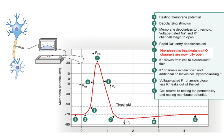
Between steps 5-7, what type of ion channel creates that part of the action potentials?
Voltage-gated K+ channels.

What is happening between steps 5-7?
Repolarization- K+ leaves the neuron.
Between steps 7 and 8, what type of ion channel creates that part of the action potential?
Voltage gated K+ channels, which are still open as the membrane hyperpolarizes.
What happens between steps 7 and 8?
Hyperpolarization- K+ continues to leave the neuron making it even more negative than resting potential.
What are the steps in an action potential?
Resting membrane potential.
Depolarizes stimulus.
Membrane depolarizes to threshold. Voltage-gated Na+ and K+ channels open.
Rapid Na+ entry depolarizes cell.
Na+ channels inactivate and K+ channels are now fully open.
K+ moves from cell to extracellular fluid.
K+ channels remain open and additional K+ leaves, hyper polarizing the cell.
Voltage-gated K+ channels close, less K+ leaks out of the cell.
Cell returns to resting ion permeability and resting membrane potential.
Voltage-Gated Na+ channels: At resting membrane potential, what is the position of the gates?
The activation gate is closed and the inactivation gate is open.
Voltage-Gated Na+ channels: What happens when the neuron is stimulated past threshold?
The activation gate opens.
Voltage-Gated Na+ channels: What happens when the activation gate opens?
Na+ enters the channel, and the neuron starts to depolarize.
Voltage-Gated Na+ channels: What happens after about 0.5 milliseconds?
The inactivation gate closes, and the channel becomes inactivated.
When do both gates return to their original positions?
About halfway through the hyperpolarization phase.
Voltage-Gated Na+ channels: What are the original positions of both gates?
The activation gate is closed, and the inactivation gate is open.
How many gates do voltage-gated channels have.
Only one
Voltage-gated K+ channels: what is the position of the K+ channel gate at resting membrane potential?
The gate is closed.
Voltage-gated K+ channels: What happens to the K+ channel gate when threshold is met?
The gate starts to slowly open.
Voltage-gated K+ channels: When are K+ channels fully open?
At peak depolarization, when Na+ channels are inactivated.
Voltage-gated K+ channels: What does K+ efflux alone create?
The repolarization phase
Voltage-gated K+ channels: What happens to K+ channels during repolarization?
They slowly close.
Voltage-gated K+ channels: What does the slow closure of K+ channels cause?
The membrane potential to hyper polarize a bit.
Voltage-gated K+ channels: When are K+ channels fully closed?
About halfway through the hyperpolarization phase.