General Bones and Spine
 Call Kai
 Learn
Learn Practice Test
 Spaced Repetition
 Match
Match Flashcards
 Knowt Play
1/69

There's no tags or description
Looks like no tags are added yet.
Name | Mastery | Learn | Test | Matching | Spaced | Call with Kai | Chat |
|---|
No analytics yet
Send a link to your students to track their progress
70 Terms
Axial skeleton includes
Skull, vertebral column, rib cage

Appendicular Skeleton
the upper and lower
extremities and hip bone

Red marrow skeleton
(hematopoietic); Humerus, Scapula, neck, metaphysis of femur, ilium
(mainly in the axial skeleton)

Yellow marrow skeleton
(mainly fat)

Skeletal Features
1. Articulations
2. Extensions and projections
3.Depressions
4. Passages
Articulations
1. Condyle
2.Facet
3.Head
Extensions and projections:
1.Crest
2.Epicondyle
3.Line
4.Process:
5. Protuberance
6.Spine:
7. Trochanter
8. Tubercle
9. Tuberosity
Depressions
Alveolus
Fossa
Fovea
Foveola
Sulcus/Groove
Passages
Canal: tubular passage or tunnel in a bone
Fissure: a slit through a bone
Foramen: a rounded hole of short length in a bone
Meatus: an opening or passage, usually air-filled
General Features of Long Bones
•Epiphyses
•Metaphysis
•Marrow (medullary) cavity
•Articular cartilage
•Diaphysis
Compact bone and spongy bone

Epiphyses
1.The end of a long bone.
2.Articulates with another bone.
3.Capped by articular cartilage.
4.During growth, it has a physis (growth cartilage/growth plate).
Metaphysis
growth zone between the epiphysis and the diaphysis during development of a long bone
2. The cone-shaped region connecting the diaphysis to the epiphysis.

Diaphysis
shaft of a long bone

Each limb is supported by a
cingulum (girdle)
Pectoral girdle
scapula + clavicle → supports the free upper limb
Hip bone supports
the free lower limb
Most joints of the appendicular skeleton are
synovial joints.
Epiphysis vs. Apophysis
Epiphysis = articulation/joint
It is involved in a joint and therefore is covered with articular cartilage.
Apophysis = attachment
Apophysis
Bones also have projections for the attachments of muscles and ligaments, but these projections do not get involved in joints and do not have articular cartilage.
During growth, an apophysis has a
has a physis or growth cartilage. Eventually over time, the physes of both epiphyses and apophyses finish growing and completely ossify (synostosis).
Both an epiphysis and an apophysis have a physis during growth. Eventually, that growth cartilage ossifies:
Epiphysis → epiphyseal line
Apophysis → apophyseal line
If the physis is interrupted by disease or malnutrition, the cells lay down bone without a filigree of new cells, producing a dense line called
lines of interrupted growth or Harris lines.
. Note the appearance of these lines extending into the metaphyses of the long bones

]fractures (fxs) occur at the ends of growing long bones through portions of the Physis called
Salter-Harris
Fractures that go through the Epiphysis are
serious/ BAD
different types of Salter-Harris fxs are based on the
The important proliferative cells are on the epiphysis side of the physis. Fxs that Epiphysis Physis Metaphysis involve the epiphysis side of the physis are more serious . fx destroys portions of the epiphysis-side of the physis are more serous

Salter-Harris Fracture Classification: Type 1
-straight across the physis, but not injuring the proliferative cell layers

Salter-Harris Fracture Classification: Type 2
the fx goes across the growth plate and away into the metaphysis
Salter-Harris Fracture Classification: Type 3
he fx goes through part of the physis and through part of the epiphysis, thus, destroying part of the proliferative cell layer

Salter-Harris Fracture Classification: Type IV (4)
the fx goes through the epiphysis, physis and metaphysis
Salter-Harris Fracture Classification; Type V (5)
proliferative cell layer is crushed. This type of fx is uncommon

the most common Salter-Harris fx in the upper limb.
Type II
the worst form of Salter-Harris fx, with the worst prognosis because of complete interruption of growth.
Type V
Structure of a typical vertebra
7 cervical
12 thoracic
5 lumbar
fused 5 sacral vertebrae into forming the sacrum
3-4 fused coccygeal vertebrae forming the coccyx
The center of gravity for the body resides slightly in front of the body of
S1
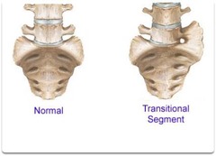
Lumbarization
Occurs when the first sacral vertebra fails to unite with the remainder of the sacrum

sacralization
L5 vertebra may fuse with the rest of the sacrum
Intervertebral disks and numbering
numbered according the the superjacent vertebra.
Ex disk connecting vertebra C5 and C6 is IVD C5
The cervical vertebrae exhibit a curve called
cervical lordotic curve

Degree of curve for cervical vertebrae
Normal 20-40 degree
The thoracic vertebrae show an anterior concave face called what curve
Thoracic kyphotic curve

Thoracic kyphotic curve degree of the curve
Normal 20-40
The lumbar vertebrae curve is called and what is the degree
lumbar lordotic curve (facing posterior)
Normal is 30-50

Fused Sacrum has a curve called
Sacral kyphotic curve
kyphosis
hunchback/ abnormal anterior bending curve
Lordosis
abnormal posterior bending
Herniated disks occur most frequently at these inflection points
IVDs C5 or C6 and IVDs L4 and L5
Noteworthy landmark on the back
The spinous process of C7
The first palpable (and longest) spinous process of the cervical vertebrae so C7 is called
vertebra prominens
Most common spinous process involved in a clay shoveler's fracture.
C7
Intervertebral foramen
each side for the emergence of a spinal nerve.

thoracic vertebrae bear facets have the same number as
respective rib of the same number.
One of the most common causes of kyphosis is
age-related degeneration of the spine
Scoliosis is classified as a Cobb angle of curvature of greater than
10°
The Cobb angle is the angle measured by
intersecting lines of the upper end-vertebra and lower end-vertebra of the structural curve.
vertebral column motion depends on
the architecture of the zygapophyseal joints which changes from one region to another.
Cervical and thoracic vertebrae motion
best at permitting rotational movements.
Lumbar vertebrae motion
do not permit much rotational movements and such movements put stress on their zygapophyseal joints.
The entire column permits lateral bending. The cervical and lumbar vertebrae are
good for flexion and extension of the body
A Risser grade of 5 means
there is not growth plate of the apophysis evident and thus, it is safe to assumes that growth has stopped and that it is unlikely for the scoliosis to progress.
Primary Curve (Scoliosis)
The largest, most rigid, and original structural curve in scoliosis that dictates the main treatment plan.
Secondary Curve (Scoliosis)
smaller, more flexible compensatory curve created naturally by the body to keep the head and torso balanced over the pelvis.
Anterior boundary of intervertebral foramen
Portions of two vertebral bodies and the intervertebral disk.
Posterior boundary of intervertebral foramen
The zygapophyseal joint and portions of the articular processes.
Superior/inferior boundaries of intervertebral foramen
Inferior vertebral notch above and superior vertebral notch below.
Location of intervertebral disk in intervertebral foramen
Located at the lower 1/3rd of the foramen.
Interlaminar space
The space between the laminae of successive vertebrae.
Interlaminar space in lower lumbar spine
Wider and less blocked compared to higher regions of the vertebral column.
Effect of flexion on interlaminar spaces
Body flexion widens the interlaminar spaces, facilitating clinical access to the vertebral canal.
Pars interarticularis (isthmus)
The portion of the lumbar vertebra lamina that anchors the superior articular process.
An important region of the lumbar vertebra
Pars interarticularis (isthmus)