Gross Anatomy: Knee
 Call Kai
 Learn
 Practice Test
 Spaced Repetition
 Match
 Flashcards
 Knowt Play
1/39

There's no tags or description
Looks like no tags are added yet.
40 Terms
patellofemoral joint
the point where the kneecap and femur are connected in the trochlear groove
not a true skeletal joint: patella separated from the femur by a synovial bursa rather than by a capsule anchored to the bones

functions of the patella
1. holding the tendon away from the joint to improve mechanical advantage in extension
2. protection of the joint
3. holding the tendon in position on the anterior of the knee
patellar tendon stabilization
1. tendon tends to pull to the outside, so the lateral condyle has more a prominent ridge to resist the movement
2. a patellar retinaculum holds tendon in place
3. lower fibers of the lateral and medial vastus muscles have an oblique orientation, placing tension on the tendon

which part of the menisci is vacularized?
the thicker outer portions meaning that the inner thin portions do not readily repair themselves
what bone are the menisci attached to?
tibia - along the outside margin of the tibial condyle and also into the fibrous joint capsule via the coronary ligament
what is the shape of the menisci?
crescent shaped
lateral vs medial coronary ligament
lateral - fibers are longer and allow more movement
medial - tighter and more vulnerable to tearing if the capsule is damaged

generally, what happens to the menisci during movement?
the free inner curvature allows for some mobility
the shift position and shape slightly during joint movement, assuring a high degree of congruence and shock absorption
what happens to the knee if the menisci are absent?
if menisci are absent due to injury, then deterioration of the knee joint is greatly accelerated
what happens to the joint/menisci during knee flexion and extension?
the femur performs a combination of rolling and gliding on the menisci
in the last 30 degrees of extension, the femur rotates medially (tibia rotates laterally) which provides a locking mechanism for the joint.
when the knee is in full extension, the joint is close-packed and will not flex until weight is removed and/or the femur rotates laterally.
*achieves stability w/o muscular effort
what muscle normally performs the unlocking rotation at the knee joint when in full extension?
popliteus muscle
development of ligaments of the knee
the fibular/tibial collateral ligaments and the anterior/posterior cruciate ligaments were arranged as collaterals of each of the two condylar joints
as the knee matures, the two synovial capsules joined into one and the two cruciate ligaments became enclosed by the fibrous capsule
medial (or tibial) collateral ligament
flat and fused into the medial side of the fibrous capsule
arises from the side of the head of the tibia well below the capsule and deep to the pes anserinus and attaches to the side of the medial condyle of the femur
tight in extension
on its deep surface, attached to the medial meniscus through capsule -- both tear

lateral (or fibular) collateral ligament
cordlike, about the diameter of a probe handle
attaches to the head of the fibula and to the side of the lateral femoral condyle
tensed in extension
normally covered by the IT band
does not attach to the fibrous capsule -- can easily fit a probe between the two

anterior and posterior collateral ligaments
have the special function of holding the femur and tibia tightly together in any position of the joint
although they become slack in flexion, tension merely shifts from one bundle of cruciate fibers to another and the knee rotates
in general, more of the anterior cruciate fibers are tense in extension while a greater portion of the posterior cruciate is tense in flexion

most common knee injury?
medial collateral ligament and medial meniscus tear due to a blow on the lateral side of an extended knee especially during a contact sport
if an extensive injury, could possibly tear the anterior cruciate ligaments too

why are other injuries less common?
in extension, the posterior cruciate ligaments in lax
blows on the medial side are less common bc your other leg is in the way
the fibular collateral and lateral coronary ligament are less tightly bound into the meniscus and capsule, thus less likely to tear
a blow to a flexed knee occurs when most ligaments are lax and the energy will probably be deflected by rotation at the hip
what is the popliteal fossa?
hollow space posterior to the knee that keeps muscles from interfering with knee flexion

what forms the popliteal fossa?
the hamstrings above and the gastroc below divide around the knee, creating a diamond-shaped fossa
what is inside the popliteal fossa?
popliteal muscle is on the floor of the fossa
popliteal artery and vein run within a thick fascial bundle
tibial nerve and common peroneal nerve
popliteus
arises from the lateral condyle of the femur inside the fibrous capsule
exits the capsule under the arcuate popliteal ligament to insert on the back of the medial head of the tibia
rotates the tibia under the femur to 'unlock' the joint from a close-packed position

fibrous capsule of the knee (and how its reinforced)
covers the sides and posterior aspects of the joint, since the front is protected by the patellar ligament and retinaculum
reinforced medially by the tibial collateral ligament, which fuses to it
strengthened posteriorly by an extension of the tendon of insertion of semimembranosus called the oblique popliteal ligament
arcuate popliteal ligament strengthens a curved free edge of the fibrous capsule surrounding the opening through which popliteus emerges
parts of gastrocnemius tendons also merge with and strengthen the capsule

synovium of the knee
formed by the fusion of separate synovia of the condyles, but also extends superiorly under the patella to lubricate the patellofemoral joint
the mature synovial capsule folds around but does not contain popliteus and the cruciate ligaments
attaches to but technically does not contain the menisci
other bursae in the area permit the smooth gliding of tendon or skin over bone

plicae ("folds")
remnants of the connections among bursae that persist in the adult as inconspicuous connective tissues
may become irritated and inflamed, causing clinical problems
suprapatellar bursa
between quadriceps tendon and femur

prepatellar bursa
superficial to the patella just below the skin

infrapatellar bursa
between the patellar ligament and the tibia

subcutaneous infrapatellar bursa
between the tibia and skin

patellar tendon (=tendon of quadriceps femoris)
tendon of vastus lateralis, vastus intermedius, vastus medialis, and rectus femoris; attaches to patella
o: muscles
i: base of patella

patellar ligament (=extension of patellar tendon beyond patella)
continuation of patellar tendon, connects patella to tibia
o: above margins/apex of patella
i: tibial tuberosity

tibiofemoral joint
knee joint

tibial (medial) collateral ligament origin and insertion
connects femur to tibia
o: medial epicondyle
i: medial meniscus and medial surface of tibia by coronary ligaments

fibular (lateral) collateral ligament origin and insertion
connects femur to tibia
o: lateral epicondyle
i: fibular head

ACL origin and insertion
prevents anterior displacement of tibia to femur, connects femur to tibia inferiomedially, tight in extension
o: anterior facet of intercondylar area
i: facet of lateral wall of intercondylar fossa

PCL origin and insertion
prevents posterior displacement of tibia to femur, connects femur to tibia, tight in flexion
o: posterior facet of intercondylar
i: facet of medial wall of intercondylar fossa

oblique popliteal ligament
reinforces fibrous membrane (capsule)
o: semimembranosus tendon
i: posterior aspect of fibrous capsule
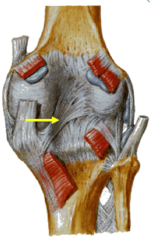
arcuate ligament
strengthens edge and protects the popliteus muscle, connects fibula and tibia
o: posterior aspect of tibia
i: fibular head

extensors of the knee
quadriceps femoris
flexors of the knee
hamstrings
sartorius
gracilis
gastrocnemius
medial rotator of tibia
popliteus