anna sec 2
 Call Kai
 Learn
 Practice Test
 Spaced Repetition
 Match
 Flashcards
 Knowt Play
1/54

There's no tags or description
Looks like no tags are added yet.
Name | Mastery | Learn | Test | Matching | Spaced | Call with Kai | Chat |
|---|
No analytics yet
Send a link to your students to track their progress
55 Terms
4 main sec for bones
Bone overall structure → microscopic bone tissue → bone formation/growth → remodeling/repair
1. Bone: big picture + gross anatomy
what actually is bone ?
how is it differnet from CT
Term |
|---|
Bone tissue |
Osteo- |
Bone functions |
Calcium reservoir |
Bone marrow |
bone classification by shape?
long bone terms?
Coverings of bone
main flow
Bone = connective tissue → bone cells + very hard extracellular matrix
bone is still organized like other CT: cells + matrix. Its unusual feature is that the matrix becomes highly mineralized and solid.
Term | Meaning / what it is | Function / importance | Quick connection |
|---|---|---|---|
Bone tissue | Specialized connective tissue with cells embedded in hard mineralized matrix | Strong support while remaining living/remodelable | Cells + collagen scaffold + mineral → strong living tissue |
Osteo- | Prefix meaning bone | Helps decode terms | osteoblast, osteocyte, osteoclast, osteon |
Bone functions | Support, protection, movement, mineral storage, blood-cell production | Skeleton supports body; protects organs; bones act as levers; stores Ca²⁺; marrow produces blood cells | Support → protect → move → store Ca²⁺ → marrow makes blood cells |
Calcium reservoir | Most body Ca²⁺ is stored in bone | Bone can release Ca²⁺ when blood/body needs it | Bone mineral ↔ blood Ca²⁺ |
Bone marrow | Soft tissue within bone spaces | Red marrow produces blood cells | Bone spaces → marrow → blood cells |
classifcation by shape:
Type | Meaning | Examples |
|---|---|---|
Long bone | Longer than wide; shaft + two ends | Humerus, radius, ulna, femur, tibia, fibula, metacarpals/metatarsals, phalanges |
Short bone | Roughly cube-shaped | Carpals, tarsals |
Sesamoid bone | Special short bone formed inside a tendon | Patella |
Flat bone | Thin, flattened, often curved | Sternum, ribs, scapula, most cranial bones |
Irregular bone | Shape doesn't fit other categories | Vertebrae, hip bones, mandible |
Don't confuse:
Shape of whole bone: long / short / flat / irregular
Type of bone tissue inside it: compact / spongy
A flat sternum still contains both compact and spongy bone.
long bone:
Term | Meaning / location | Function / connection | Quick flow |
|---|---|---|---|
Diaphysis | Shaft/body of a long bone | Long central portion | middle = diaphysis |
Epiphysis | Proximal or distal end | Usually participates in joints | ends = epiphyses |
Metaphysis | Junction between diaphysis and epiphysis | Contains growth region in growing bone | epiphysis → metaphysis → diaphysis |
Epiphyseal plate | Hyaline cartilage growth plate in young bone | Bone grows in length here | cartilage growth → replacement by bone → length ↑ |
Epiphyseal line | Remnant after growth plate closes | Indicates longitudinal growth is finished | plate → ossifies → line |
Medullary cavity | Hollow cavity in diaphysis | Contains marrow; reduces weight | shaft → cavity → marrow |
Articular cartilage | Hyaline cartilage covering joint surfaces | Reduces friction + absorbs compression | bone end → cartilage → joint |
Coverings of bone
Term | Meaning | Function | Quick flow |
|---|---|---|---|
Periosteum | CT membrane covering outside of bone, except articular cartilage | Anchors tendons/ligaments; carries vessels/nerves; contains bone-forming cells | peri = around → periosteum = around bone |
Fibrous layer of periosteum | Outer dense CT portion | Mechanical strength + tendon/ligament attachment | outer → strong/fibrous |
Osteogenic layer | Inner cellular periosteal layer | Contains osteoprogenitor cells/osteoblasts | inner → bone growth/remodeling |
Perforating (Sharpey's) fibers | Collagen fibers extending periosteum into bone | Strongly anchor periosteum and tendons/ligaments to bone | periosteum → Sharpey's fibers → bone |
Endosteum | Thin CT lining internal bone surfaces | Contains cells involved in remodeling | endo = inside → endosteum = inside bone |
Tendon/ligament → periosteum → Sharpey's fibers → bone
That is why muscle force can be transmitted strongly into the skeleton.
Bone tissue: matrix, cells, compact vs spongy
bone matrix
Component |
|---|
Osteoid |
Hydroxyapatite |
Mineralization / calcification |
flow
bone cells
Cell |
|---|
Osteoprogenitor cell |
Osteoblast |
Osteocyte |
Osteoclast |
flow
bone matrix:
Component | What it is | What it gives bone | Quick flow |
|---|---|---|---|
Osteoid | Organic bone matrix made mainly of type I collagen plus ground-substance molecules | Tensile strength + some flexibility | collagen scaffold → resists pulling |
Hydroxyapatite | Calcium-phosphate mineral crystals | Hardness + resistance to compression | mineral → hardness |
Mineralization / calcification | Deposition of calcium salts into osteoid | Converts flexible osteoid into hard bone | osteoid first → Ca²⁺ salts deposited → hard bone |
Collagen → tensile strength/flexibility
Hydroxyapatite → hardness/compression resistance
The instructor uses a useful construction analogy:
Collagen/osteoid = reinforcing framework → hydroxyapatite = concrete
The osteoblast first creates the organic scaffold and then mineral is deposited into it.
bone cells:
Cell | Meaning / origin | Main function | Quick flow |
|---|---|---|---|
Osteoprogenitor cell | Mesenchymal stem-like precursor | Gives rise to osteoblasts | mesenchymal cell → osteoprogenitor → osteoblast |
Osteoblast | Bone-forming cell | Secretes osteoid; promotes mineral deposition | BLAST = builds |
Osteocyte | Mature bone cell trapped in matrix | Maintains bone matrix | osteoblast → trapped in matrix → osteocyte |
Osteoclast | Large multinucleated, monocyte-lineage cell | Resorbs/breaks down bone | CLAST = crushes |
flow: Mesenchymal precursor → osteoblast → secretes osteoid → matrix mineralizes → osteoblast trapped in lacuna → osteocyte
Monocyte-lineage cells fuse → multinucleated osteoclast → acid + enzymes → bone resorption → Ca²⁺ released
Osteoclast identification
Large + multinucleated + ruffled border → bone destruction/resorption
The ruffled border increases the surface area facing bone; the cell secretes acid and enzymes there to dissolve mineral and organic matrix.
C. Lacunae, canaliculi, lamellae
Term | Meaning | Why needed | Quick connection |
|---|---|---|---|
Lacuna | Small cavity containing an osteocyte | Houses mature bone cell | osteocyte lives in lacuna |
Canaliculi | Tiny channels connecting lacunae | Osteocytes exchange nutrients/wastes because solid bone matrix blocks ordinary diffusion | lacuna → canaliculi → lacuna |
Lamella(e) | Layer(s) of mineralized bone matrix | Organize bone for strength | lamella = layer |
Collagen orientation | Fibers run in different directions in adjacent lamellae | Greatly resists twisting/torsion | alternating collagen directions → torsional strength |
mineralized matrix is too solid for nutrients simply to diffuse through, so osteocytes remain connected through canaliculi.
flow:
Blood vessel → canaliculi → osteocytes in lacunae → neighboring osteocytes
compact vs spongy bone
Compact bone | Spongy / cancellous bone | |
|---|---|---|
Appearance | Dense, solid | Open, honeycomb-like |
Main structure | Osteons | Trabeculae |
Osteons? | YES | NO |
Lamellae? | Yes | Yes |
Osteocytes/lacunae? | Yes | Yes |
Main location | Outer bone; especially thick in shafts | Interior of bones/epiphyses |
Blood supply | Through canals inside osteons | From vessels in marrow/endosteum surrounding trabeculae |
Big advantage | Very strong | Strong but lightweight; marrow spaces |
Mechanical design | Resists high stress | Trabeculae align with stress lines |
Dense outer compact bone + open inner spongy bone → strong without being excessively heavy.
Spongy bone flow
Trabeculae → arranged along lines of stress → transmit force efficiently → strength with little weight
Trabecula = little beam/strut of bone
The instructor compares them to flying buttresses in a cathedral.
Compact bone: osteon system
Term | Meaning | Function / recognition |
|---|---|---|
Osteon / Haversian system | Cylindrical structural unit of compact bone | Looks like rings around a central hole |
Central / Haversian canal | Canal through center of osteon | Blood vessels + nerves |
Concentric lamellae | Circular layers around central canal | Alternating collagen orientation → strength |
Lacunae | Spaces between lamellae | Contain osteocytes |
Canaliculi | Tiny channels between lacunae | Nutrient/waste communication |
Perforating / Volkmann's canal | Canal running across/perpendicular to osteons | Connects central canals with each other/periosteal vessels |
Interstitial lamellae | Incomplete lamellae between osteons | Remnants of old remodeled osteons |
Circumferential lamellae | Lamellae around entire inner/outer circumference of bone | Reinforce whole bone against twisting |
Nutrient foramen | Opening through bone surface | Entry for nutrient artery |
Build an osteon from the center outward:
Central/Haversian canal → concentric lamellae → lacunae with osteocytes → canaliculi connecting cells
And:
Central canal ↔ Volkmann canal ↔ neighboring central canal
Textbook adds that adjacent lamellae have collagen fibers running in roughly opposite directions, which is why osteons resist twisting so well.
Histology shortcut
Lots of circular bull's-eyes → compact bone → osteons
Irregular branching beams + open marrow spaces → spongy bone → trabeculae
3. Bone formation + growth
Ossification
A. Intramembranous ossification
Ossification
Ossification / osteogenesis = formation of bone tissue
There are two routes, but both ultimately make bone.
Type | Starting tissue | Main bones | Quick flow |
|---|---|---|---|
Intramembranous ossification | Mesenchyme directly | Most skull flat bones + clavicle | mesenchyme → osteoblast → osteoid → woven bone → remodeled bone |
Endochondral ossification | Hyaline cartilage model | Most bones of body | cartilage model → cartilage dies/calcifies → osteoblasts replace it with bone |
A. Intramembranous ossification
Don't overcomplicate it:
Mesenchymal cells → osteoblasts → osteoid → mineralization → woven bone/trabeculae → remodeling → compact outside + spongy inside
Important:
There is NO cartilage model first.
Textbook adds that woven bone initially forms around embryonic blood vessels and is later reorganized into mature compact/spongy bone.
B. Endochondral ossification
Hyaline cartilage model → bone gradually replaces cartilage
Long-bone sequence
1. Hyaline cartilage model → 2. bone collar around diaphysis → 3. cartilage in center calcifies; chondrocytes die → 4. periosteal bud enters → 5. primary ossification center forms in diaphysis → 6. medullary cavity develops → 7. secondary ossification centers form in epiphyses → 8. cartilage remains only at articular surfaces + epiphyseal plates
The instructor specifically emphasizes primary center = diaphysis and secondary centers = epiphyses. bone trans- Anna
Primary vs secondary
Primary ossification center → diaphysis
Secondary ossification centers → epiphyses
At the end:
Epiphysis | epiphyseal plate | metaphysis | diaphysis
Epiphyseal plate: how bones grow longer
This is a good book detail to know because it is very testable.
From epiphysis → toward diaphysis:
Zone | What happens | Memory |
|---|---|---|
Resting zone | Small relatively inactive cartilage cells | Rest |
Proliferation zone | Chondrocytes undergo mitosis | Produce more cells |
Hypertrophic zone | Older chondrocytes enlarge | Huge cells |
Calcification zone | Matrix calcifies; chondrocytes die | Calcify + die |
Ossification zone | Osteoblasts replace cartilage with bone | Bone replaces cartilage |
One-line plate flow
Rest → proliferate → hypertrophy → calcify/die → ossify
This explains how length increases:
Cartilage added on epiphyseal side → older cartilage replaced by bone on diaphyseal side → bone gets longer while plate stays relatively thin.
Eventually:
cartilage production slows → bone replacement catches up → epiphyseal plate closes → epiphyseal line → length growth stops.
Length vs width
Length → epiphyseal plate → endochondral growth
Width/thickness → appositional bone growth from periosteum
That distinction is worth memorizing exactly.
Remodeling, mechanical stress + repair
Bone is NOT static
The instructor emphasizes this heavily:
Osteoclast resorption ↔ osteoblast deposition = continuous bone remodeling
Process | Cell | What happens |
|---|---|---|
Bone deposition | Osteoblast | Osteoid deposited → mineralized |
Bone resorption | Osteoclast | Acid dissolves mineral + enzymes digest organic matrix |
Remodeling | Both | Old bone removed and new bone deposited |
Mechanical stress
More mechanical load → osteoblast activity/deposition ↑ → bone strengthens
Less mechanical load → resorption predominates → bone weakens
This is why weight-bearing exercise strengthens bone and prolonged unloading, such as in microgravity, promotes bone loss. The lecture explicitly uses this loading/unloading idea.
Calcium connection
Bone = Ca²⁺ reservoir
Need blood Ca²⁺ → bone resorption can release Ca²⁺
Excess deposition → Ca²⁺ incorporated into bone
Important transcript correction
There are two verbal slips later in the transcript that I would not memorize:
It temporarily reverses osteoblast/osteoclast roles. Correct is osteoblast BUILDS, osteoclast BREAKS DOWN.
It says calcitonin tells bone to release calcium. Chapter 6 instead identifies parathyroid hormone (PTH) as stimulating bone resorption and Ca²⁺ release.
Use:
BLAST = builds
CLAST = crushes
Fracture repair
This is much easier if memorized as one sequence:
Fracture → hematoma → fibrocartilaginous (soft) callus → bony (hard) callus → remodeling
Stage | What happens |
|---|---|
1. Hematoma | Broken blood vessels → clot + inflammation |
2. Fibrocartilaginous callus | Fibrous CT/cartilage bridges broken ends |
3. Bony callus | Woven/spongy bone replaces soft callus |
4. Remodeling | Excess bone removed; compact/spongy architecture restored according to stress |
So:
temporary CT/cartilage bridge → temporary woven bone bridge → mature remodeled bone
mega flow bone tissue
bone organizes as flow
Mesenchymal precursor → osteoblast → osteoid (type I collagen) → hydroxyapatite deposited → osteoblast trapped → osteocyte in lacuna → canaliculi connect osteocytes
bone organizes as:
Lamellae → either trabeculae in spongy bone OR osteons in compact bone
throughout life:
Osteoclast removes old bone ↔ osteoblast replaces bone → remodeling adapts bone to mechanical stress
During development:
Mesenchyme directly → intramembranous bone
Hyaline cartilage model → endochondral bone
fracture repair:
hematoma → soft callus → bony callus → remodeled mature bone
dont confuse bones
Osteoblast = builds bone
Osteocyte = maintains bone
Osteoclast = breaks bone
Lacuna = room containing osteocyte
Canaliculus = tiny channel connecting rooms
Lamella = layer of matrix
Osteon = whole circular unit of compact bone
Haversian canal = through center of osteon
Volkmann canal = connects Haversian canals
Compact bone = osteons
Spongy bone = trabeculae, NO osteons
Diaphysis = shaft
Epiphysis = end
Metaphysis = junction/growth region
Primary ossification center = diaphysis
Secondary ossification center = epiphysis
Length growth = epiphyseal plate
Width growth = appositional growth
Skeletal muscle
Whole muscle → fascicle → muscle fiber (cell) → myofibril → sarcomere → myofilaments → actin + myosin
Then the functional pathway is:
nerve signal → muscle action potential → Ca²⁺ release → actin–myosin interaction → sarcomeres shorten → muscle fiber shortens → CT layers transmit force → tendon → bone moves
1. Three types of muscle tissue
Feature | Skeletal muscle | Cardiac muscle | Smooth muscle |
|---|---|---|---|
Location | Attached mainly to skeleton | Heart only | Walls of hollow organs, blood vessels, GI tract, airways, etc. |
Cell shape | Long cylindrical fibers | Short, branched cells | Spindle-shaped cells |
Nuclei | Many, peripheral | Usually 1, sometimes 2; central | 1 central |
Striations | Yes | Yes | No |
Sarcomeres | Yes | Yes | No |
Control | Somatic / voluntary | Autonomic / involuntary; autorhythmic | Autonomic / involuntary |
Major special feature | Large multinucleated fibers | Intercalated discs + gap junctions | Filaments arranged irregularly; contracts slowly and can hold shape |
Main job | Move skeleton / generate force | Squeeze heart chambers → pump blood | Squeeze or change diameter of hollow organs/tubes |
The instructor specifically contrasts striated skeletal/cardiac muscle with nonstriated smooth muscle.
Quick histology IDs
Skeletal → long + striped + many peripheral nuclei
Cardiac → striped + branching + central nucleus + intercalated discs
Smooth → NO stripes + spindle shape + one central nucleus
2. Skeletal muscle organization: large → small
Level | What it is | Wrapped/surrounded by | Quick meaning |
|---|---|---|---|
Whole muscle | Entire organ | Epimysium | epi = around whole muscle |
Fascicle | Bundle of muscle fibers | Perimysium | peri = around bundle |
Muscle fiber / myofiber | One skeletal muscle cell | Endomysium | endo = around individual fiber |
Myofibril | Contractile organelle inside fiber | — | Made of repeating sarcomeres |
Sarcomere | Basic contractile unit | Z disc → Z disc | Unit that shortens |
Myofilaments | Protein filaments within sarcomere | — | Thick + thin |
Thick filament | Myosin | — | Pulling motor |
Thin filament | Actin + troponin + tropomyosin | — | Structure myosin pulls |
The transcript repeatedly builds muscle from the entire muscle into fascicles and then individual fibers, each with its own CT covering.
Structural flow
Muscle → fascicle → muscle fiber → myofibril → sarcomere → thick/thin myofilaments
Connective-tissue flow
Muscle fiber → endomysium → fascicle → perimysium → whole muscle → epimysium → tendon → periosteum → bone
That is also how force gets transmitted:
sarcomere contraction → fiber pulls endomysium → perimysium → epimysium → tendon → bone
The professor emphasizes that these CT layers are not just wrapping; they also carry blood vessels, lymphatics, nerves, and contractile force
3. Skeletal muscle fiber terminology
Term | Meaning / function | Quick connection |
|---|---|---|
Muscle fiber / myofiber | One skeletal muscle cell | long + multinucleated |
Sarcolemma | Muscle cell plasma membrane | cell membrane |
Sarcoplasm | Muscle cell cytoplasm | cytoplasm |
Myofibril | Long contractile organelle filling the fiber | many sarcomeres end-to-end |
Sarcoplasmic reticulum (SR) | Specialized smooth ER | stores Ca²⁺ |
T tubule | Invagination of sarcolemma | carries electrical signal deep into fiber |
Terminal cisternae | Enlarged SR sacs beside T tubule | store/release Ca²⁺ |
Triad | 1 T tubule + 2 terminal cisternae | couples electrical signal → Ca²⁺ release |
Skeletal muscle fibers form by fusion of many precursor cells, explaining why one fiber has many nuclei. The transcript also identifies the sarcolemma and sarcoplasm as muscle-specific terms
Myofiber = whole muscle cell
Myofibril = contractile organelle inside that cell
So:
one fiber contains MANY myofibrils.
Sarcomere: know this diagram conceptually
Sarcomere = Z disc → Z disc
Inside it:
Z disc | I band | A band [H zone — M line — H zone] A band | I band | Z disc
Structure | What is there? | What to remember |
|---|---|---|
Z disc | Anchors thin actin filaments | Boundary of sarcomere |
I band | Thin filaments only | Light |
A band | Entire length of thick/myosin filament; much overlaps actin | Dark |
H zone | Thick filament only, no actin overlap | Lighter center of A band |
M line | Center; anchors thick filaments | Middle of sarcomere |
Titin | Elastic protein from Z disc toward thick filament | Keeps myosin aligned + recoil/prevents overstretch |
The lecture defines a sarcomere as the repeating Z-line-to-Z-line unit and builds the striation pattern from I and A bands
Why skeletal muscle looks striped
Thin only → I band = lighter
Thick + overlap → A band = darker
Thick only in center → H zone = somewhat lighter
Because all the myofibrils line up their bands with one another:
aligned sarcomeres → aligned light/dark bands → visible striations
The transcript directly links the arrangement of thick and thin filaments to the striped appearance
What changes during contraction?
Structure | During contraction |
|---|---|
Sarcomere | Shortens |
Z discs | Move closer together |
I band | Shortens |
H zone | Shortens / may disappear |
A band | STAYS SAME LENGTH |
Actin filament | Same length |
Myosin filament | Same length |
Overlap | Increases |
Why?
The filaments themselves do NOT shorten.
Thin actin slides toward M line between thick myosin → overlap increases → sarcomere shortens
So:
Sliding ≠ shrinking proteins
The lecture explicitly emphasizes that the A band remains relatively constant while the I band and H zone decrease.
Best exam shortcut
A stays; I + H shrink.
Proteins of contraction
Protein | Location | Function | Quick memory |
|---|---|---|---|
Actin | Thin filament | Contains binding sites for myosin | rope being pulled |
Myosin | Thick filament | Motor protein; heads bind actin and pull | puller |
Tropomyosin | Along actin | Covers myosin-binding sites at rest | blocker |
Troponin | Attached to tropomyosin | Binds Ca²⁺ → shifts tropomyosin | Ca²⁺ switch |
Titin | Z disc → thick filament | Elastic alignment/recoil | spring |
The thin filament contains actin with myosin-binding sites; tropomyosin covers those sites, and troponin controls tropomyosin in response to calcium
Regulatory flow
No Ca²⁺ → tropomyosin blocks actin → myosin cannot bind → relaxed
Ca²⁺ → binds troponin → tropomyosin moves → actin sites exposed → myosin binds → contraction
Cross-bridge cycle: how myosin actually pulls actin
Core sequence
ATP hydrolyzed → myosin head becomes energized → myosin binds actin → power stroke pulls actin → new ATP binds myosin → myosin releases actin → ATP hydrolyzed → head resets
So ATP has two crucial roles:
ATP → energizes/cocks myosin head
and
ATP binding → allows myosin to detach from actin
Big result
Repeated cross-bridge cycles → actin slides inward → sarcomere shortens
Calcium turns contraction ON/OFF
The sarcoplasmic reticulum = Ca²⁺ storage tank.
State | Calcium | Result |
|---|---|---|
Contraction | Ca²⁺ released into sarcoplasm | Ca²⁺ binds troponin → tropomyosin moves → contraction |
Relaxation | Ca²⁺ pumped back into SR using ATP | Tropomyosin blocks actin again → relaxation |
Core flow
SR releases Ca²⁺ → Ca²⁺ + troponin → tropomyosin moves → cross-bridges → contraction
Then:
Ca²⁺ pumped back into SR → troponin loses Ca²⁺ → tropomyosin reblocks actin → relaxation
The lecture repeatedly reduces this to the useful rule:
Ca²⁺ present → contraction
Ca²⁺ removed → relaxation
9. How the nerve causes Ca²⁺ release
This connects the nervous system all the way to the sarcomere.
Excitation–contraction flow
Motor neuron action potential → ACh released → ACh binds receptors on motor end plate → muscle action potential → travels along sarcolemma → down T tubules → SR terminal cisternae release Ca²⁺ → Ca²⁺ binds troponin → tropomyosin moves → actin–myosin cross-bridges → contraction
Neuromuscular junction terminology
Term | Meaning |
|---|---|
Motor neuron | Somatic nerve cell controlling skeletal muscle |
Neuromuscular / myoneural junction | Junction between motor neuron and muscle fiber |
Axon terminal | End of motor neuron releasing neurotransmitter |
Synaptic cleft | Gap between neuron and muscle |
Acetylcholine (ACh) | Neurotransmitter released onto skeletal muscle |
Motor end plate | Specialized receiving region on muscle sarcolemma |
Acetylcholinesterase | Breaks down ACh → signal stops |
Neuromuscular junction mini-flow
Neuron AP → Ca²⁺ enters axon terminal → ACh exocytosis → ACh crosses cleft → receptors bind ACh → muscle AP
Then:
AChE breaks down ACh → stimulation stops
10. Motor units
Motor units = how muscle controls force
Motor unit = ONE motor neuron + ALL skeletal muscle fibers it innervates
One motor neuron can branch and control multiple fibers.
Why it matters
Few motor units recruited → small force
More motor units recruited → greater force
So a whole muscle is not simply:
OFF / ON
It can produce graded force:
small recruitment → medium recruitment → large recruitment
The instructor connects this directly to why you can pick up something light versus something heavy.
Skeletal muscle fiber types
Fiber | Metabolism / structure | Contraction | Fatigue | Best for |
|---|---|---|---|---|
Slow oxidative (SO / Type I) | Many mitochondria, capillaries, myoglobin; aerobic | Slow | Very resistant | Posture, endurance |
Fast oxidative (FO) | Many mitochondria/capillaries; oxidative | Fast | Fairly resistant | Sustained locomotion |
Fast glycolytic (FG / Type II) | Large fibers, fewer mitochondria, mainly glycolysis | Fast + powerful | Fatigues quickly | Sprinting, heavy brief force |
Fast recognition
Slow oxidative → endurance
Fast oxidative → fast + endurance-ish
Fast glycolytic → power burst
The instructor uses the same distinction: oxidative fibers provide more sustainable activity, whereas fast glycolytic fibers provide a large burst but tire quickly.
Cardiac muscle — only the essentials for now
Feature | Cardiac muscle |
|---|---|
Shape | Short + branched |
Nuclei | 1–2 central |
Striated? | Yes |
Sarcomeres? | Yes |
Control | Involuntary / autonomic modulation |
Special junction | Intercalated discs |
Gap junction function | Electrical signal spreads cell → cell |
Mechanical junction function | Cells stay physically attached during forceful contraction |
Special property | Autorhythmicity — heart can initiate contractions without somatic nerve input |
Intercalated disc logic
Mechanical junctions → keep cells physically connected
Gap junctions → electrically connect cells
Therefore:
one region depolarizes → signal spreads through cardiac network → coordinated contraction
Cardiac and skeletal muscle both use sarcomeres and actin–myosin sliding, but cardiac cells are short/branched and connected into networks. The lecture emphasizes intercalated discs and gap junctions for coordinated contraction.
Important transcript correction
At one point the video says cardiac cells “don't have myofibrils.” Do not memorize that literally. Cardiac muscle does have myofibrils containing sarcomeres; their organization is simply less uniformly bundled than in skeletal muscle.
Smooth muscle — only the essentials
Feature | Smooth muscle |
|---|---|
Shape | Spindle-shaped |
Nucleus | 1 central |
Striated? | NO |
Sarcomeres? | NO |
Actin/myosin? | YES |
Filament arrangement | Irregular, crisscrossing network |
Anchor structures | Dense bodies / dense plaques |
Troponin? | No troponin regulatory system |
Main Ca²⁺ regulator | Uses calmodulin-related pathways instead |
Control | Autonomic nerves, hormones, local chemical signals |
Function | Slow, sustained squeezing of tubes/hollow organs |
The lecture describes smooth cells as elongated, spindle-shaped, nonstriated cells with one central nucleus. skeletal muscle trans
Smooth contraction idea
Instead of neat sarcomeres:
irregular actin + myosin anchored to dense bodies → Ca²⁺ activates contraction → filaments pull in multiple directions → whole cell becomes shorter and thicker
That is why:
skeletal/cardiac → organized sarcomeres → stripes
but
smooth → no sarcomeres → no stripes
Unitary vs multi-unit smooth muscle
Type | Connection | Result |
|---|---|---|
Unitary / visceral | Cells connected by gap junctions | Sheet contracts together |
Multi-unit | Few/no gap junctions | Individual cells controlled more independently |
final flow
Brain/spinal cord → somatic motor neuron → AP reaches axon terminal → Ca²⁺ enters terminal → ACh released → ACh binds motor end plate → muscle AP → sarcolemma → T tubule → terminal cisternae/SR release Ca²⁺ → Ca²⁺ binds troponin → tropomyosin moves → myosin binds actin → ATP-powered cross-bridge cycling → thin filaments slide inward → I band + H zone shrink → sarcomere shortens → myofibril shortens → muscle fiber shortens → endomysium → perimysium → epimysium → tendon → periosteum → bone moves
And relaxation is basically the reverse regulatory step:
nerve signal stops → ACh removed → Ca²⁺ pumped back into SR → tropomyosin covers actin → cross-bridges stop → muscle relaxes
That gives her the molecular → cellular → tissue → whole-muscle → bone movement connection the professor keeps emphasizing, rather than memorizing a pile of unrelated muscle terms.
overall view integument
Skin organization → epidermis → dermis/hypodermis + color → hair/nails → glands
1. Integument: big picture
Outside → inside:
Epidermis → Dermis → Hypodermis/subcutaneous tissue → Deep fascia → Muscle
Important distinction:
Skin proper = epidermis + dermis
Hypodermis/subcutaneous tissue lies beneath skin; the textbook treats it as part of the integumentary system but not skin proper. Human anatomy (Ninth Edition) (…
Region | Tissue | Main job | Quick ID |
|---|---|---|---|
Epidermis | Keratinized stratified squamous epithelium | Protective/water-resistant barrier | No blood vessels; many keratinocytes |
Dermis | Connective tissue | Strength, elasticity, vessels, nerves; supports epidermis | Collagen + elastic fibers |
Hypodermis | Mainly adipose + areolar CT | Cushion, insulation, fat storage, anchors skin | Lots of fat cells |
Main skin functions
Protection → temperature regulation → excretion → vitamin D production → sensation
The epidermis protects against abrasion, microorganisms, water loss, and UV, while sweat glands and dermal blood vessels help regulate temperature.
epidermis
2. EpidermisEpidermal cell types
Cell | Main function | Easy association |
|---|---|---|
Keratinocyte | Makes keratin; main epidermal cell | Keratin → protection |
Melanocyte | Produces melanin | Pigment + UV protection |
Dendritic / Langerhans cell | Immune surveillance; phagocytosis/antigen presentation | Defense |
Tactile epithelial / Merkel cell | Light-touch receptor | Touch |
The lecture emphasizes that keratinocytes dominate the epidermis, while Langerhans cells provide immune defense, melanocytes produce pigment, and Merkel cells detect light touch. integument trans integument trans
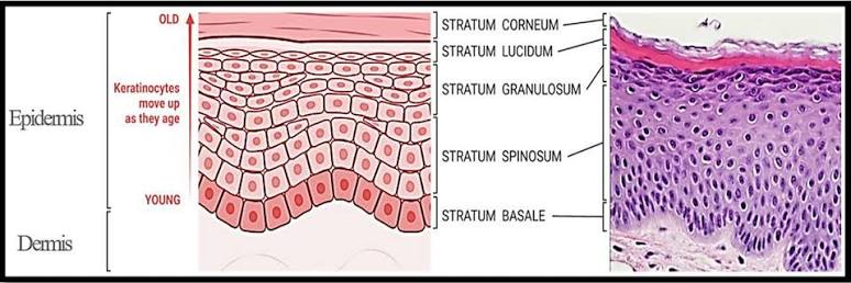
Epidermal layers — deepest → superficial
This is probably the most important integument flow:
Basale → Spinosum → Granulosum → Lucidum → Corneum
Mnemonic: B S G L C
Layer | What happens | Quick flow/ID |
|---|---|---|
Stratum basale / germinativum | Deepest; mitosis makes new keratinocytes; melanocytes & Merkel cells here | Birth of cells |
Stratum spinosum | Strong desmosomes + pre-keratin; cells look “spiny” | Spines → strength |
Stratum granulosum | Keratinization increases; keratohyalin + lipid/waterproofing granules; cells begin dying | Granules → keratin + waterproofing |
Stratum lucidum | Clear layer of dead keratinocytes | ONLY thick skin |
Stratum corneum | Many layers of dead, flat, keratin-filled cells | Barrier + shedding |
specifies that stratum lucidum occurs only in thick skin of the palms and soles.
Keratinocyte life flow
Basale mitosis → cells pushed upward → keratin increases → cells flatten → cells die → corneum → shed/desquamate
The lecture describes continual migration from the basale toward the surface followed by death and shedding.
Desquamation = shedding of dead surface keratinocytes.
Thick vs thin skin
Thick skin | Thin skin | |
|---|---|---|
Where? | Palms + soles | Rest of body |
Epidermal strata | 5 | 4 |
Lucidum | Present | Absent |
Corneum | Very thick | Thinner |
Hair | No hair | Usually hair present |
Friction ridges | Very prominent | Less prominent |
Important source clarification
The transcript briefly suggests lucidum might be present but simply hard to see in thin skin. For the textbook/test answer, use: Lucidum = thick skin only.
3. Dermis, hypodermis + skin color
Dermis
The dermis has two layers:
Layer | Tissue | Function | Fast ID |
|---|---|---|---|
Papillary dermis | Areolar CT | Nutrient exchange with epidermis; dermal papillae; sensory structures | Papillary = superficial + loose |
Reticular dermis | Dense irregular CT | Strength + stretch/recoil | Reticular = deep + tough |
Quick flow
Papillary → loose/areolar → exchange + papillae
Reticular → dense irregular → collagen in many directions → strength
The papillary layer sends dermal papillae upward into the epidermis, increasing surface contact and strengthening the epidermal-dermal junction. These ridges contribute to fingerprints. Human anatomy (Ninth Edition) (…
Fingerprints
Dermal papillae/ridges → epidermis follows their contours → friction ridges → fingerprints
Dermatoglyphs = fingerprint/palm/sole ridge patterns.
Hypodermis
Adipose predominates → cushioning + insulation + energy storage
Also:
Loose attachment → skin can move over deeper structures
Histology shortcut:
Large empty-looking fat cells underneath dermis → hypodermis
Skin color
Three major contributors from the textbook:
Factor | Effect |
|---|---|
Melanin | Brown/black/red-yellow pigmentation; major source of variation |
Carotene | Yellow-orange pigment |
Hemoglobin/blood flow | Pink-red color depending on blood flow + oxygenation |
Human anatomy (Ninth Edition) (…
Melanin flow
Tyrosine → melanocyte makes melanin → packages melanin granules → transfers them to keratinocytes → pigment moves upward with keratinocytes → absorbs UV
The lecture places melanocytes in the stratum basale and explains that they transfer pigment granules into neighboring keratinocytes. integument trans
Two melanins emphasized
Eumelanin → brown/black
Pheomelanin → red/orange
Skin tone depends largely on the type + amount of melanin produced, not simply whether melanocytes are present.
Why melanin matters
More melanin → more UV absorption → greater protection of living epidermal cells
But UV is also needed for:
UV → epidermal vitamin D production → helps intestinal Ca²⁺ absorption → bone health
The lecture explicitly connects melanin's UV protection with vitamin D production
4. Hair + nailsHair
Hair = dead cells filled with hard keratin
Part | Meaning/function |
|---|---|
Shaft | Portion above skin |
Root | Portion inside skin |
Hair follicle | Epidermal tube surrounding/growing the hair |
Hair bulb | Enlarged base of follicle |
Hair matrix | Dividing cells that produce hair |
Hair/dermal papilla | CT projection with capillaries supplying matrix |
Arrector pili | Smooth muscle → pulls hair upright → goosebumps |
Cuticle | Outer hair layer |
Cortex | Main middle layer |
Medulla | Central core, when present |
Human anatomy (Ninth Edition) (…
Hair-growth flow
Stem cells → hair matrix mitosis → cells move upward → hard keratinize + die → hair shaft grows outward
And:
Dermal papilla blood vessels → nourish matrix → matrix makes hair
The instructor specifically emphasizes the matrix as the growth area and the dermal papilla as its blood supply.
Piloerection
Cold/fear → sympathetic stimulation → arrector pili contracts → hair stands up → goosebumps
Nails
Nail = epidermal modification made of hard keratin
Structure | What it is |
|---|---|
Nail plate/body | Visible hard keratin nail |
Free edge | Distal portion extending past finger/toe |
Nail root | Proximal portion under skin |
Nail bed | Epidermis beneath nail plate |
Nail matrix | Growth zone producing nail |
Lunula | Visible pale/white crescent over matrix |
Nail folds | Skin bordering nail |
Eponychium / cuticle | Proximal fold extending onto nail |
Nail growth flow
Nail matrix mitosis → hard keratin produced → nail plate pushed distally
Matrix = grows nail is the single most important nail distinction.
5. Skin glands
This table is worth memorizing because it connects back to the gland classifications from Chapter 4.
Gland | Structure | Secretion | Opens where? | Main function |
|---|---|---|---|---|
Sebaceous | Simple branched/alveolar | Holocrine → sebum/oil | Usually hair follicle | Lubricates hair/skin; slows water loss; antibacterial |
Eccrine sweat | Simple coiled tubular | Merocrine/exocytosis; watery sweat | Skin surface | Thermoregulation |
Apocrine sweat | Simple coiled tubular; larger | Protein/lipid-rich secretion | Usually hair follicle | Body-odor-associated secretion; active from puberty |
Ceruminous | Modified apocrine | Cerumen | Ear canal | Earwax |
Mammary | Modified sweat gland | Milk | Nipple | Nourishes infant |
Sebaceous glands use holocrine secretion, meaning whole secretory cells break down to release lipid-rich sebum. Human anatomy (Ninth Edition) (…
Sebaceous flow
Androgens/puberty → sebaceous activity ↑ → sebum → hair follicle → skin/hair lubricated
Blocked gland:
sebum trapped → blockage + inflammation/bacteria → acne
Eccrine vs apocrine — know this distinction
Eccrine | Apocrine | |
|---|---|---|
Distribution | Widespread; especially palms/soles/forehead | Mainly axilla + anal/genital/perineal areas |
Opens | Directly onto skin | Usually into hair follicle |
Secretion | Watery, salts | Thicker; proteins + lipids |
Main association | Cooling | Body odor |
Active | Shortly after birth | Puberty |
Odor? | Usually little | Bacterial breakdown → odor |
Eccrine cooling flow
Body hot → eccrine sweat ↑ → sweat reaches skin → water evaporates → heat leaves body → cooling
The lecture describes eccrine glands as simple coiled tubular glands whose watery secretion primarily functions in thermoregulation.
Apocrine odor flow
Apocrine secretion → proteins/lipids reach follicle/skin → skin bacteria break them down → body odor
general skin
Repeated/chronic friction → basale mitosis ↑ + keratin ↑ → epidermis thickens → CALLUS
Rapid severe friction → epidermis separates from dermis + fluid accumulates → BLISTER
The lecture directly contrasts slow friction producing calluses with acute friction producing fluid-filled blisters. integument trans integument trans
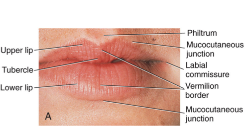
Mucocutaneous junction = transition from external skin → mucous membrane, e.g. lips and certain genital/anal regions
mega flow integument
Epidermis = keratinized epithelium → protection
Basale → makes cells → Spinosum → strengthens → Granulosum → keratinizes/waterproofs → Lucidum in thick skin → Corneum → dead protective cells shed
Then underneath:
Papillary dermis = areolar + papillae → exchange/fingerprints
Reticular dermis = dense irregular CT → strength/stretch
Hypodermis = adipose → cushion + insulation
Appendages:
Hair matrix → hair → sebaceous gland lubricates it → arrector pili raises it
Nail matrix → nail plate
Glands:
Sebaceous → sebum → holocrine
Eccrine → watery sweat → skin surface → cooling
Apocrine → protein/lipid secretion → hair follicle → bacterial breakdown → odor
And color:
Melanocyte → melanin → keratinocyte → UV protection
1. Joints: the big picture
Joint = articulation = place where skeletal elements meet.
Arthrology = study of joints.
A joint can be classified in two different ways at the same time:
STRUCTURE = what physically connects the bones
FUNCTION = how much movement it allows
Chapter 9 classifies function as synarthrosis, amphiarthrosis, diarthrosis, and structure as fibrous, cartilaginous, synovial. Human anatomy (Ninth Edition) (…
Functional classification
Functional type | Meaning | Quick meaning |
|---|---|---|
Synarthrosis | Essentially immovable | syn = together tightly |
Amphiarthrosis | Slightly movable | little movement |
Diarthrosis | Freely movable | most limb joints |
Flow
No movement → synarthrosis
Some movement → amphiarthrosis
Free movement → diarthrosis
The lecture uses exactly this movement-based system before introducing structural joint types. joints trans
2. Structural classification
Structural type | What connects bones? | Joint cavity? | Main subtypes | Typical movement |
|---|---|---|---|---|
Fibrous | Dense fibrous CT | No | Suture, syndesmosis, gomphosis | Mostly none/slight |
Cartilaginous | Cartilage | No | Synchondrosis, symphysis | None/slight |
Synovial | Bones separated by fluid-filled cavity | YES | 6 shape types | Freely movable |
Master structural flow
Fibrous CT between bones → fibrous joint
Cartilage between bones → cartilaginous joint
Fluid-filled cavity between articular surfaces → synovial joint
3. Fibrous + cartilaginous joints
Fibrous joints
Dense regular CT connects bones → strong connection → little/no movement
Type | Structure | Example | Functional class | Fast ID |
|---|---|---|---|---|
Suture | Very short interlocking fibrous tissue | Skull sutures | Synarthrosis | skull seams |
Syndesmosis | Bones connected by a ligament/interosseous membrane | Distal tibiofibular joint | Usually little movement | ligament joint |
Gomphosis | Peg-in-socket + periodontal ligament | Tooth in socket | Synarthrosis in textbook classification | tooth joint |
The transcript emphasizes the distal tibiofibular syndesmosis and periodontal ligament of a gomphosis.
Important course/book distinction
The lecture describes teeth as having a tiny amount of “give,” but the Chapter 9 classification table lists gomphosis as immobile/synarthrotic. For an exam classification question, I would use:
Gomphosis → fibrous → synarthrosis
Cartilaginous joints
Type | Material connecting bones | Example | Movement | Quick flow |
|---|---|---|---|---|
Synchondrosis | Hyaline cartilage | Epiphyseal plate; first rib → sternum | Synarthrosis | hyaline → no movement |
Symphysis | Fibrocartilage | Pubic symphysis; intervertebral disc | Amphiarthrosis | fibrocartilage → strength + slight movement |
Chapter 9 specifically distinguishes synchondrosis = hyaline cartilage from symphysis = fibrocartilage, and describes symphyses as shock-absorbing, slightly movable joints.
Easy distinction
SynCHONDrosis → CHOND = cartilage → HYALINE
SymPHYSIS → fibrocartilage → pubic symphysis/intervertebral disc
Intervertebral disc
The instructor spends extra time on this, so I would know the parts.
Structure | What it is | Function |
|---|---|---|
Annulus fibrosus | Outer rings of fibrocartilage | Contains/resists expansion |
Nucleus pulposus | Gel-like central core | Compresses → shock absorption |
Flow
Compression on spine → nucleus pulposus compressed → force spreads outward → annulus fibrosus contains it → shock absorbed
The transcript describes the nucleus pulposus as a gel-like center surrounded by fibrocartilage rings.
A herniated disc occurs when the annulus is disrupted and nucleus material protrudes; posterior/posterolateral protrusion can irritate nearby nerves.
4. Synovial joints — most important movable joints
Synovial joint = freely movable = diarthrosis
The instructor's general picture is:
bone end + articular cartilage → fluid-filled cavity → articular cartilage + bone end → entire joint enclosed by capsule.
General synovial-joint anatomy
Structure | What it is | Function |
|---|---|---|
Articular cartilage | Hyaline cartilage covering bone ends | Reduces friction + absorbs compression |
Joint/articular cavity | Space between articulating bones | Contains synovial fluid |
Articular capsule | Envelope surrounding joint | Holds joint together |
Fibrous layer | Outer dense irregular CT of capsule | Strength/stability |
Synovial membrane | Inner loose CT layer | Produces synovial fluid |
Synovial fluid | Viscous blood filtrate + lubricating molecules | Lubricates + nourishes avascular cartilage |
Ligament | Dense regular CT, bone → bone | Reinforces/stabilizes joint |
Articular disc / meniscus | Fibrocartilage within some joints | Improves fit + distributes load/shock |
Bursa | Synovial-fluid-filled sac | Reduces friction between structures |
Tendon sheath | Long tubular bursa around tendon | Lets tendon glide smoothly |
Chapter 9 emphasizes the cavity, two-layer capsule, synovial membrane/fluid, articular cartilage, menisci, bursae and tendon sheaths.
One distinction she absolutely needs
Ligament = bone → bone
Tendon = muscle → bone
The instructor stresses this because both can be seen around the same joint but have different jobs.
Synovial fluid
Blood filtrate (formed when fluid, water, electrolytes, and small molecules filter out of the tiny blood vessels (capillaries) in the synovial membrane and into the joint space) → synovial membrane modifies it → synovial fluid → lubrication + cartilage nutrition
Because:
Articular cartilage is avascular → nutrients must diffuse from synovial fluid
The textbook calls movement-driven movement of fluid into/out of cartilage weeping lubrication.
5. Six types of synovial joints
Synovial type | Axis | Main movement | Best example | Shape idea |
|---|---|---|---|---|
Plane / gliding | Nonaxial | Gliding/sliding | Intercarpal, intertarsal | flat surfaces slide |
Hinge | Uniaxial | Flexion + extension | Elbow, interphalangeal | door hinge |
Pivot | Uniaxial | Rotation | Proximal radioulnar, atlantoaxial | axle rotates |
Condylar / ellipsoid | Biaxial | Flex/ext + abd/add → circumduction | MCP/knuckles, wrist | oval surface |
Saddle | Biaxial | Flex/ext + abd/add; opposition at thumb | 1st carpometacarpal | saddle-shaped |
Ball-and-socket | Multiaxial | Flex/ext + abd/add + rotation + circumduction | Shoulder, hip | ball in cup |
Chapter 9's six structural types follow exactly this nonaxial → uniaxial → biaxial → multiaxial organization.
Axes shortcut
Plane → no fixed axis
Hinge + pivot → ONE axis
Condylar + saddle → TWO axes
Ball-and-socket → THREE axes / multiaxial
Shape determines movement
This connection matters more than memorizing names:
Joint surface shape → determines axes → determines possible movement
For example:
Elbow trochlea shaped like hinge → one axis → flexion/extension
Shoulder spherical head + shallow glenoid → multiaxial → huge range of motion
Hip spherical head + deeper acetabulum → multiaxial but more stable / less mobile than shoulder

6. Movement terminology
Movement | Meaning | Do this to remember |
|---|---|---|
Flexion | Decrease joint angle | Bend elbow |
Extension | Increase joint angle | Straighten elbow |
Hyperextension | Extension beyond anatomical position | Look upward |
Abduction | Move away from midline | Raise arm laterally |
Adduction | Move toward midline | Bring arm back |
Circumduction | Flex + abduct + extend + adduct in sequence | Draw a circle with arm |
Rotation | Bone turns around its longitudinal axis | Turn head left/right |
The lecture defines flexion as decreasing the angle and extension as increasing it.
It then defines abduction/adduction and circumduction as a combination of several angular movements.
7. Lab bridge: joint → movement → muscle
Joint | Bones articulating | Joint type | Main movements | What that tells you in muscle lab |
|---|---|---|---|---|
Shoulder / glenohumeral | Scapula + humerus | Ball-and-socket | Flex/ext, abd/add, rotation, circumduction | Many muscles can act in many directions |
Elbow | Humerus + ulna/radius | Hinge | Flex/ext | Muscle anterior/posterior to joint often flexes/extends |
Proximal radioulnar | Radius + ulna | Pivot | Pronation/supination | Think forearm rotators |
Wrist / radiocarpal | Radius + carpals | Condylar | Flex/ext, abd/add, circumduction | Wrist muscles named flexor/extensor often reveal action |
Thumb CMC | Trapezium + metacarpal I | Saddle | Flex/ext, abd/add, opposition | Explains special thumb muscles |
Hip | Os coxae + femur | Ball-and-socket | Flex/ext, abd/add, rotation, circumduction | Many powerful multidirectional muscles |
Knee | Femur + tibia | Modified hinge | Mainly flex/ext + some rotation | Think quadriceps vs hamstrings |
Ankle | Tibia/fibula + talus | Hinge | Dorsiflexion/plantar flexion | Anterior leg vs posterior leg actions |
Intercarpal/intertarsal | Adjacent carpals/tarsals | Plane | Gliding | Small movements combine into larger hand/foot motion |
Atlantoaxial | C1 + C2 | Pivot | Rotation | Turn head “no” |
Important transcript/book correction
The transcript loosely mentions the knee and TMJ while discussing condyloid joints. For the Chapter 9 classification, use:
Knee = modified hinge
TMJ = modified hinge
The true high-yield condylar examples are MCP joints and wrist/radiocarpal joint.
8. How joint names work in lab
Name | Decode it |
|---|---|
Atlantoaxial | atlas + axis |
Costovertebral | rib/costal + vertebra |
Radioulnar | radius + ulna |
Radiocarpal | radius + carpals |
Carpometacarpal | carpal + metacarpal |
Metacarpophalangeal | metacarpal + phalanx |
Interphalangeal | between phalanges |
Intercarpal | between carpals |
Intertarsal | between tarsals |
Sternoclavicular | sternum + clavicle |
Acromioclavicular | acromion + clavicle |
See two bones → identify bones → combine their names → often you already know the joint name.
The transcript explicitly uses atlantoaxial, costovertebral, MCP and intertarsal examples for this. joints trans
9. Joint stability
A joint must balance:
Mobility ↔ stability
Generally:
more mobility → less inherent stability
more structural restriction → greater stability
Chapter 9 emphasizes three stabilizers:
Articular surface shape + ligaments + muscle tone
That connects directly to what she will see in lab:
Bone shape determines where movement can occur
Ligaments prevent too much movement
Muscles/tendons actively stabilize and move the joint
A sprain involves ligament injury/stretching, which can temporarily increase abnormal joint movement; the lecture connects ligament damage with instability.
mega review
BONES MEET → JOINT / ARTICULATION
↓ classify by FUNCTION
Synarthrosis = none
Amphiarthrosis = slight
Diarthrosis = free
↓
classify by STRUCTURE
Fibrous → dense CT → suture / syndesmosis / gomphosis
Cartilaginous → cartilage → synchondrosis (hyaline) / symphysis (fibrocartilage)
Synovial → articular cartilage + cavity + synovial fluid + capsule + ligaments → freely movable
↓
if synovial, look at SHAPE
Plane → glide
Hinge → flex/extend
Pivot → rotate
Condylar → flex/ext + abd/add
Saddle → flex/ext + abd/add + thumb opposition
Ball-and-socket → flex/ext + abd/add + rotate + circumduct
↓
then in muscle lab
Identify muscle → identify which joint it crosses → ask which side of the joint it passes → predict movement → check muscle name/action
So the full system is:
Bone shape → joint structure → permitted axis → permitted movement → muscle crosses joint → muscle contracts → bone moves
That is the connection I would want her to have in her head before memorizing individual muscles, because then a name like flexor, extensor, adductor, or pronator is describing a movement she already understands rather than being another isolated term.
Heart = 2 pumps in series → right heart sends blood to lungs → left heart sends blood to body → valves keep flow one-way → conduction system times the contractions.
the right and left sides function as two pumps, with the right ventricle sending blood into the pulmonary circuit
and the much stronger left ventricle generating pressure for systemic circulation.
1. Heart location, orientation + coverings
Term | Meaning / importance | Quick ID |
|---|---|---|
Mediastinum | Region between the two pleural cavities/lungs | Heart is in middle mediastinum |
Diaphragm | Muscle forming floor of thorax | Heart rests on its central tendon |
Apex | Pointed inferior end of heart | Points inferiorly/anteriorly/left; formed mainly by LV |
Base | Broad posterior surface | Mainly associated with atria, especially LA; great vessels nearby |
Great vessels | Large vessels entering/leaving heart | SVC, IVC, pulmonary trunk/arteries, pulmonary veins, aorta |
The instructor repeatedly emphasizes: between lungs → on diaphragm → middle mediastinum, with the great vessels superior to the heart. heart trans heart trans
Lab orientation shortcut
Apex points left → base is posterior → great vessels are superior → heart rests on diaphragm
And if you can see the SVC and IVC entering the same chamber, you have found the right atrium.
2. Pericardium vs heart wall
These are easy to mix up, so keep them separate.
Pericardium = sac AROUND heart
Outside → inside:
Fibrous pericardium → parietal serous pericardium → pericardial cavity + fluid → visceral serous pericardium/epicardium → heart
Structure | What it is | Function |
|---|---|---|
Fibrous pericardium | Tough outer dense CT | Protects/anchors heart; prevents excessive expansion |
Parietal serous pericardium | Serous layer lining inner fibrous sac | Helps produce lubricating fluid |
Pericardial cavity | Small potential space | Contains serous fluid → reduces friction |
Visceral pericardium | Serous layer directly on heart | Same structure as epicardium |
The instructor stresses that the lubricated pericardial sac allows the continuously beating heart to move without rubbing against surrounding structures. heart trans
Heart wall = tissue OF heart
Epicardium → Myocardium → Endocardium
Layer | Meaning | Function |
|---|---|---|
Epicardium | Outer heart layer = visceral pericardium | Smooth protective external covering |
Myocardium | Cardiac muscle | Contracts → pumps blood |
Endocardium | Smooth endothelial inner lining | Lines chambers + covers valves |
Fast distinction
PERIcardium = around heart
EPIcardium = outside heart wall
MYOcardium = muscle
ENDOcardium = inside lining
The transcript shows this exact outside-to-inside relationship.
3. External + internal heart anatomy
External landmarks
Structure | Meaning / function |
|---|---|
Auricle | Ear-like extension of an atrium; increases atrial volume |
Coronary sulcus | Groove between atria and ventricles |
Anterior interventricular sulcus | Groove between ventricles anteriorly |
Posterior interventricular sulcus | Groove between ventricles posterior/inferiorly |
Interatrial septum | Wall between atria |
Interventricular septum | Wall between ventricles |
Sulcus = groove.
Coronary vessels run in these grooves, so:
Sulcus → identifies chamber boundary + gives vessels protected route across heart surface
4. Four chambers
Chamber | Receives blood from / sends blood to | Structures to know | Big idea |
|---|---|---|---|
Right atrium (RA) | Receives SVC + IVC + coronary sinus → sends to RV | Pectinate muscles, auricle, fossa ovalis, crista terminalis | Receives oxygen-poor systemic blood |
Right ventricle (RV) | RA → pulmonary trunk | Trabeculae carneae, papillary muscles, chordae tendineae, tricuspid valve | Pulmonary pump |
Left atrium (LA) | Receives 4 pulmonary veins → LV | Mostly smooth wall; pectinate muscles mainly in auricle | Receives oxygen-rich lung blood |
Left ventricle (LV) | LA → aorta | Trabeculae carneae, papillary muscles, chordae tendineae, mitral valve | Systemic pump; thickest myocardium |
Why is LV so thick?
Lungs are nearby → RV needs relatively low pressure
Whole body is far away → LV needs much higher pressure → thicker myocardium
This pressure difference is a major structure-function point in the lecture.
Internal structures
Term | Meaning / function |
|---|---|
Pectinate muscles | Parallel ridges in atrial walls; help atrial contraction |
Trabeculae carneae | Irregular muscular ridges inside ventricles |
Papillary muscles | Ventricular muscles attached to chordae |
Chordae tendineae | Tendinous “heart strings” connecting papillary muscles → AV valve cusps |
Fossa ovalis | Depression in RA septum; remnant of fetal foramen ovale |
Moderator band | RV structure carrying conducting fibers toward papillary region |
Do not confuse
Pectinate = ATRIA
Trabeculae carneae = VENTRICLES
Papillary muscle + chordae = support AV valves
Blood flow — memorize this exactly
This is probably the single most important flow of the whole cardiovascular unit.
Complete blood-flow sequence
Body → SVC/IVC + coronary sinus → RIGHT ATRIUM → tricuspid valve → RIGHT VENTRICLE → pulmonary semilunar valve → pulmonary trunk → R/L pulmonary arteries → LUNGS → pulmonary veins → LEFT ATRIUM → mitral/bicuspid valve → LEFT VENTRICLE → aortic semilunar valve → aorta → BODY
Two circuits
Right heart → pulmonary circuit → lungs
Left heart → systemic circuit → body
Very important vessel rule
Artery = carries blood AWAY from heart
Vein = carries blood TOWARD heart
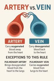
Not:
❌ artery = oxygenated
❌ vein = deoxygenated
The instructor specifically warns about this.
Therefore:
Pulmonary arteries = deoxygenated but still arteries

Pulmonary veins = oxygenated but still veins
6. Heart valves = ONE-WAY gates
Valve | Between | Prevents backflow into |
|---|---|---|
Tricuspid / right AV | RA → RV | Right atrium |
Pulmonary semilunar | RV → pulmonary trunk | Right ventricle |
Mitral / bicuspid / left AV | LA → LV | Left atrium |
Aortic semilunar | LV → aorta | Left ventricle |
Easy order
Tri → Pulmonary → Mitral → Aortic
following blood flow from RA onward.
AV vs semilunar
AV valves | Semilunar valves | |
|---|---|---|
Where? | Atrium → ventricle | Ventricle → artery |
Names | Tricuspid + mitral | Pulmonary + aortic |
Chordae tendineae? | YES | NO |
Papillary muscles? | YES | NO |
Purpose | Prevent ventricular blood → atrium | Prevent arterial blood → ventricle |
Valve function flow
Valves don't actively decide to open.
Pressure behind valve > pressure ahead → valve opens
Pressure ahead > pressure behind → valve closes
For an AV valve:
Ventricle contracts → pressure rises → AV valve closes → papillary muscles tighten chordae → cusps cannot evert into atrium
The textbook specifically emphasizes the four valves, their backflow-prevention roles, and that the cardiac skeleton surrounds and anchors them. heart trans
Cardiac skeleton
This is a nice Chapter 19 detail.
Dense CT rings around valve openings → cardiac skeleton
Functions:
anchors valves → prevents openings from overstretching → anchors myocardium → electrically separates atria from ventricles
That last function connects directly to the conduction system:
atria cannot simply electrically stimulate ventricles directly → impulse must use AV pathway


7. Coronary circulation = heart feeds itself
The blood sitting inside the chambers cannot adequately nourish the thick myocardium.
So:
Aorta → coronary arteries → myocardium → cardiac veins → coronary sinus → right atrium
Major coronary arteries
Vessel | Where / what to know |
|---|---|
Right coronary artery (RCA) | Runs in coronary sulcus; supplies much of right heart |
Right marginal artery | Branch of RCA along right margin |
Posterior interventricular artery / PDA | Runs in posterior interventricular sulcus |
Left coronary artery (LCA) | Short trunk from aorta |
Anterior interventricular artery / LAD | Runs toward apex in anterior interventricular sulcus |
Circumflex artery | Curves around left side in coronary sulcus |
Coronary artery shortcut
LCA → LAD + circumflex
RCA → marginal + usually PDA
Cardiac veins
Great cardiac vein → anterior
Middle cardiac vein → posterior interventricular region
Small cardiac vein → right side
Most →
coronary sinus → right atrium
Lab connection
If you see a vessel in:
Anterior interventricular sulcus → think LAD + great cardiac vein
Posterior interventricular sulcus → think PDA + middle cardiac vein
Coronary sulcus → think coronary arteries/circumflex + coronary sinus posteriorly

8. Cardiac muscle
This should connect directly back to the muscle module.
Feature | Cardiac muscle |
|---|---|
Cell shape | Short + branched |
Nuclei | Usually 1 central, sometimes 2 |
Striated? | Yes |
Sarcomeres? | Yes |
Control | Involuntary |
Mitochondria | Very abundant |
Cell connection | Intercalated discs |
Special property | Autorhythmicity |
Intercalated discs
Two main jobs:
Mechanical connection + electrical connection
Junction | Function |
|---|---|
Fascia adherens / desmosome-like junctions | Hold cells together during strong contraction |
Gap junctions | Let ions/electrical signals move cell → cell |
Therefore:
one cardiac cell depolarizes → gap junctions → neighboring cells depolarize → chamber contracts as coordinated network
That is why cardiac muscle acts like a functional syncytium even though it consists of individual cells.
9. Electrical conduction system
This is the key sequence to memorize:
SA node → atrial myocardium → AV node → AV bundle → R/L bundle branches → Purkinje/subendocardial conducting fibers → ventricular myocardium
Structure | Job |
|---|---|
SA node | Pacemaker; starts heartbeat |
Atrial myocardium | Signal spreads → atria contract |
AV node | Briefly delays impulse |
AV bundle / bundle of His | Only normal electrical route from atria → ventricles |
R/L bundle branches | Carry signal down interventricular septum |
Purkinje fibers | Spread signal through ventricular walls |
Cardiac skeleton | Electrically insulates atria from ventricles |
Why the AV delay?
Atria contract first → finish filling ventricles → THEN ventricles contract
Why signal goes toward apex first?
The great arteries leave superiorly.
So:
signal reaches apex → ventricles contract bottom → upward → blood pushed toward pulmonary trunk + aorta
That is an excellent structure-function connection from the lecture.

10. Electrical signal → pumping cycle
Now connect conduction to actual blood movement.
Step 1 — Filling / ventricular diastole
Ventricles relaxed → AV valves open → blood flows atria → ventricles
Most ventricular filling happens passively.
Step 2 — Atrial systole
SA node fires → atria contract → small final amount of blood pushed into ventricles
The instructor calls this essentially “topping off” the ventricles.
Step 3 — Ventricular systole begins
AV node → bundle → branches → Purkinje fibers → ventricular myocardium contracts
Pressure rises →
AV valves CLOSE
This creates:
S1 = “LUB” → AV valves close
Step 4 — Ejection
Once:
ventricular pressure > artery pressure
→ semilunar valves open
→ RV ejects into pulmonary trunk
→ LV ejects into aorta
Step 5 — Ventricular diastole
Ventricles relax →
arterial pressure > ventricular pressure → semilunar valves CLOSE
This creates:
S2 = “DUB” → semilunar valves close
Pressure falls further →
AV valves reopen → filling begins again
Heartbeat flow in one line
Fill → atria contract/top off → ventricles contract → AV valves shut = LUB → semilunar valves open/ejection → ventricles relax → semilunar valves shut = DUB → AV valves reopen → fill again
Terminology
Systole = contraction
Diastole = relaxation/filling
So:
ventricular systole → eject
ventricular diastole → fill
11. ECG — only the basic connections
ECG part | Electrical event | What follows mechanically |
|---|---|---|
P wave | Atrial depolarization | Atrial contraction |
QRS complex | Ventricular depolarization | Ventricular contraction |
T wave | Ventricular repolarization | Ventricular relaxation |
12. Lab orientation: fastest ways to identify structures
What you see | What it tells you |
|---|---|
Very thick ventricular wall | Left ventricle |
Thinner ventricular wall | Right ventricle |
Apex | Mainly LV |
Most anterior heart surface | Mainly RV |
Four pulmonary veins entering | Left atrium |
SVC + IVC entering | Right atrium |
Fossa ovalis | Right atrium/interatrial septum |
Pectinate muscles | Atrium |
Trabeculae carneae | Ventricle |
Papillary + chordae | Ventricle + AV valve |
3 cusps AV valve | Tricuspid → right |
2 cusps AV valve | Mitral → left |
Coronary sulcus | Separates atria from ventricles |
Interventricular sulcus | Separates R/L ventricles externally |
Pulmonary trunk | Leaves RV |
Aorta | Leaves LV |
Cadaver trick
If she's staring at a heart and completely lost:
Find apex → find thick LV → opposite thinner chamber = RV → vessels entering atria tell you right vs left → grooves orient chamber boundaries → then identify valves/vessels.
Final mega-flow: everything connected
This is the one I would put at the end of her notes.
BODY
↓ deoxygenated blood
SVC + IVC + coronary sinus
↓
RIGHT ATRIUM
↓ tricuspid
RIGHT VENTRICLE
↓ pulmonary valve
pulmonary trunk → pulmonary arteries
↓
LUNGS: CO₂ out / O₂ in
↓
pulmonary veins
↓
LEFT ATRIUM
↓ mitral
LEFT VENTRICLE
↓ aortic valve
AORTA
↓
BODY
And controlling that pump:
SA node fires
→ atria depolarize/contract
→ ventricles topped off
→ AV node delays
→ AV bundle
→ bundle branches
→ Purkinje fibers
→ contraction starts near apex
→ ventricles squeeze upward
→ AV valves close = LUB
→ semilunar valves open
→ blood ejected
→ ventricles relax
→ semilunar valves close = DUB
→ AV valves reopen
→ filling begins again
And supplying the pump itself:
Aorta → coronary arteries → myocardium → cardiac veins → coronary sinus → RA
So the entire chapter connects as:
anatomical location → chambers → valves → one-way blood flow → cardiac muscle → conduction → pressure changes → heart sounds → continuous circulation.
That is the framework I would have her memorize first; then all of the small structures like fossa ovalis, trabeculae carneae, papillary muscles, sulci, LAD, Purkinje fibers, etc. have an obvious place instead of becoming an isolated list.