CHEM 3653: Exam 2 practice
 Call Kai
 Learn
 Practice Test
 Spaced Repetition
 Match
 Flashcards
 Knowt Play
1/282

There's no tags or description
Looks like no tags are added yet.
283 Terms
Match the descriptions below with the correct type of protein structure. Some of the choices may be used more than once or not at all.
The amino acid sequence of a protein
primary structure
Match the descriptions below with the correct type of protein structure. Some of the choices may be used more than once or not at all.
Alpha-helices
secondary structure
Match the descriptions below with the correct type of protein structure. Some of the choices may be used more than once or not at all.
Beta-sheets
secondary structure
Match the descriptions below with the correct type of protein structure. Some of the choices may be used more than once or not at all.
3D shape of a folded polypeptide
tertiary structure
Match the descriptions below with the correct type of protein structure. Some of the choices may be used more than once or not at all.
3D shape of an oligomeric protein
quaternary structure

Primary structure
the order of amino acids in a protein (sequence)

secondary structure
parts of the amino acid chains form a alpha-helices or a beta-sheet (helices and sheets)

tertiary structure
the entire polpypetide folds into a 3D shape (one folded chain)

quaternary structure
two or more polypeptide chains come together (multiple chains)
pentenary structure
how proteins interact with other molecules in cellular environment
A protein consists of four polypeptide chains joined together. Which level of protein structure describes how these chains are arranged?
Quaternary structure

The adjacent Ramachandran plot indicates allowed combinations of psi and phi bond angles in the contoured regions. Would a phi angle of −60 degrees and a psi angle of −50 degrees fall within an allowed region?
Yes, that particular phi and psi combination would fall within an allowed region.

The adjacent Ramachandran plot indicates allowed combinations of psi and phi bond angles in the contoured regions. Would a phi angle of −120 degrees and a psi angle of +120 degrees fall within an allowed region?
Yes, within the beta-sheet region.

The adjacent Ramachandran plot indicates allowed combinations of psi and phi bond angles in the contoured regions. Would a phi angle of 0 degrees and a psi angle of 0 degrees fall within an allowed region?
No, not within a typical allowed region for most amino acids.

The adjacent Ramachandran plot indicates allowed combinations of psi and phi bond angles in the contoured regions. Would a phi angle of -120 degrees and a psi angle of -120 degrees fall within an allowed region?
Yes

Phi and psi are inside the allowed region
Backbone angles are favored since atoms wont clash

Phi and psi are outside the allowed region
Backbone angles are unfavored because atoms can clash (steric clash)

Steric clash
atoms get to close to each other, causing repulsion
Match the descriptions below with the correct number. You may use the numerical choices more than once or not at all. Note: Mb represents myoglobin, and Hb represents hemoglobin.
Number of Fe2+ ions found in Mb
1
Match the descriptions below with the correct number. You may use the numerical choices more than once or not at all. Note: Mb represents myoglobin, and Hb represents hemoglobin.
Number of Fe²⁺ ions found in Hb
4
Match the descriptions below with the correct number. You may use the numerical choices more than once or not at all. Note: Mb represents myoglobin, and Hb represents hemoglobin.
Number of coordination sites on an Fe²⁺ (in both Mb and Hb)
6
Match the descriptions below with the correct number. You may use the numerical choices more than once or not at all. Note: Mb represents myoglobin, and Hb represents hemoglobin.
Number of Fe²⁺ coordination sites taken up by heme atoms (both Mb and Hb)
4
Match the descriptions below with the correct number. You may use the numerical choices more than once or not at all. Note: Mb represents myoglobin, and Hb represents hemoglobin.
Number of protein subunits in Mb
1
Match the descriptions below with the correct number. You may use the numerical choices more than once or not at all. Note: Mb represents myoglobin, and Hb represents hemoglobin.
Number of protein subunits in Hb
4
Number of oxygen molecules that can bind to one fully saturated myoglobin molecule
1
number of oxygen molecules that can bind to one fully saturated hemoglobin molecule
4
number of Fe²⁺ coordination sites occupied by oxygen when oxygen is bound
1
Number of histidine residues directly coordinating the heme Fe²⁺ in myoglobin
1
number of heme groups found in hemoglobin
4
number of total coordination sites available on Fe²⁺
6
what does myoglobin (Mb) consist of
1 protein subunit + 1 heme group + 1 Fe2+ ions
what does hemoglobin (Hb) consist of
4 protein subunit + 4 heme group + 4 Fe2+ ions
what does Fe2+ consist of
6 coordination sites
what does the 6 coordination sites consist of
4 heme + 1 protein + 1 O2
How many coordination sites on the Fe²⁺ ion are occupied by nitrogen atoms from the heme group?
4
Myoglobin isolated from whales has a P50 of 0.26 kPa. Myoglobin isolated from a deep-sea worm binds to O₂ with a P50 of 0.05 kPa. Does this represent tighter or weaker O₂ binding than in whale myoglobin?
tighter binding, because half saturation of the myoglobin with O₂ is reached at a lower pO₂.
Myoglobin isolated from seals has a P50 of 0.20 kPa. A myoglobin is isolated from a fish that binds to O₂ with a P50 of 0.50 kPa. Does this represent tighter or weaker binding of O₂ than is seen with the seal myoglobin?
Weaker binding, because half saturation occurs at a higher pO₂.
Myoglobin isolated from dolphins has a P50 of 0.30 kPa. A myoglobin is isolated from a diving bird that binds to O₂ with a P50 of 0.08 kPa. Does this represent tighter or weaker binding of O₂ than is seen with the dolphin myoglobin?
Tighter binding, because half saturation occurs at a lower pO₂.
Myoglobin: Lower P50
Tighter O2 binding and higher affinity
Myoglobin: Higher P50
Weaker O2 binding and lower affinity
Hemoglobin A has a P₅₀ of 20 mmHg, while hemoglobin B has a P₅₀ of 30 mmHg. Which has tighter oxygen binding?
Hemoglobin A because it has the lower P₅₀
Hemoglobin: Higher P50
Weaker O2 binding and lower affinity
Hemoglobin: Lower P50
Tighter O2 binding and higher affinity
Hemoglobin A has a P₅₀ of 60 mmHg, while hemoglobin B has a P₅₀ of 30 mmHg. Which has tighter oxygen binding?
Hemoglobin B because it has the lower P₅₀
Which of the statements about enzymes are true? Select all that apply:
A) A substrate must bind to the active site before catalysis can occur.
B) Generally, an enzyme is specific for a particular substrate. For example, trypsin catalyzes the hydrolysis of the peptide bond following arginine and lysine residues (provided that proline does not follow these residues).
C) Nonbiological catalysts and enzymes tend to have a similar degree of reaction specificity.
D) Catalysis occurs at the active site, which usually consists of a pocket on the surface of the enzyme.
E) None of the given statements is true.
Options A, B, and D
Enzymes
proteins that speed up a reaction
Substrate
molecule the enzyme acts on
Active site
specific region of the enzyme where the substrate binds and the reaction occurs
Where must a substrate bind for enzyme catalysis to occur?
enzyme’s active site
What happens after a substrate binds to the active site?
catalysis will occur
How specific are ensymes?
Highly specific, because they are better at selecting particular substrates and reactions.
Where does a catalysis reaction occur?
active site, which is a pocket on the enzyme’s surface
What happens to enzymes during a chemical reaction?
It will increase the speed
Biological catalyst
an enzyme that speeds up a reaction

trypsin
an enzyme that cuts peptide bonds after Arg or Lys

chymotrypsin
an enzyme that cuts peptide bonds, usually after aromatic amino acids
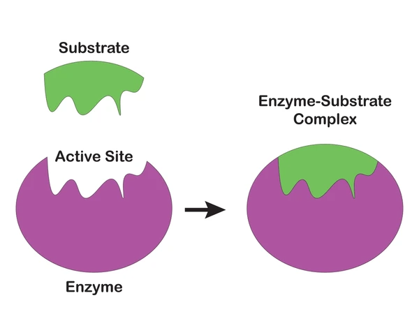
substrate
the molecule an enzyme acts on

active site
where the substrate binds and catalysis happens

nonbiological catalyst
a catalyst that isn’t an enzyme
Which is generally more specific for its substrate: a biological catalyst or a nonbiological catalyst?
Biological catalyst (enzyme).
Which interactions can contribute to the intrinsic binding energy during enzymatic catalysis? Select all that apply.
A) electrostatic interactions
B) permanent covalent bonding
C) hydrogen bonding
D) London dispersion interactions
E) nucleophilic attack by serine
Options A, C, and D
Electrostatic interactions
Attraction between opposite electrical charges (+ and −)
Permanent covalent bonding
When atoms share electrons to form a lasting bond
Hydrogen bonding
A weak attraction involving hydrogen and another electronegative atom, such as oxygen or nitrogen
London dispersion interactions
Weak attractions caused by temporary changes in electron distribution
Nucleophilic attack by serine
When serine attacks a substrate and forms a new chemical bond
Intrinsic binding energy
Energy released when an enzyme binds to its substrate through favorable interactions.
Why does electrostatic interactions contribute to intrinsic binding energy during enzymatic catalysis?
opposite electrical charges attract each other (positive attracts negative)
Why does hydrogen bonding contribute to intrinsic binding energy during enzymatic catalysis?
helps the substrate interact with the active site
Why does london dispersion interactions contribute to intrinsic binding energy during enzymatic catalysis?
very weak attarctions caused my temporary charges in electron distribution
How does permanat covalent bonding NOT contribute to intrinsic binding energy during enzymatic catalysis?
attaches the substrate to the enzyme instead of allowing it to bind temporarily and be released
How does nucleophilic attack by serine NOT contribute to intrinsic binding energy during enzymatic catalysis?
serine chemically attacks the substrate to form a bond, rather than simply helping the enzyme and substrate stick together through weak attractions.
Which of the following are examples of noncovalent interactions involved in substrate binding? Select all that apply.
A) Hydrogen bonds
B) Ionic attractions
C) Covalent bond formation
D) Van der Waals interactions
E) Nucleophilic attack
options A, B, and D
noncovalent interactions
Weak attractions between atoms or molecules that do not involve sharing electrons to form a new covalent bond; helps enzymes hold onto their substrate
substrate binding
When a substrate (molecule) attaches to an enzyme's active site, the enzyme can perform a chemical reaction; the substrate attaches to the enzyme’s active site
Which of the following does NOT directly contribute to intrinsic binding energy through noncovalent interactions?
Permanent covalent bonding
During chymotrypsin catalysis, Ser195 attacks the carbonyl carbon of a substrate. This is an example of which process?
Covalent catalysis
An enzyme's active site contains a positively charged amino acid that attracts a negatively charged substrate. What type of interaction is occurring?
Electrostatic interaction
Which interaction results from temporary fluctuations in electron distribution between atoms?
London dispersion interaction
Which of the following interactions help stabilize an enzyme-substrate complex through intrinsic binding energy? Select all that apply.
A) Attraction between opposite electrical charges
B) Hydrogen bonding between enzyme and substrate
C) Permanent covalent bonding between enzyme and substrate
D) London dispersion forces
options A, B, and D
Match the disaccharides sucrose below with the correct descriptions. Note that the given descriptions may occur more than once or not at all.
A) D-glucopyranosyl-(β1→2α)-D-fructofuranose
B) D-glucopyranosyl-(α1→4)-D-glucopyranose
C) D-glucopyranosyl-(α1→2β)-D-fructofuranose
D) D-galactopyranosyl-(β1→4)-D-glucopyranose
E) D-glucopyranosyl-(α1→4α)-D-glucopyranose
F) D-galactopyranosyl-(β1→4β)-D-glucopyranose
option C
Match the disaccharides maltose below with the correct descriptions. Note that the given descriptions may occur more than once or not at all.
A) D-glucopyranosyl-(β1→2α)-D-fructofuranose
B) D-glucopyranosyl-(α1→4)-D-glucopyranose
C) D-glucopyranosyl-(α1→2β)-D-fructofuranose
D) D-galactopyranosyl-(β1→4)-D-glucopyranose
E) D-glucopyranosyl-(α1→4α)-D-glucopyranose
F) D-galactopyranosyl-(β1→4β)-D-glucopyranose
option B
Match the disaccharides lactose below with the correct descriptions. Note that the given descriptions may occur more than once or not at all.
A) D-glucopyranosyl-(β1→2α)-D-fructofuranose
B) D-glucopyranosyl-(α1→4)-D-glucopyranose
C) D-glucopyranosyl-(α1→2β)-D-fructofuranose
D) D-galactopyranosyl-(β1→4)-D-glucopyranose
E) D-glucopyranosyl-(α1→4α)-D-glucopyranose
F) D-galactopyranosyl-(β1→4β)-D-glucopyranose
option D
What is a disaccharide?
Two simple sugars joined together by a glycosidic bond; sucrose, maltose, and lactose

What is sucrose?
glucose + fructose (α1→2β); found in regular sugar, fruits, and sweets

What is maltose?
glucose + glucose (α1→4); found in germinating grains (barley)
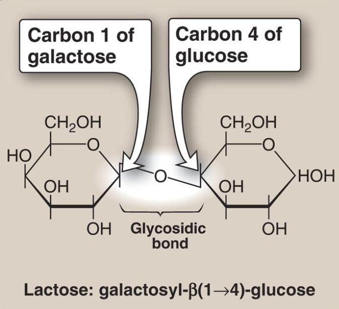
What is lactose?
galactose + glucose (β1→4); found in milk and dairy products
α (alpha) bond
One orientation of the glycosidic bond
β (beta) bond
the other orientation of the glycosidic bond
1 → 4 bond
Carbon 1 of the first sugar connects to carbon 4 of the second sugar
1 → 2 bond
Carbon 1 of the first sugar connects to carbon 2 of the second sugar.

What is cellobiose?
Glucose + Glucose (β1→4)

What is isomaltose?
Glucose + Glucose (α1→6)
What is trehalose?
Glucose + Glucose (α1→1α)
What is a polysaccharide?
large carbohydrate polymers made of ten or more simple sugar (monosaccharide) units joined by glycosidic bonds; glycogen, amylose, amylopectin, and cellulose.
What are is glycogen main bonds?
α1→4, branches α1→6
What are is amylose main bonds?
α1→4
What are is amylopectin main bonds?
α1→4, branches α1→6
What are is cellulose main bonds?
β1→4
D-glucopyranosyl-(β1→4)-D-glucopyranose describes which disaccharide?
Cellobiose