Respiratory Quiz -Health Assessment Lecture
 Call Kai
 Learn
 Practice Test
 Spaced Repetition
 Match
 Flashcards
 Knowt Play
1/164

There's no tags or description
Looks like no tags are added yet.
165 Terms
The thorax or thoracic cavity
commonly called the chest, is a closed cavity of the body, containing structures needed for respiration.
• The thorax is surrounded by ribs and muscles and extends from the base of the neck to the diaphragm.
Mediastinum:
Contains the heart, trachea, esophagus, and major blood vessels of the body.
Right and left pleural cavity:
Contains a lung
external respiration
The exchange of oxygen and carbon dioxide at the alveoli level of the lung
internal respiration
Gases are transported from the lungs via the blood to the cells of the body. As the gases move across the systemic capillaries, exchange of oxygen and carbon dioxide occurs at the cellular level
The upper respiratory tract includes:
– Nose
– Mouth
– Sinuses
– Pharynx
– Larynx
– Proximal portion of the trachea
The lower respiratory tract includes:
The distal portion of the trachea
– Bronchi
– Lungs
– Pleural membranes, the muscles of respiration, and
the mediastinum complete the lower respiratory tract.
The trachea is located
in the mediastinum, and descends
from the larynx in the neck to the main bronchi at the
distal point.
The trachea is a
flexible and mobile structure, bifurcating
anteriorly at about the sternal angle and posteriorly at
about vertebrae T3 to T5.
The trachea contains
16 to 20 rings of hyaline cartilage
that help to maintain the shape of the trachea and
prevent its collapse during inspiration and expiration.
The carina
Just above the point of bifurcation, the last tracheal
cartilage, known as the carina, is expanded. The carina
separates the openings of the two main bronchi.
The trachea, like other structures of the respiratory tract,
is lined with
a mucus-producing membrane that traps
dust, bacteria, and other foreign bodies.
Anteriorly, the trachea bifurcates at about the level of the
sternal angle, forming the right and left main bronchi.
• The main bronchus enters each lung at the hilus (medial
depression) and maintains an oblique position in the
mediastinum
The right main bronchus
is shorter, wider, and more
vertical than the left main bronchus; therefore, aspirated
objects are more likely to enter the right lung
The bronchi and the many branches
continue to warm
and moisten air as it moves along the respiratory tract to
the alveoli in the lungs
The left lung
has two lobes (upper and lower) and tends
to be longer and narrower than the right lung
The apex and base of the lungs
The apex of each lung is slightly superior to the inner
third of the clavicle, and the base of each lung is at the
level of the diaphragm
The pleura
is a thin, double-layered, serous membrane
that lines each pleural cavity.A pleural fluid is produced by these membranes that acts
as a lubricant, allowing the lung to glide during the
respiratory cycle of inspiration and expiration
The surface tension created by the fluid and the negative
pressure between the membranes
helps keep the lungs expanded.
• As the negative pressure changes, one is able to move air into and out of the lungs
The mediastinum
is the middle section of the thoracic cavity and is surrounded by the right and left pleural cavities.
• The mediastinum contains the heart, the trachea, the esophagus, the proximal portion of the right and left main bronchi, and the great vessels of the body.
Respiratory process
is a general term that encompasses the structures and activities of respiration.
• The respiratory process is dependent on the muscles of the thorax, the structures of the thoracic cage, and the ability of air to move in and out of the body
Respiratory Muscles
The muscles of the thoracic cage (internal and external intercostals) and the diaphragm assist in the breathing process.
• The accessory muscles of the neck (trapezius, scalenes,
and sternocleidomastoid), abdomen (rectus), and chest
(pectorals) assist the respiratory cycle as necessary.
• Accessory muscles play a major role in the respiratory
cycle during distress and pathology.
The thoracic cage
consists of the bones, cartilage, and
muscles of the thorax.
• The sternum (breastbone) is located in the anterior
midline of the thorax.
• The 12 pairs of ribs circle the body, form the lateral
aspects of the thorax, and are attached to the vertebrae
and sternum.
For air to enter the body,
the respiratory muscles contract, the chest expands, alveolar pressure decreases, and the negative intrapleural pressure increases. These combined activities allow air to enter the expanded lungs
During expiration
the passive phase of the process, the
activities reverse themselves: the lungs recoil, and air
leaves the body.
eupnea
The regular, even depth, rhythmic pattern of inspiration and expiration describes eupnea
dyspnea
A change in this pattern, producing shortness of breath or difficulty in breathing
The balance of gas exchange between oxygen and
carbon dioxide is influenced by
the nervous system, the
cardiovascular system, and the musculoskeletal system.
The respiratory system has a major role in helping the
body to maintain
acid–base balance.
• The concentration of carbon dioxide in the blood directly influences the blood concentration of carbonic acid and hydrogen ions.
• The respiratory system responds to the needs of the body to either retain or excrete carbon dioxide
The first bony landmark to be considered is the
sternum, commonly called the breastbone. It is a flat, elongated bone located in the midline of the anterior thoracic cage and consists of three parts: the manubrium, the body, and the xiphoid process.
The depression at the superior border
is called the suprasternal notch or jugular notch. This becomes a
primary landmark used to identify and locate other landmarks.
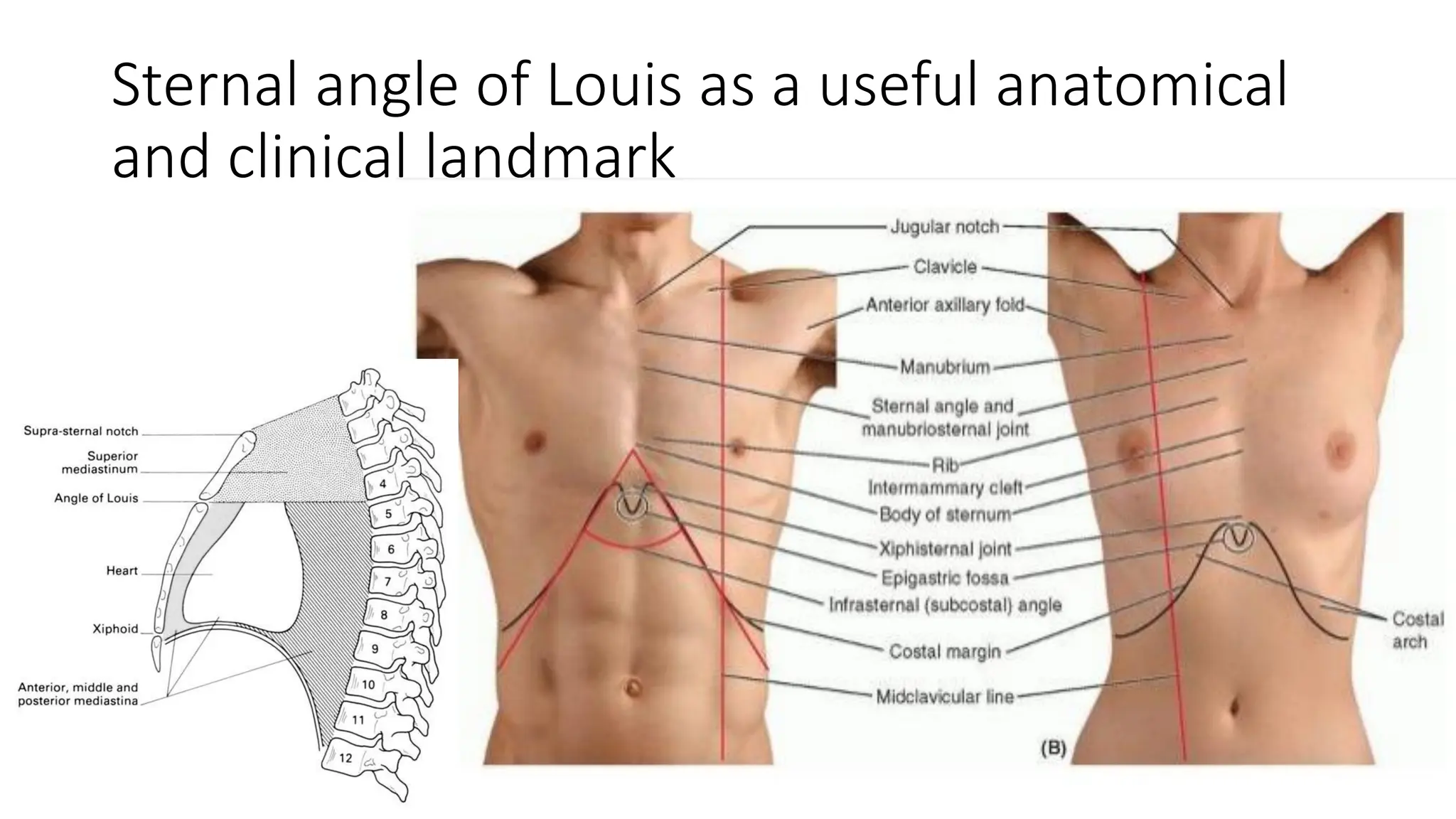
angle of Louis
As the clavicles and sternum meet, a horizontal ridge is formed,
referred to as the sternal angle or angle of Louis. The
second rib and the second intercostal space are at this
level of the sternum
costal angle.
The xyphoid process and the inferior borders of the
seventh ribs form a triangle
Bilaterally, the first seven ribs
attach to the sternum and are called true ribs
Ribs 8, 9, and 10 attach to
cartilage of the superior rib, and ribs 11 and 12 are free floating and do not articulate anteriorly
The seventh cervical vertebra (C7)
is most visible at the base of the neck.
• This prominent C7 vertebra is used to count and locate other spinous processes. When two spinous processes are equally prominent, they are C7 and T1
The midclavicular line
begins at the midpoint of the clavicle and descends to the level of the 12th rib. The
nipples of the breast are slightly lateral to this line. This line subdivides the right and left thoracic cage into two equal parts
The anterior axillary line (AAL)
is another line drawn parallel to the sternal line. It begins at the anterior fold of
the axillae and descends along the anterior lateral aspect of the thoracic cage to the 12th rib
landmarks on the posterior aspect of the thoracic cage
The vertebral line, the right and left scapular lines, and the right and left posterior axillary lines are used as landmarks
The vertebral or midspinous line
commences at C7 and descends through the spinous process of each thoracic vertebra. It divides the vertebral column in half, forming the posterior right and left thoracic cage
The posterior axillary line (PAL)
is parallel to the vertebral line. It starts at the posterior axillary fold and descends along the lateral aspect of the thoracic cage to the 12th rib
Three imaginary lines identify the lateral aspect of the
thoracic cage
he imaginary lines include the anterior axillary, posterior axillary, and midaxillary lines.
• The midaxillary line is parallel to the anterior and posterior axillary lines. This line descends from the middle of the axillae to the level of the 12th rib. It forms the frontal plane, dividing the thorax into the anterior and posterior portions
The trachea bifurcates
forming the right and left main bronchi. Anteriorly, this occurs at the level of the angle of Louis or sternal angle. Posteriorly, this bifurcation occurs between the third and fifth thoracic vertebrae
The apices of the lung extend
2 cm to 4 cm (0.78–1.57
in.) above the inner third of the clavicle anteriorly.
• Posteriorly, the apices of the lungs are located superior to
the scapula between the vertebral line and midscapular
line.
The base of the lung has three reference points.
The lung is cone-shaped, and the base of the lung is located at the sixth intercostal space at the midclavicular line.
• At the midaxillary line, the base of the lung is at the eighth intercostal space.
• At the scapular line on the posterior thorax, the base of the lung is at the 10th intercostal space.
This triangle, also known as
The clavicle, the scapula, and the lateral base of the neck form a triangle at the superior aspect of the thorax.
Kronig’s area is used for palpation of muscles and lymph nodes and for percussion and auscultation of the apex and the lungs
Patients with allergies or asthma should be encouraged
to explore
the possibility of allergens in their work or
home environment.
Secondhand smoke at home or work environment
can
also lead to respiratory distress
Exposure to large amounts of dust
in a granary or mine
may lead to the development of silicosis.
Workers in some industries may be exposed to
substances that are hazardous to their respiratory health,
such as caustic fumes, fungi, asbestos, coal tar, nickel,
silver, textile fibers, chromate, and vinyl chlorides.
• Coal miners are susceptible to pneumoconiosis, a form of
black lung disease.
People working in an office building may need to be
concerned with
air conditioners and forced hot-air heat.
The ducts of the cooling and heating systems can carry
airborne organisms, increasing the risk for respiratory
infections.
Newborns have a
high respiratory rate
Pediatric patients have
respirations that are abdominal in
nature.
In pregnant patients,
as the uterus enlarges, increasing pressure in the abdominal cavity limits diaphragmatic excursion and can result in more rapid and shallow respirations.
Stress, anxiety, pain, and fatigue may
exacerbate
respiratory problems
Patients experiencing acute or chronic respiratory
problems
will have a physiologic alteration with gas exchange.
• This alteration may limit or restrict the individual’s ability to independently perform the activities of daily living and to participate in activities, exercise, and sports.
Limitations that affect a patient’s ability to independently
function on a daily basis contribute to
social isolation,
changes role activities, lower self-esteem, and increases
the dependency factor with support systems
Certain drugs, such as bronchodilators
are used in the treatment of respiratory conditions and may cause the hands to tremble visibly. This is not to be confused with nervousness
Parents of young children who have experienced severe
asthmatic attacks in the past
may be extremely anxious
any time the child develops a cold, seasonal allergy, or
any other respiratory problem
The geographical location of an individual’s environment
will also influence respiratory health. Factors to be considered are temperature, moisture, altitude, and pollution.
• Factors within the home and social environment will influence respiratory health. Forced hot-air heat is very drying to the membranes of the body.
The focused interview for the respiratory system is
concerned with data related to the structures and
functions of that system.
• Subjective data related to respiratory status are gathered
during the focused interview.
• Be prepared to observe the patient and listen for cues
related to the function of the respiratory system.
What to consider when asking questions
age, gender, race, culture, environment, health
practices, past and concurrent problems, and therapies
when framing questions and using techniques to elicit
information
If a patient is experiencing dyspnea, cyanosis, difficulty
with speech, and the anxiety that accompanies any of
these problems
attention must focus on relief of
symptoms and restoration of oxygenation
Focused interview questions include:
– General Questions
– Questions related to illness or infection
– Questions related to symptoms, pain, and behaviors
– Questions related to age
Health records, the results of laboratory tests, and x-rays
are
important secondary sources to be reviewed and
included in the data-gathering process.
Equipment includes:
– Examination gown and drape
– Examination gloves
– Examination light
– Stethoscope
– Tissues
– Face mask for nurse, if indicated
Normal breath sounds include:
– Eupnea
– Eupnea with a sigh
Asthma
A chronic hyperreactive condition resulting in bronchospasm, mucosal edema, and increased mucus secretion. This condition usually occurs in response to inhaled irritants or allergens.
• Subjective findings:
– Dyspnea (shortness of breath)
– Anxiety
– Chest pain
Asthma
• Objective findings:
– Wheezing
– Diminished breath sounds
– Absent breath sounds (with severe asthma)
– Increased respiratory rate
– Increased use of accessory muscles
– Decreased oxygen saturation
Atelectasis
A collapse or impaired inflation of one or more areas of
the lung (NHLBI, 2013).
• During atelectasis, the alveoli or an entire lung may
collapse from airway obstruction, such as a mucous plug,
lack of surfactant, or a compressed chest wall.
Atelectasis
• Subjective findings:
– Absence of symptoms, if only a small portion of the lung is affected.
– Dyspnea, if significant portions of the lung are affected.
Atelectasis
• Objective findings:
– Decreased or absent breath sounds over the affected
area
– Increased respiratory rate
– Decreased oxygen saturation
– Cyanosis (if severe)
Chronic Bronchitis
Chronic inflammation of the tracheobronchial tree leads to increased mucus production and blocked airways.
• A productive cough is present.
Chronic Bronchitis
• Subjective findings:
– Dyspnea
– Fatigue (related to increased work of breathing)
Chronic Bronchitis
• Objective findings:
– Chronic productive cough
– Increased respiratory rate
– Use of accessory muscles
– Wheezes
– Rhonchi
Emphysema
A condition in which chronic inflammation of the lungs leads to destruction of alveoli and decreased elasticity of the lungs. As a result, air is trapped and lungs hyperinflate.
Subjective findings of emphysema
– Dyspnea, especially upon exertion
– Air hunger (related to hypoxemia, CO2 retention, and air trapping).
Emphysema
• Objective findings:
– Barrel chest and decreased chest expansion
– Cyanosis
– Hypercarbia (increased blood concentration of CO2)
– Clubbing of fingers
– Use of accessory muscles
– Diminished breath sounds
Lobar Pneumonia
An infection that causes fluid, bacteria, and cellular debris to fill the alveoli.
• Subjective findings:
– Dyspnea
– Fatigue
– Chills
Lobar Pneumonia
• Objective findings:
– Increased respiratory rate
– Fever
– Productive cough
– Decreased oxygen saturation
– Bronchial breath sounds and crackles
Pleural Effusion
A fluid accumulation in the pleural space. Pleural effusion may be asymptomatic, or it could present with common signs and symptoms.
• Subjective findings:
– Dyspnea
– Occasional sharp, nonradiating chest pain
Pleural Effusion
• Objective findings:
– Cough
– Diminished or absent breath sounds
– Decreased or absent tactile fremitus
– No voice transmission
Pneumothorax
A condition in which air moves into the pleural space and causes partial or complete collapse of the lung. Pneumothorax can be spontaneous, traumatic, or tension.
Subjective findings:
– Dyspnea
– Sharp chest pain with inspiration
– Anxiety
Pneumothorax
• Objective findings:
– Increased respiratory rate
– Decreased oxygen saturation
– Diminished or absent breath sounds over the affected area
– Decreased chest wall expansion on the affected side
– Tracheal deviation to the unaffected side
Left Heart Failure
Heart failure occurs when the heart is unable to effectively pump oxygen-rich blood.
• Heart failure may affect one or both sides of the heart. Most often, both the left and right sides of the heart are affected (NHLBI, 2014).
• Left heart failure typically produces respiratory symptoms as blood that is not ejected by the left ventricle backs up into the lungs.
Right heart failure typically produces
peripheral edema,
as well as other manifestations
with left heart failure
increased pressure in the pulmonary veins causes interstitial edema around the alveoli and may cause edema of the bronchial mucosa.
left heart failure Subjective findings:
– Dyspnea, especially upon exertion
– Orthopnea (shortness of breath that happens when you lie flat and goes away when you sit or stand up)
– Anxiety
Left Heart Failure
• Objective findings:
– Increased respiratory rate
– Decreased oxygen saturation
– Pulmonary congestion with auscultation of wheezes or crackles in lung bases
– Pallor
– Decreased chest wall expansion on the affected side
– Tracheal deviation to the unaffected side
During development, the fetus receives
its nutrients and oxygen from its mother. The lungs are nonfunctional, and oxygen is carried in blood from the placenta to the right side of the heart.
Compared with adults, children have much
smaller, more compliant airways until early adolescence.
children are more prone to
airway collapse and blockage. Oxygen needs are higher in small children because their increased metabolic rates result in higher oxygen consumption.
The foramen ovale
The majority of this blood passes through the foramen ovale to the left side of the heart, then into the aorta to enter the systemic circulation. The foramen ovale is a passageway for blood between the right and left atria.
ductus arteriosus
The rest of the blood passes through the pulmonary artery and ductus arteriosus and enters the aorta (Figure 26.7). The ductus arteriosus is an opening between the pulmonary artery and the descending aorta.
The risk of respiratory failure is greatest in
infants, toddlers, and preschoolers, but children of all ages experience respiratory failure much more quickly than adults.
The foramen ovale closes shortly after
after birth because of increased pulmonary vascular return and decreased pressure in the right side of the heart.
The ductus arteriosus closes within
24 to 48 hours in response to multiple physiologic events, including decreased pulmonary resistance and decreased pressure in the right atrium versus increased pressure in the left atrium.
what happens if the foramen ovulae and the ductus arteriosus remain patent
Murmurs may be auscultated if these openings remain patent. However, if a ventricular septal defect is present, the murmur it causes may not be auscultated until week 4 to week 6 after birth.
As individuals age
The lungs lose their elasticity, the skeletal muscles begin to weaken, and bones lose their density. As a result, it becomes more difficult for the older adult to expand the thoracic cage and take a deep breath.
The appearance of a barrel chest and calcification of cartilage contribute to
the decrease in the circumference of the chest (thoracic excursion). Thus, the older adult inhales and exhales smaller amounts of air.