MSK/PAED practice exam
 Call Kai
 Learn
 Practice Test
 Spaced Repetition
 Match
 Flashcards
 Knowt Play
1/48

There's no tags or description
Looks like no tags are added yet.
49 Terms
What are the attachments of the supraspinatus, infraspinatus, subscapularis and teres minor tendons of the shoulder?
Muscle | Origin | Insertion |
|---|---|---|
Supraspinatus | Supraspinous fossa | Superior facet of greater tuberosity |
Infraspinatus | Infraspinous fossa | Middle facet of greater tuberosity |
Teres minor | Lateral border of scapula | Inferior facet of greater tuberosity |
Subscapularis | Subscapular fossa | Lesser tuberosity |
What are the attachments of the long & short head biceps brachii of the shoulder?
Long head of biceps: Supraglenoid tubercle and superior glenoid labrum (origin); tendon passes through the bicipital groove.
Short head of biceps: Coracoid process; shares its origin with coracobrachialis (origin).
Distal insertion: Radial tuberosity
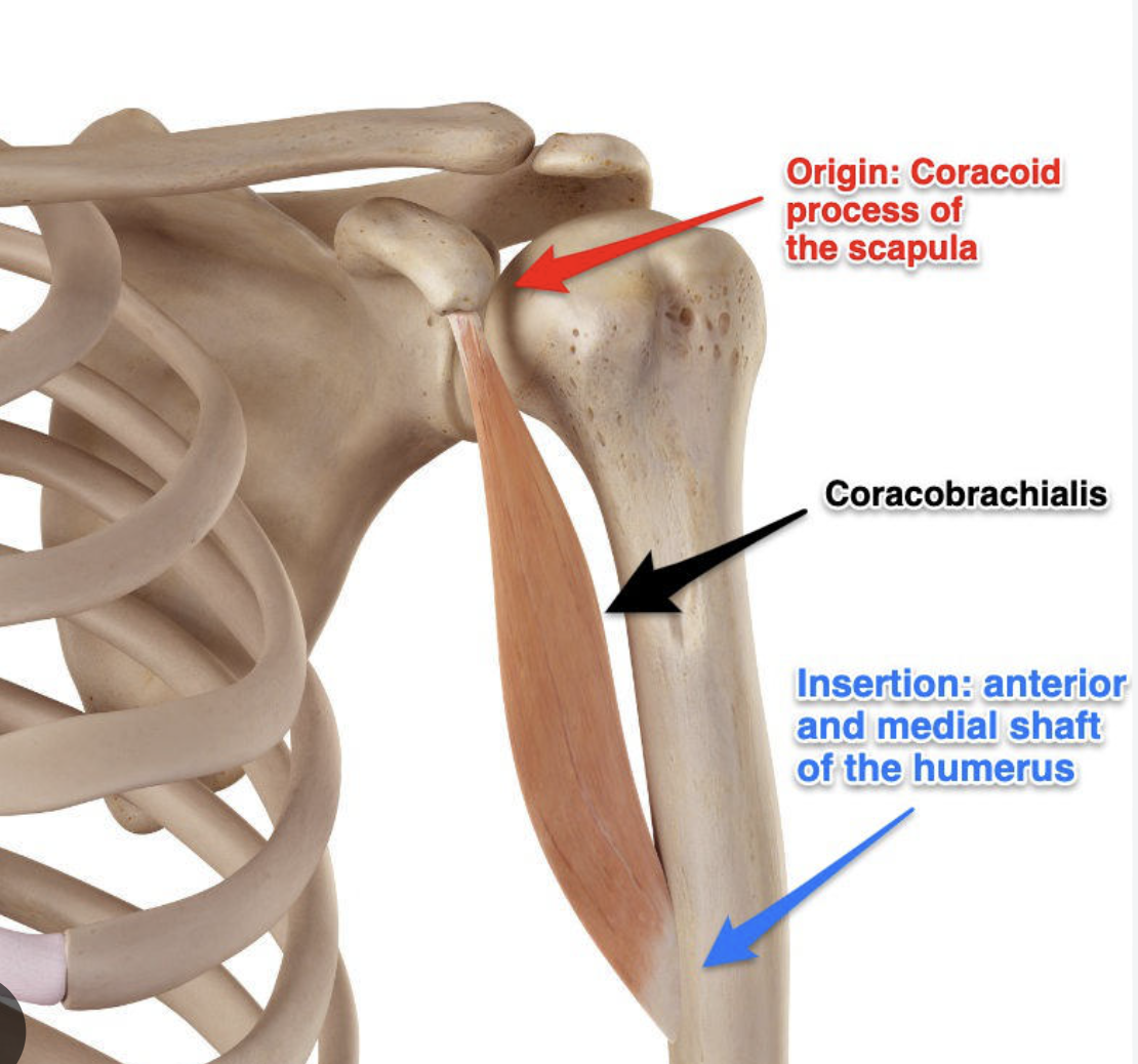
What are the attachments of the coracobrachialis?
Origin: Coracoid process of the scapula (often sharing a conjoined tendon with the short head of the biceps brachii)
Insertion: Middle third of the anteromedial/medial surface of the humeral shaft
What are the attachments of the deltoid?
Origin
Anterior part: Lateral third of the clavicle (anterior surface)
Middle part: Acromion process of the scapula
Posterior part: Spine of the scapula
Insertion
Deltoid tuberosity of the humerus

What are the attachments of the triceps?
Origins
Long Head: Infraglenoid tubercle of the scapula
Lateral Head: Posterior surface of the humerus, superior to the radial groove
Medial Head: Posterior surface of the humerus, inferior to the radial groove
Insertion
Olecranon Process

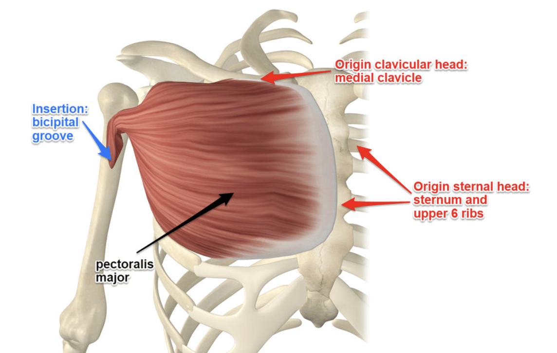
What are the attachments of the Pectoralis Major?
Origin
The pectoralis major originates from three main regions:
Clavicular Head
Sternocostal Head: The sternum and the costal cartilages of ribs 1-6
Abdominal Head: Rectus sheath of the abdominal wall.
Insertion
Humerus: Bicipital groove
What are the attachments of the Teres Major?
Origin
Superior to the inferior angle of the scapula
Insertion
Medial lip (crest) of the intertubercular groove of the anterior humerus

What are the attachments of the Latissimus Dorsi?
Origin
Spinous processes of T7 to L5
Posterior third of the iliac crest.
Scapula: Inferior angle of the scapula
Insertion: The bicipital groove

Dynamic assessment involving shoulder abduction. What features would you be looking for? (4 marks)
Subacromial-subdeltoid (SASD) bursa and potentially supraspinatus impingement under the acromion. Observing if this reproduces the patient's characteristic pain.
The inserting fibres of the distal biceps tendon may appear hypoechoic. Name and describe the artefact. (3 marks)
Anisotropy is an ultrasound artefact in which a structure appears artificially hypoechoic because the ultrasound beam is not perpendicular to its reflective fibres.
How to correct anisotropy artefact?
Heel-toe the transducer.
Adjust the transducer angle.
Rotate or reposition the probe to obtain perpendicular insonation.
Assess the tendon in multiple planes.
List the important structures to be imaged when assessing the lateral elbow. (3 marks)
Common extensor tendon origin (CETO): Originates from the lateral epicondyle, particularly the extensor carpi radialis brevis.
Radial collateral ligament (RCL): Extends from the lateral epicondyle towards the annular ligament.
Annular ligament: Surrounds and stabilises the radial head.
Exam pearl: Lateral epicondylitis is primarily associated with pathology of the common extensor tendon, particularly the ECRB component.
Define and list the sonographic appearances of De Quervain's tenosynovitis
Stenosing tenosynovitis affecting the Compartment 1 of the wrist, involving the abductor pollicis longus (APL) and extensor pollicis brevis (EPB). Usually associated with repetitive thumb and wrist movements.
Sonographic appearances:
Thickening of the APL and EPB tendons.
Hypoechoic and heterogeneous tendon changes.
Thickening of the overlying extensor retinaculum.
Fluid within the tendon sheath.
Synovial hypertrophy.
Increased colour Doppler vascularity.
Possible compartmental septation separating APL and EPB.

Define and list the sonographic appearances of Trigger finger
Definition: Stenosing tenosynovitis involving the flexor tendon and A1 pulley at the level of the metacarpophalangeal joint.
The thickened pulley restricts normal gliding of the flexor tendon, resulting in catching or locking during finger flexion and extension.
Sonographic appearances:
Thickened, hypoechoic A1 pulley.
Thickened and potentially heterogeneous flexor tendon.
Possible tendon sheath fluid.
Synovial hypertrophy and hypervascularity.
Impaired tendon gliding during dynamic examination.
Sudden movement or snapping of the tendon through the pulley.

Define and list the sonographic appearances of Stener lesion
Definition: A complete rupture of the ulnar collateral ligament (UCL) of the thumb MCP joint, where the distal ligament becomes displaced superficial to the adductor aponeurosis.
The interposed adductor aponeurosis prevents the ruptured ligament from returning to its normal insertion, impairing spontaneous healing.
Sonographic appearances:
Disrupted or absent normal UCL fibres.
Retracted, thickened and hypoechoic distal ligament.
Mass-like appearance of the retracted ligament proximal to its normal insertion.
Adductor aponeurosis interposed between the ligament and its insertion.
Possible associated avulsion fracture at the base of the proximal phalanx.

Define and list the sonographic appearances of Cubital tunnel syndrome
Definition: Compression or irritation of the ulnar nerve as it passes through the cubital tunnel at the medial elbow, commonly behind the medial epicondyle.
Sonographic appearances:
Increased cross-sectional area of the ulnar nerve.
Hypoechoic and swollen nerve.
Loss of normal fascicular/honeycomb appearance.
Flattening of the nerve at the compression site.
Proximal nerve enlargement.
Increased intraneural vascularity on colour Doppler.
Possible dynamic subluxation or dislocation of the nerve over the medial epicondyle during elbow flexion.
A cross-sectional area above approximately 10 mm² at the medial epicondyle is commonly considered suspicious.

Define and list the sonographic appearances of an Umbilical hernia
Protrusion of abdominal contents through a defect in the umbilical ring.
May contain preperitoneal fat, omentum or bowel.
Demonstrates a fascial defect and hernia sac.
Define and list the sonographic appearances of an Epigastric hernia
Define and list the sonographic appearances of an Epigastric hernia
Occurs through the linea alba between the xiphoid process and umbilicus.
Commonly contains preperitoneal fat.
Appears as a focal protrusion through a defect in the linea alba.
Define and list the sonographic appearances of an Incisional hernia
Occurs through a previous surgical incision due to weakness or disruption of the abdominal wall.
Demonstrates protrusion of fat or bowel through the surgical fascial defect.
Define and list the sonographic appearances of a Spigelian hernia
It occurs through the Spigelian fascia along the linea semilunaris, lateral to the rectus abdominis muscle.
What are the general ultrasound findings for abdominal wall hernias?
Focal fascial defect.
Protrusion of fat, omentum or bowel through the defect.
Hernia sac and neck.
Increased protrusion with coughing or Valsalva.
Assess reducibility with gentle transducer pressure.
Assess bowel peristalsis and vascularity if bowel is present.
Draw the anatomical structures of the medial ankle. (4 marks)
The tarsal tunnel is located posterior and inferior to the medial malleolus. It is covered by the flexor retinaculum, which attaches between the medial malleolus and calcaneus.
Remember the mnemonic:
Tom, Dick And Very Nervous Harry
From anterior to posterior:
Tom – Tibialis posterior tendon.
Dick – Flexor digitorum longus tendon.
And – Posterior tibial artery.
Very – Posterior tibial veins.
Nervous – Tibial nerve.
Harry – Flexor hallucis longus tendon.
What is additional medial ankle anatomy?
Medial malleolus.
Talus.
Calcaneus.
Sustentaculum tali.
Navicular bone.
Flexor retinaculum.
Deltoid ligament.
Spring ligament (plantar calcaneonavicular ligament).
What are the tendon insertions of the medial ankle tendons?
Tibialis posterior: Mainly navicular tuberosity, with expansions to adjacent tarsal bones.
Flexor digitorum longus: Bases of distal phalanges of toes 2–5.
Flexor hallucis longus: Base of distal phalanx of the great toe.
Describe a hip joint effusion
Anechoic or hypoechoic fluid within the anterior joint recess.
Distension of the joint capsule, lifting it away from the femoral neck.
Possible internal debris or synovial thickening.
Describe an Iliopsoas bursa
Fluid distension of the bursa.
Anechoic or hypoechoic fluid surrounding the iliopsoas tendon.
Possible synovial thickening and Doppler hypervascularity.
Describe a Trochanteric bursa
Fluid distension.
Bursal wall thickening.
Possible internal debris.
Increased Doppler vascularity.
Associated gluteus medius/minimus tendinopathy may be present.
Describe the sonographic appearance of patellar tendinopathy. (4 marks)
Definition: Degenerative pathology of the patellar tendon due to repetitive mechanical loading, commonly associated with jumping activities. Often called jumper's knee.
Sonographic appearances:
Thickening of the patellar tendon, particularly at its proximal attachment to the inferior patellar pole.
Hypoechoic regions within the tendon.
Loss of normal fibrillar echotexture.
Heterogeneous tendon architecture.
Increased colour Doppler vascularity/neovascularisation.
Possible intratendinous calcification or enthesophytes at the patellar insertion.
Possible associated partial-thickness tearing.
Define Paratenonopathy
Inflammation or degeneration of the paratenon, the connective tissue surrounding certain tendons, particularly the Achilles tendon.
Sonographic appearances:
Thickening of the paratenon.
Hypoechoic changes surrounding the tendon.
Anechoic or hypoechoic fluid around the tendon.
Increased colour Doppler vascularity.
Peritendinous oedema.
The tendon itself may remain relatively normal, or may demonstrate associated tendinopathy.
Describe Plantar fasciitis
Definition: Degeneration and inflammation-like changes of the plantar fascia, most commonly at its origin from the medial calcaneal tubercle.
Sonographic appearances:
Thickening of the proximal plantar fascia.
Measurement greater than approximately 4 mm near its calcaneal origin is commonly considered abnormal.
Hypoechoic and heterogeneous echotexture.
Loss of normal fibrillar appearance.
Blurring of the normal fascial margins.
Increased Doppler vascularity.
Possible adjacent soft tissue oedema.
Possible calcaneal enthesophyte.
Define Haglund's deformity
Definition: Haglund's deformity is a bony prominence of the posterosuperior calcaneus. When associated with surrounding soft tissue inflammation, retrocalcaneal bursitis or insertional Achilles tendinopathy, it is commonly described as Haglund's syndrome.
Sonographic appearances:
Prominent posterosuperior calcaneal cortex.
Irregularity of the calcaneal insertion.
Retrocalcaneal bursal distension with fluid.
Thickened, heterogeneous distal Achilles tendon.
Hypoechoic changes in the Achilles tendon insertion.
Possible intratendinous calcification or enthesophytes.
Increased Doppler vascularity.
Possible changes to the adjacent Kager's fat pad.

Draw the anatomical structures of the lateral knee. (4 marks)
1. Iliotibial band (ITB)
Thick fascial band running down the lateral thigh.
Inserts at Gerdy's tubercle on the anterolateral proximal tibia.
Passes over the lateral femoral epicondyle.
2. Lateral collateral ligament (LCL)
Origin: Lateral femoral epicondyle.
Insertion: Head of fibula.
Appears as a thick echogenic fibrillar structure on ultrasound.
3. Biceps femoris tendon
Distal tendon inserts onto the head of the fibula.
Lies superficial to the LCL near its insertion.
4. Popliteus tendon
Originates from the lateral femoral condyle.
Travels through the popliteal hiatus and inserts on the posterior proximal tibia.
Whilst scanning a tendon you are looking for neovascularisation. List four factors you would consider changing to improve its detection. (2 marks)
Reduce PRF / velocity scale: Allows detection of slower blood flow.
Reduce wall filter: Prevents low-velocity flow signals from being filtered out.
Increase colour Doppler gain: Increase until background noise appears, then reduce slightly.
Use power Doppler: More sensitive to low-velocity flow and less dependent on flow direction.
Other important considerations:
Apply minimal transducer pressure because compression can obliterate small vessels.
You are scanning a foreign body. Describe all the information required to report its location. (2 marks)
Anatomical location: Specific body region and side.
Depth: Distance from the skin surface to the foreign body.
Position: Relationship to nearby anatomical landmarks.
Orientation: Longitudinal, transverse or oblique orientation.
Dimensions: Length and width, and thickness if measurable.
Relationship to important structures: Nearby tendons, muscles, fascia, vessels, nerves and bone.
Number: Single or multiple foreign bodies.
Also describe whether there is associated:
Surrounding inflammatory reaction.
Soft tissue oedema.
Granuloma formation.
Fluid collection or abscess.
With reference to the supraspinatus tendon, describe the types of tears possible. (5 marks)
1. Articular-sided partial-thickness tear
Involves the deep surface of the tendon adjacent to the humeral head.
Appears as a focal hypoechoic or anechoic defect extending from the articular surface.
Does not involve the full thickness of the tendon.
2. Bursal-sided partial-thickness tear
Involves the superficial surface of the tendon adjacent to the SASD bursa.
Appears as a focal defect extending from the bursal surface.
The deeper articular fibres remain intact.
3. Intrasubstance / interstitial tear
Occurs within the substance of the tendon.
Appears as a focal hypoechoic or anechoic defect within the tendon.
Does not communicate with either the articular or bursal surface.
4. Full-thickness tear
Extends from the articular surface through to the bursal surface.
May demonstrate a focal fluid-filled defect.
There is communication between the glenohumeral joint and subacromial-subdeltoid bursa.
May be associated with tendon retraction.
5. Complete or extensive tear
A complete rupture involves the entire width of the tendon, with possible retraction of the tendon stump towards the musculotendinous junction.
Chronic overuse injuries account for the majority of tendon disease. Explain the process of tendinosis. (4 marks)
Definition: Tendinosis is a chronic degenerative condition of a tendon resulting from repetitive mechanical loading and failed tendon healing.
Pathophysiology:
Repetitive mechanical loading produces microscopic damage to the collagen fibres.
Repeated injury exceeds the tendon's ability to repair itself.
Normal organised type I collagen becomes disrupted and is replaced by less organised collagen, with increased type III collagen.
Increased ground substance and mucoid degeneration develop, resulting in tendon thickening and disorganisation.
Neovascularisation may occur, with small blood vessels developing within the degenerated tendon.
The tendon becomes mechanically weaker and more susceptible to partial or complete tearing.
Describe the appearances of a muscle tear. (2 marks)
Sonographic appearances:
Disruption of normal muscle architecture and fibrillar pattern.
Focal hypoechoic or anechoic defect within the muscle.
Haematoma or fluid accumulation between muscle fibres.
Muscle swelling and oedema.
Retraction or separation of torn muscle fibres in more severe tears.
Possible increased vascularity during healing.
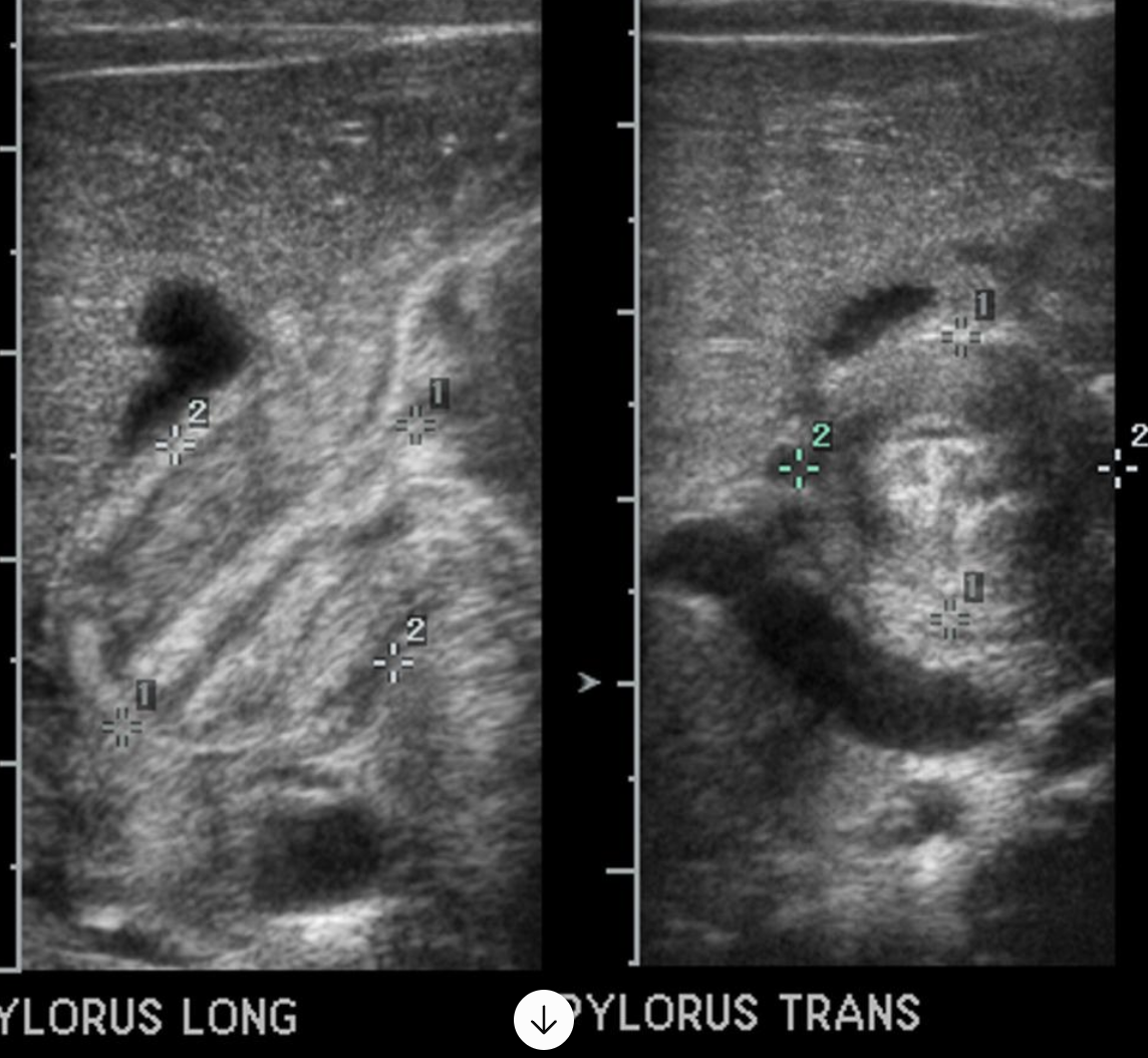
List your primary criteria for diagnosing infantile hypertrophic pyloric stenosis (IHPS). (4 marks)
1. Pyloric muscle thickness
Commonly accepted diagnostic threshold: approximately 3 mm or greater.
Measure the hypoechoic muscular layer, excluding the echogenic mucosa.
2. Pyloric canal length
Commonly used traditional threshold: greater than approximately 17 mm.
Some references and institutions use different thresholds.
List your secondary criteria for diagnosing infantile hypertrophic pyloric stenosis (IHPS). (4 marks)
Secondary sonographic criteria
Increased pyloric muscle echogenicity is not typical; the hypertrophied muscle is predominantly hypoechoic.
Elongated pyloric canal.
Transverse target or doughnut sign due to circumferential hypoechoic muscular thickening.
Longitudinal cervix sign / shoulder sign.
Failure of the pylorus to relax and open.
Failure of gastric contents to pass through the pyloric canal.
Gastric distension and retained contents.
Increased gastric peristalsis, particularly in earlier stages.
Describe the sonographic findings of an obstructed duplex renal collecting system. Discuss further imaging that may be required and why. (6 marks)
Definition: A duplex collecting system occurs when a kidney has two separate pelvicalyceal collecting systems, usually arising from duplication of the ureteric bud.
The Weigert-Meyer rule is important:
Upper pole moiety: usually associated with ectopic ureteric insertion or ureterocele and obstruction.
Lower pole moiety: more commonly associated with vesicoureteric reflux (VUR).
Sonographic appearances
1. Duplicated collecting system
2. Obstruction of the upper pole moiety
3. Ureterocele - a cystic dilatation of the distal ureter within the bladder.
Discuss further imaging that may be required of an obstructed duplex renal collecting system.
1. Voiding cystourethrogram (VCUG / MCUG)
Used to assess:
Vesicoureteric reflux.
Reflux into the upper or lower moiety.
Bladder neck obstruction.
Urethral abnormalities.
Degree of ureterocele prolapse where relevant.
2. MAG3 diuretic renogram
Assesses:
Renal function.
Differential function of the two kidneys.
Drainage and possible functional obstruction.
Whether there is impaired drainage from the affected moiety.
3. DMSA renal scintigraphy
Assesses:
Cortical function.
Differential functioning renal tissue.
Renal scarring.
4. MR urography
Provides detailed anatomical information about:
Duplicated collecting systems.
Course of the ureters.
Ectopic ureteric insertion.
Upper and lower moiety anatomy.
Relative renal function when functional MR urography is used.
What are the sonographic features of paediatric spinal cord tethering associated with spinal dysraphism? (3 marks)
Definition: Tethered cord syndrome occurs when the spinal cord is abnormally anchored, restricting its normal movement and potentially causing progressive neurological dysfunction as the child grows.
Sonographic features:
1. Low-lying conus medullaris
Normally terminates around L1–L2, generally at or above the L2–L3 disc space.
A conus terminating abnormally low is suspicious for tethering.
2. Thickened filum terminale
Normal filum is generally less than 2 mm.
A filum greater than 2 mm may indicate pathology, particularly if it is echogenic due to fat.
3. Reduced mobility of the cord and nerve roots
Reduced normal pulsatile movement.
Restricted movement of the cauda equina.
Cord may appear fixed posteriorly rather than centrally positioned within the spinal canal.
Associated findings:
Lipoma of the filum terminale.
Spinal lipoma.
Myelomeningocele or other dysraphic abnormalities.
Possible dermal sinus tract.
Describe the cranial ultrasound findings in a premature infant (32 weeks) with known haemorrhage. How would you classify the intracranial haemorrhage? (5 marks)
Premature infants are particularly susceptible because the germinal matrix contains fragile, immature blood vessels.
The germinal matrix is located near the caudothalamic groove, adjacent to the lateral ventricles.
Sonographic appearances
1. Germinal matrix haemorrhage (GMH)
Focal echogenicity at the caudothalamic groove.
Typically seen in the region of the head of the caudate nucleus.
Appears as an echogenic focus adjacent to the lateral ventricle.
May be unilateral or bilateral.
2. Intraventricular haemorrhage (IVH)
If bleeding extends into the ventricular system:
Echogenic blood or clot within the lateral ventricles.
Increased echogenicity of the ventricular contents.
Possible echogenic clot within the dependent portion of the ventricle.
Possible ventricular dilatation in more severe haemorrhage.
3. Periventricular haemorrhagic infarction
Increased echogenicity of the adjacent periventricular brain parenchyma.
May occur alongside severe IVH.
Can subsequently develop into cystic changes or tissue loss.

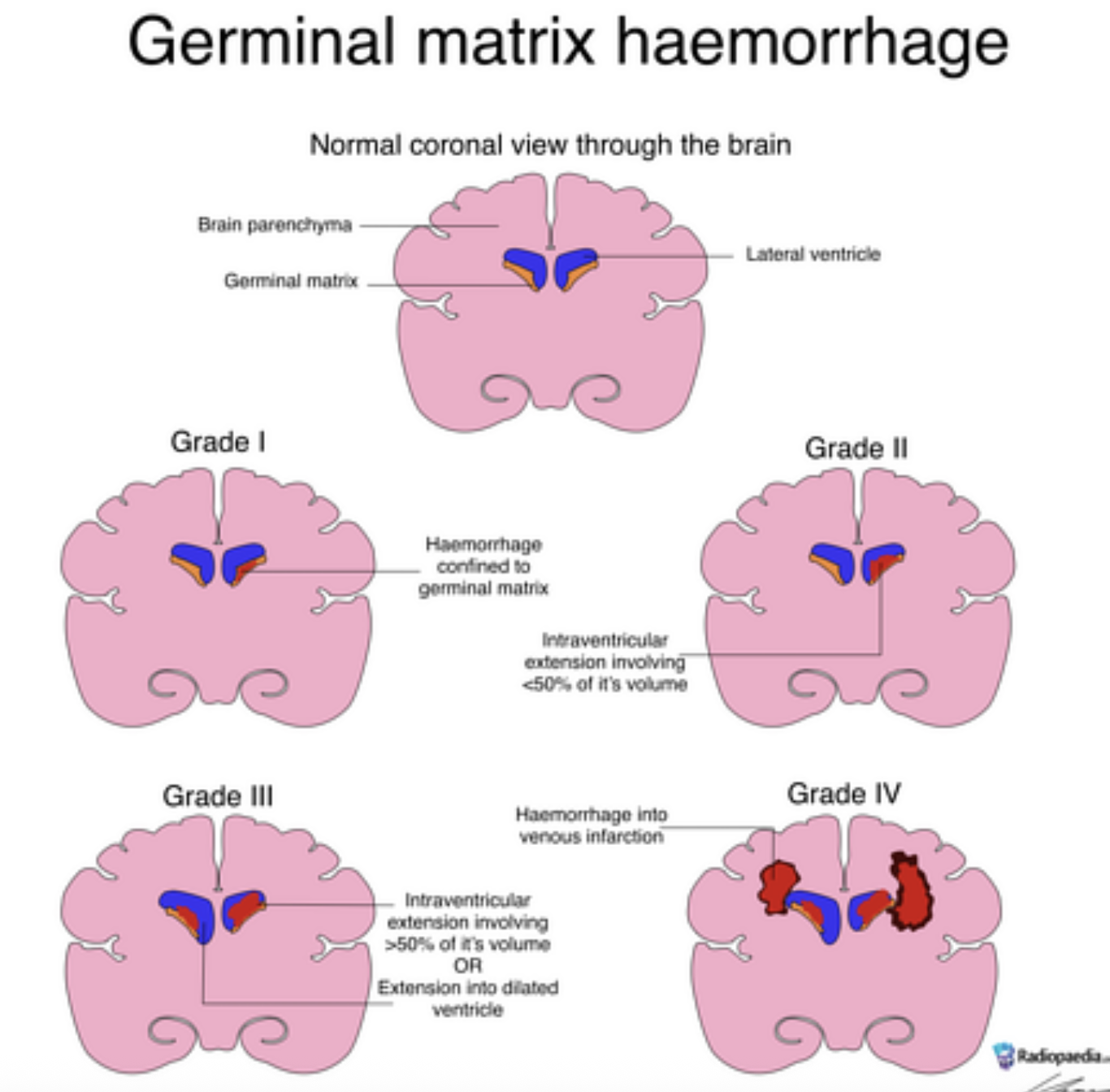
How would you grade the GMHs?
Grade I – Germinal matrix haemorrhage
Haemorrhage confined to the germinal matrix/subependymal region.
Echogenic focus at the caudothalamic groove.
No significant intraventricular haemorrhage.
Grade II – Intraventricular haemorrhage without ventricular dilatation
Blood extends into the lateral ventricles.
No associated ventricular dilatation.
Grade III – Intraventricular haemorrhage with ventricular dilatation
Extensive intraventricular blood.
Associated ventricular distension/dilatation.
Grade IV – Traditionally described as intraventricular haemorrhage with parenchymal extension
Haemorrhage associated with periventricular parenchymal involvement.
Modern terminology recognises this as periventricular haemorrhagic infarction (PVHI), rather than simply blood extending directly into the brain tissue.
Discuss the sonographic appearances of multicystic dysplastic kidney (MCDK). (3 marks)
Definition: MCDK is a congenital renal abnormality in which normal renal parenchyma is replaced by multiple non-functioning cysts of varying sizes.
Sonographic appearances:
Multiple cysts of varying sizes replacing the renal parenchyma.
Cysts do not communicate with one another.
No normal renal collecting system or normal central renal sinus architecture.
Echogenic dysplastic tissue between cysts.
Normal renal architecture is absent or severely distorted.
The affected kidney may be enlarged initially and may involute over time.

Differentiate MCDK and hydronephrosis
MCDK | Hydronephrosis |
|---|---|
Multiple non-communicating cysts | Communicating dilated collecting system |
Disorganised renal architecture | Dilated renal pelvis and calyces |
No normal central renal sinus | Dilated central collecting system |
You are performing an abdominal ultrasound on an 18-month-old. Discuss patient and equipment preparation, approach and modifications if distressed. (5 marks)
A. Patient preparation
Review the clinical indication and previous imaging.
Confirm the child's identity and relevant clinical history.
Explain the examination to the parent and child using age-appropriate language.
Ask about fasting requirements, particularly if the gallbladder and upper abdomen are being assessed.
Follow departmental fasting protocols, avoiding unnecessary prolonged fasting in a toddler.
Consider hydration and bladder filling if the urinary tract is part of the examination.
Allow the parent or caregiver to remain present.
B. Equipment preparation
Select an appropriate paediatric ultrasound preset.
Use a low-frequency curvilinear transducer for deeper abdominal structures.
Use a high-frequency linear transducer for superficial structures and detailed assessment.
Select an appropriately sized probe for the child's body habitus.
Optimise gain, depth, focus and frequency.
Use sufficient warm ultrasound gel.
Maintain a warm, comfortable examination room.
C. Approach to the patient
Introduce yourself and establish rapport with the child and parent.
Allow the child to become familiar with the equipment.
Use simple language and explain what the probe is doing.
Encourage the child to lie comfortably, ideally with the parent nearby.
Begin with the easiest or most important part of the examination.
Use distraction techniques such as toys, videos or talking.
Use gentle transducer pressure and avoid causing discomfort.
Acquire images opportunistically when the child is settled.
D. If the child becomes distressed
Pause the examination and allow the child to settle.
Encourage the parent to hold or comfort the child.
Use toys, videos, songs or a favourite comfort object.
Consider scanning the child while sitting on the parent's lap.
Reduce the duration of individual scanning sessions.
Prioritise clinically important anatomy and pathology.
Modify the position, such as scanning in a lateral decubitus or sitting position.
If necessary, stop and arrange an alternative examination time rather than causing unnecessary distress.
Young female hockey player with a lump on her calf. Primary differential? (1 mark)
A muscle hernia is a plausible examination answer in a young athlete presenting with a focal lump.
Definition: Protrusion of muscle tissue through a defect or weakness in the overlying deep fascia.
Sonographic appearances:
Focal protrusion of muscle through a fascial defect.
Continuity with the underlying muscle.
The bulge may become more prominent with muscle contraction or standing.
Dynamic ultrasound helps demonstrate the abnormality.