Biomaterial Implantation and Acute Inflammation
 Call Kai
 Learn
Learn Practice Test
 Spaced Repetition
 Match
Match Flashcards
 Knowt Play
1/61

There's no tags or description
Looks like no tags are added yet.
Name | Mastery | Learn | Test | Matching | Spaced | Call with Kai | Chat |
|---|
No analytics yet
Send a link to your students to track their progress
62 Terms
Biomaterials-Tissue Interactions
local and systemic
Local Biomaterial-Tissue Interactions
at the biomaterial-tissue interface
effect of material on host tissues → blood/material interactions, toxicity, mods of healing, inflammaion, infection, and tumorigenesis
effect of environment on materials → physical/mechanical effects (wear, fatigue, corrosion, and stress-corrosion cracking) and biological effects (adsorption of tissue constituients by implant, enzymatic degradation, and calification)
Systemic Biomaterial - Tissue Interactions
embolization (device can release particles that can block an artery), hypersensitivity, elevation of implant elements in blood, and lymphatic particle transport (small particles can be picked up by macrophages)
Device-Associated Complications - Biomaterial-Tissue Interactions
thrombosis/thromboembolism, infection, exuberant or defective fealing, biomaterials failure, adverse local tissue reaction, and adverse systemic effect
Squence/Continuum of Host Reaction Following Implantation of Medial Devices
injury
blood-material interaction
provisional matrix formation
acute inflammation
chronic inflammation
granulation tissue
foreign-body reaction
fibrosis/fibrous capsule development
Provisional Matrix
imeediate development of this happens when there is an injury to vascularized tissue during implantation
consists of fibrins, inflammatory products/cells, activated platelets, and endothelial cells
provides structural/biochemical components to the process of wound healing
viewed as a naturally derived biodegrdable sustained release system
Fibrins
produced by the activation of coagulation and thrombosis
What can the provisional matrix be viewed as?
a naturally derived biodegrdable sustained release system
What does the provisional matrix release and why?
mitogens, chemoattractants, cytokines, and GFs to control subsequent wound healing
Inflammation
a localized protective reaction of tissue to irritation, injury, or infection
serves to contain, neutralize, or ‘wall off’ injurious agents/processes
acute or chronic
goal is to kill infectious agent or warn off other things??
Acute Inflammation
first hrs to days after assault (mins to days)
exudation of fluid and plasma proteins (edema)
emigration/localization of leukocytes at implant site
neutrophils get there first
monocytes recruited 6 hrs later by signals released from neutrophils and then converted to macrophages
over days to weeks
macrophages become dominant → phagocytosis but more powerful
chemical mediators
Over Days to Weeks - Acute Inflammation
phagocytosis (similar to neutrophils) but more powerful
chemical mediators (inflammatory and adaptive responses and systemic effects (fevers))
Macrophages become Dominant (Days to Weeks) - Acute Inflammation
particle-sized biomaterials sequestered in macrophage → some degraded and some resist so re-released after macrophage dies and lyses and then sequestered again
majority of biomaterials: engulfment and degradation may not occur due to large size → frustrated phagocytosis (bad bc could break down biomaterial or harm the tissue instead) → adehsion and release of lysosomal enzymes and other products → ???? (depending on size, macrophages will have diff. methods of phagocytosis)
Chemical Mediators (Days to Weeks) - Acute Inflammation
IL-1 and TNF-alpha (inflammatory response)
IL-1 and TNF-alpha - Acute Inflammation
inflammatory response
promotes migration of inflammatory cells → fight against invasion and clear up foreign material and tissue debris
promotes clotting → recover homeostasis
IL-1 and IL-6 - Acute Inflammation
adapative immune response
Termination of Acute Inflammation
production of IL-1 receptor antagonist (IL-1ra) by the same cells
production of TGF-beta that inhibits activation of cells involved in the inflammatory response
chemical mediators being inactivated or destroyed quickly in solution (short half-life so event is contained and localized)
Inflammation Timeline
acute → chronic → persistent chronic (very bad and can cause pathilogical issues; may have to remove implant) or granulation tissue and foreign body reaction

Chronic Inflammation
persist over weeks to months
Clinical Signs of Inflammation
redness, tissues heating, swelling, and pain
Inflammation Injury
vasodilation of vascular wall → redness/heating
permeability increase → exudation → swelling
kinins released from blood clotting cascade (neurotransmitter that causes feeling of pain) → pain
Exudation
when fluid, proteins, and blood cells from the blood enter the tissue
Plasma Cells
many different leukocyte types (polymorphonuclear leukocytes/granulocytes, monocytes, lymphocytes, and platelets)
Polymorphonuclear Leukocytes (PMN or PML) or Granulocytes
type of leukocyte with mutiple sub-nuclei linked together
neutraphils - highly destructive of microorganisms; reacts to acidic/basic stains
eosinophils - less phagocytotic; attach to/destroy parasites; reacts to acidic stains
basophils - release histamine, heparin, bradykinin, mediate inflammation, but most important in allergic reactions; reacts to basic stains
Lymphocytes
type of leukocyte
T cell and B cells → part of adaptive immunity
Platelets
type of leukocyte
blood clotting
Neutrophils
type of leukocyte (more specifically an abundant type of granulocyte)
highly destructive of microorganisms (first in cellular defense, formed in bone marrow (lives for 4-8 hrs in blood), 4-5 days in target tissue)
are activated and migrate
Activation and Migration of Neutrophils
rolling, activation and adhesion, and diapedesis
then in tissue (phagocytosis, respiratory burst, → macrophage, secretion of chem. mediators)

Rolling - Activation and Migration of Neutrophils
loose binding between selectin on endothelial cells and proteoglycan on neutrophils; can still move
Activation and Adhesion - Activation and Migration of Neutrophils
chemoattractant IL-8 and MIP-1beta induce conformational change in integrin of neutrophils to have a higher affinity to ICAM or VCAM on EC
cell gets stuck and chemoattractant induces confirmational change in integrin to achieve a higher affinity
Diapedesis - Activation and Migration of Neutrophils
PECAM-1; migrare out of vascular and squeeze between cells into the tissue
Once in Tissue - Activation and Migration of Neutrophils
express high levels of receptors for chemoattractants to find target area (phagocytosis, respiratory burst, secretion of chemical mediators, → macrophage)
Actions of Neutrophils
phagocytosis, respiratory burst, and secretion of chemical mediators
Phagocytosis - Action of Neutrophils
express greater # of receptors for antibody/complement-caoted foreign particles to help with removal (englufs invasive species)
phagosome, phagolyosome, and granules

Phagosome
type of neutrophil
large vesicles that ingest large particles
Phagolysosome
type of neutrophil
fusion of a phagosome wih a lysome
Granules
type of neutrophil
contain bactericidal agents and enzymes
Respiratory Burst - Action of Neutrophils
glucose metabolism increases up to 10-fold (use a lot of energy)
oxygen consumption increases 2-3 fold
formation of reactive oxygen and nitrogen spcies intracellular granules (destructive and kill cells)
essentially chemical reactions that produce reactive species
Secretion of Chemical Mediators - Action of Neutrophils
MIP-1alpha and MIP-1beta to recruit monocytes
IL-8 to attract more neutrophils
Monocytes and Macrophages
monocytes turn into macrophages by emigrating from the vasculature (migrate out of blood vessel and into tissue) and then differentiating into macrophages in the tissue (long lived, up to months)
changes during transition: cell enlarge (5-10x), intracellular organelles increase in # and complexity, cells aquire increased phagocytic ability, and increased secretion of many soluble factors
Changes in Monocytes → Macrophages
cell enlarge (5-10x)
intracellular organelles increase in # and complexity
cells aquire increased phagocytic ability
increased secretion of many soluble factors
In Vitro Assays for Inflammatory Response
leukocyte assays - adhesion, spreading, migration, cytokine release, and cell surface markers
Adhesion - Leukocyte Assays
allows the cells to attach to the testing material for a set amount of time and tne rinse away non-adhered cells (in a controlled manner) then count adhered ones (stain, image, and quantify)
essentially looks at how the cells adhere to the biomaterial surface
Spreading - Leukocyte Assays
determine the surface area of the cells
Migration - Leukocyte Assays
capillary tube test and ring test
Boyden chamber assay (soluble factor effect)
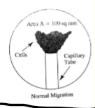
Capillary Tube Test
type of migration leukocyte assay
how well the cells migrate through a capillary tube
Ring Test
type of migration leukocyte assay
put a ring around the cells and let it grow to there and then take it out and see how much they migrate past it

Boyden Chamber Assay
type of migration leukocyte assay
uses the soluble factor effect (chemoattractant)
2 chambers and membrane allows the cells to migrate through

Cytokine Release - Leukocyte Assays
interleukins and TNF-alpha using ELISA; helps to find the phenotypic function of the cell
Cell Surface Markers - Leukocyte Assays
using fluorescence-activated cell sorted (FACS or flow cytometry)
Enzyme-Linked Immunosorbent Assay (ELISA)
plate coated w/ a capture antibody
sample added and any antigen present binds to capture antibody
detecting antibody added and binds to antigen
enzyme-linked secondary antibody is added and binds to detecting antibody
substrate added and converted by enzyme to detectable form

Flow Cytometer
a technique for counting, examining, and sorting microscopic particles suspended in a stream fluid
allows simultaneous multi-parametric analysis of the physial and/or chemical characteristics of single cells flowing through an optical and/or electronic detection apparatus
can use scattered light to differentiate size/shape of cells/aprticles
can use fluorescence to detect immunochemically labeled cells or proteins
can sort sort cells using electrostatic deflection

What does flow cytometry allow for?
it allows for the simultaneous multi-parametric analysis of the physial and/or chemical characteristics of single cells flowing through an optical and/or electronic detection apparatus
Electrostatic Deflection
a technique used during flow cytometry to sort cells by employing charged plates to change the path of the cell
Cytotoxicity Assay
most often use established cells line
has + and - controls
ISO standards are legally required by the FDA
3 morphological assays (direct contact, agar diffusion, and elution or extract dilution)
Cytotoxicity
causes toxic effects (death, alterations in cellular membrane permeability, enzymatic inhibition, etc.) at the cellular level
Positive Controls - Cytotoxicity Assay
samples that are known to be toxic and so if cells do not die, know something is wrong; ex. low molecular weight organotin stabilized PVC, gum rubber, and dilute solutions of toxic chemicals
Negative Controls - Cytotoxicity Assay
samples known to cause no toxic effect; ex. high density polyethylene (inert polymer)
Direct Contact - Morophological Cytotoxicity Assays
place sample directly in the culture → observe under microscope → live/dead stain, MTT assay, etc.
advantages - mimics clinical use
disadvantages - risk of physical trauma, influenced by the diffusion rate of the leachable
Agar Diffusion Test - Morphological Cytotoxicity Assay
contains a smaple of + control material; discoloration that extends outward from materual indicated presence of the sample has caused cells to lyse (so lose the vital stain incorporated in the agar layer)
advantages - better conc. gradient and can test one side of the material (side touching agar)
disadvantages - requires flat surface, limited by solubility of toxicant in agar, risk of thermal shock (from pouring agar soln onto the cells), and risk of absorbing water from agar (can dehydrate cells)

Elution Test - Morphological Cytotoxicity Assay
determines the cytotoxicity of leachables
soak material in MEM (minimum essential medium) and apply extract media at diff. doses to culture
advantages - separate extraction from testing, can test dose response, has a choice on extract conditions
disadvantages - additonal time and steps

In vivo Examination of Acute Inflammation
