A&P3 Lab Activity 4
 Call Kai
 Learn
 Practice Test
 Spaced Repetition
 Match
 Flashcards
 Knowt Play
1/68

There's no tags or description
Looks like no tags are added yet.
69 Terms

A
sublingual gland

B
submandibular gland

C
esophagus

D
liver

E
gallbladder

F
transverse colon

G
ascending colon

H
appendix

I
rectum

J
sigmoid colon

K
descending colon

L
pancreas

M
stomach

N
parotid gland

O
anus

P
cecum
this structure hangs off the cecum and contains helpful bacteria
appendix
this muscular tube squeezes food from the throat to the stomach
esophagus
this portion of the small intestine receives bile and pancreatic juices for chemical digestion
duodenum
chemical digestion finishes here and most of absorption takes place in these structures
jejunum & ileum
this organ solidifies feces by absorbing water
large intestine
this organ produces bile and stores lots of nutrients such as glycogen
liver
fecal matter is stored here until defecation
rectum
these glands moisten food and release enzymes to start chemical digestion
salivary glands
this structure mixes food with acid and other secretions to create chyme
stomach
this structure produces insulin and glucagon, as well as digestive enzymes and bicarbonate
páncreas

A
lower esophageal sphincter

B
cardiac region

C
pyloric sphincter

E
body

F
fundus
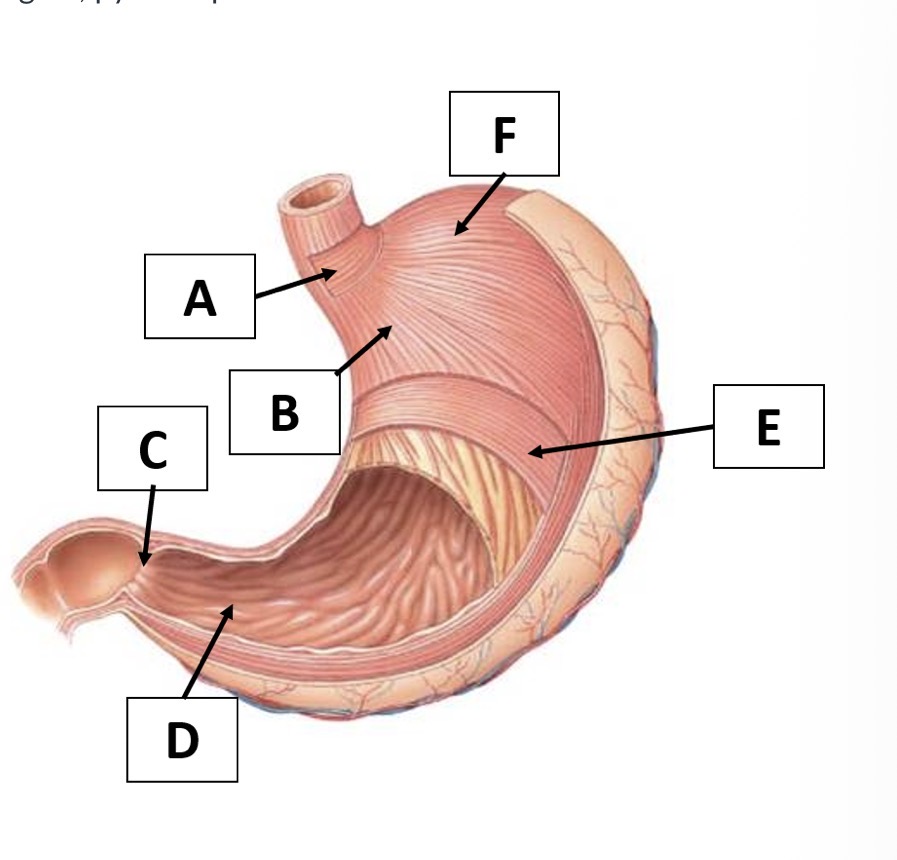
D
Pyloric area

A
appendix

B
ascending colon

C
duodenum

D
transverse colon

E
jejunum

F
descending colon

G
ileum

H
sigmoid colon

I
rectum

A
mucosa

B
submucosa

C
circular muscle

D
longitudinal muscle

E
serosa

F
lymphoid nodule

G
villus

H
brush border cell

I
blood capillary

J
lacteal
this blood vessel absorbs nutrients, vitamins and minerals
capillary
this circular band of smooth muscle pushes food forward
circular muscle layer
this structure absorbs fats bound to chylomicrons
lacteal
this longitudinal smooth muscle contracts in front of food to open the tube
longitudinal muscle layer
this lymphatic tissue monitors the intestines for pathogens
lymphoid nodule
this innermost layer is next to the lumen and is made of epithelium
mucosa layer
this outermost layer is the visceral peritoneum
serosa layer
this layer is underneath the mucosa and contains blood vessels and nerves
submucosa layer
these projections are covered with brush border cells
villus

A
hepatic portal vein

B
superior mesenteric vein

C
inferior mesenteric vein
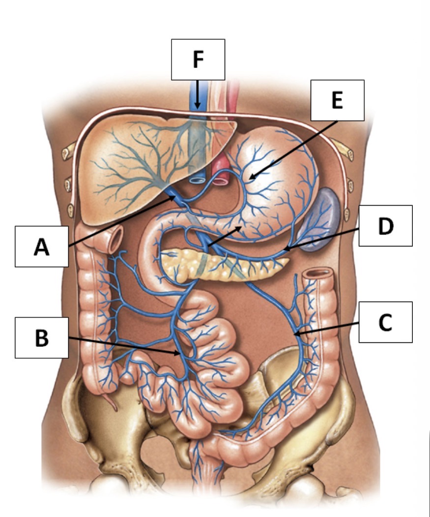
D
splenic vein

E
gastric veins

F
inferior vena cava
the most common type of IBD that affects the large intestine and/or rectum, usually continuous inflammation of one region
ulcerative colitis
can affect any part of the GI tract, but commonly affects the ilieum and first portion of the large intestine, often spread out in patches
Crohn's disease
autoimmune condition caused by a gluten allergy, typically resolves when gluten is removed from the diet
celiac's disease