Immune suppression in tumor-draining lymph nodes
 Call Kai
 Learn
Learn Practice Test
 Spaced Repetition
 Match
Match Flashcards
 Knowt Play
1/16

There's no tags or description
Looks like no tags are added yet.
Name | Mastery | Learn | Test | Matching | Spaced | Call with Kai | Chat |
|---|
No analytics yet
Send a link to your students to track their progress
17 Terms
What is a sentinal lymph node
First tumour draining LN/ The first lymph node that drains lymph from the primary tumor site.
DC subset | what function? |
|---|---|
cDC1 | |
cDC2 | |
Migratory DCs (migDCs) | |
Plasmacytoid DCs (pDCs) | |
Monocyte-derived DCs (moDCs) |
DC subset | Main function |
|---|---|
cDC1 | Cross-presentation of antigens to CD8+ T cells |
cDC2 | Antigen presentation to CD4+ T cells |
Migratory DCs (migDCs) | Travel to lymph nodes and present antigen to T cells |
Plasmacytoid DCs (pDCs) | Produce type I interferons |
Monocyte-derived DCs (moDCs) | Promote inflammatory responses; can also be influenced by tumor-derived tolerogenic signals |
What are the 5 key activities of DC?
Migration: DCs must reach the lymph node draining the tumor.
Antigen presentation: MHC molecules present antigen-derived peptides to T cells.
Co-stimulation: DCs provide additional signals needed for effective T-cell activation.
Effector-activating cytokines: for example, IL-12 and type I interferons.
Memory-inducing cytokines: the slide identifies IL-1β as an example.
Roles of migratory cDC1 and resident (in LN) cDC1
Migratory cDC1 transport tumor antigens from the tumor to the lymph node.
These DCs stimulate CD8+ T cells.
The activated CD8+ T cells produce XCL1, which attracts lymph-node-resident cDC1 through XCR1.
Migratory cDC1 transfer tumor antigen to resident cDC1.
Resident cDC1 are particularly potent at stimulating the anti-tumor CD8+ T-cell response.
Where are the LN important in the mellman cancer immunity cycle
Antigen presentation and T cell priming
How to identify the sentinel LN
Can be identified using the triple-technique (radioactive tracer/blue-dye tracer/hand-held detector to localize the radioactive LNo
Injecting a radioactive tracer near the primary tumor.
Injecting blue dye at the tumor site.
Identifying and removing the lymph node that is radioactive (“hot”) and blue.
Progenitor-exhausted T cells are not equivalent to terminally exhausted T cells.
Progenitor-exhausted cells retain the capacity for self-renewal and further differentiation.
How cancer cells can regulate immune cell behavior / Tumor cells can manipulate immune cells through four main mechanisms.
Through secreted factors (cytokines, chemokines, growth factors)
Through extracellular vesicles
By changing the metabolic environment through oxygen and nutrient consumption
Via direct cell-cell contact
Tumor cells secrete suppressive factor: cytokines, chemokines, growth factors and extracellular vesicles
These factors can suppress locally, in the tumor, but also drain to the SLN
DC development, migration and activation are hampered by many of
these factors
These factors induce Treg formation, which hampers T cell and NK cell functions in tumor-draining LNs
This can form a metastatic niche within the SLN and downstream LNs
Tumor-derived exosomes/extracellular vesicles can suppress the SLN environment and promote tumor metastasis. How
Tumor cells can secrete exosomes (or extracellular vesicles – (EVs))
These EVs can contain suppressive surface molecules (like PD-L1 or TGFbeta) that can change immune cells
EVs can also contain mRNAs or miRNAs that can alter immune cell behavior and make them suppressive
This can create a metastatic niche in the SLN

Tumor-secreted cytokines, like IL-10, IL-6 and PGE2 hamper DC activation. How
Myeloid cell have a high plasticity
Their phenotype, and subsequent functioning, can be altered by suppressive cytokines
The STAT3 and p38 pathways when activated, hamper DC development and maturation.
This suppression of DC development can result downstream in Treg induction, anergic/suppressed T cells, promotion of angiogenesis, promotion of tumor invasion
Immune status in tumor- draining LNs at least is influenced by the following anatomical, histological or immunological factors:
Factor | Type | Main implication |
|---|---|---|
Proximity to the tumor | Anatomical | Determines exposure to tumor-derived signals |
Size of the primary tumor | Histological / tumor-related | Can influence migratory DC activation |
Tumor-secreted suppressive factors | Immunological | Can impair DC maturation and alter immune-cell function |
Tumor metastasis in the lymph node | Histological | Can disrupt resident DCs and favour suppressive populations |
3 types of TME based on T cell infiltation

Type 1 IFN is a crucial player in antitumour response. List its functions
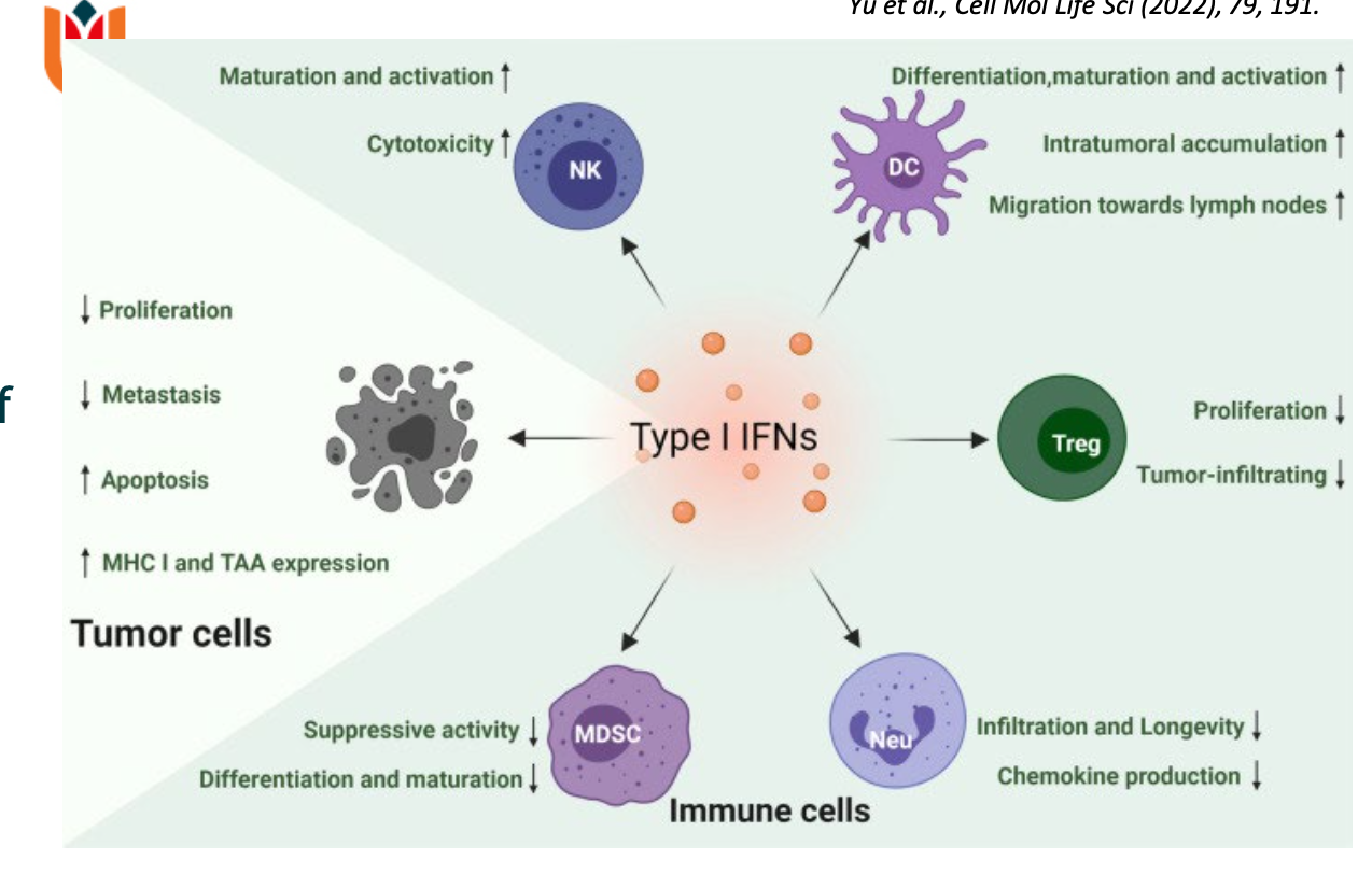
Functions of IFN 1 as a tumour progressor when the immune balance is lost

Who secretes type 1 IFN
PDC . Plasmacytoid DC in LN
GM-CSF
CpG
list their function
GM-CSF — recruits and activates conventional DCs
CpG — stimulates TLR9
CpG is a ligand for Toll-like receptor 9 (TLR9). Plasmacytoid DCs can respond to TLR9 stimulation by producing large amounts of type I interferons.
How can we restore the working of the SLN?
locally injecting immune activating compounds (CpG ± GM-CSF)
anti-CTLA-4 checkpoint inhibition
Early-stage disease: DC activation may be an appropriate strategy to strengthen the initiation of anti-tumor immunity.
Later-stage disease: combining DC activation with checkpoint inhibition may be needed to address both inadequate immune priming and established immune suppression.
The SLN is important to initiate an anti-tumor immune response, but tumors suppress their functioning
To overcome immune suppression in tumor-draining LNs we aim to:
Activate LNR-cDC and –pDC in SLN/tumor-draining LNs using stimuli that will promote a type I interferon response and skew towards a Th1 T cell response. At the same time we want to reduce Treg frequencies.
Combine this treatment with checkpoint inhibitors (anti-CTLA4, anti-PD(L)1, and/or others) to counteract tumor- and immune activation induced T cell inhibition in the LNs and the tumor.
Deliver these agents locally to reduce systemic toxicity, while providing systemic protection.