MLS 4510: Analysis of Body Fluids - Urinalysis and Renal Function
 Call Kai
 Learn
 Practice Test
 Spaced Repetition
 Match
 Flashcards
 Knowt Play
1/118

Earn XP
Description and Tags
Comprehensive practice vocabulary flashcards covering renal anatomy and physiology, physical, chemical, and microscopic urinalysis, renal diseases, metabolic disorders, and renal function testing.
119 Terms
Nephron
The structural and functional unit of the kidney, numbering approximately 1 to 1.5 million per kidney.
Cortical Nephrons
Nephrons making up approximately 85% of all nephrons, situated primarily in the renal cortex; their main functions are removing waste products and reabsorbing nutrients.
Juxtamedullary Nephrons
Nephrons characterized by long Loops of Henle extending deep into the renal medulla, primarily responsible for concentrating urine.
Renal Blood Flow Rate
The total volume of blood flowing through both kidneys, measuring approximately 1,200mL/min (total renal plasma flow is approximately 600–700mL/min).
Glomerular Filtration Barrier
The nonselective filtering unit composed of fenestrated capillary endothelium, the basement membrane (basal lamina), and podocyte foot processes forming filtration slits.
Shield of Negativity
A negative electrical charge present on the glomerular filtration barrier that electrostatically repels negatively charged plasma molecules, particularly albumin.
Juxtaglomerular Apparatus
A regulatory structure comprising juxtaglomerular cells in the afferent arteriole and the macula densa in the distal convoluted tubule that monitors blood pressure and sodium content to control renal hemodynamics.
Renin-Angiotensin-Aldosterone System (RAAS)
A systemic hormonal cascade initiated by macula densa sensing of low sodium/decreased blood pressure, triggering renin secretion, conversion of angiotensinogen to angiotensin I, and production of active angiotensin II to induce vasoconstriction, sodium retention, and ADH release.

Countercurrent Mechanism
The selective exchange of water and solutes in the descending and ascending Loops of Henle of juxtamedullary nephrons that maintains the hypertonic osmotic gradient of the renal medulla.
Maximal Reabsorptive Capacity (Tm)
The upper concentration limit at which active tubular transport mechanisms can reabsorb a substance from the filtrate back into the peritubular capillary blood before it spills into the urine.
Renal Threshold
The plasma concentration of a substance at which active tubular reabsorption reaches capacity and the substance begins to appear in the urine (e.g., 160–180mg/dL for glucose).
Antidiuretic Hormone (ADH / Vasopressin)
A hormone released by the posterior pituitary that regulates water reabsorption in the distal convoluted tubules and collecting ducts by altering the water permeability of the luminal membranes.
Renal Tubular Secretion in Acid-Base Balance
The active secretion of hydrogen ions (H+) by renal tubular epithelial cells into the tubular lumen coupled with the generation and reabsorption of bicarbonate (HCO3−) and excretion of ammonium (NH4+).
Uroscopy
The historic diagnostic examination of urine appearance, color charts, and sediment, systematically documented by early physicians including Hippocrates.
Oliguria
A marked decrease in total urine output, defined quantitatively as less than 400mL/day in adults.
Anuria
The complete cessation of urine flow, indicative of severe kidney damage or a drastic reduction in renal blood flow.
Nocturia
An increase in nocturnal excretion of urine, exceeding the normal daytime-to-nighttime excretion ratio of 2:1 to 3:1.
Polyuria
An abnormal increase in daily urine volume, defined in adults as greater than 2,500mL/day.
First Morning Void
An ideal, concentrated specimen collected immediately upon waking, optimal for routine screening, pregnancy testing, and evaluating orthostatic proteinuria.
24-Hour Urine Specimen Container
A specialized, large-volume container used for timed quantitative analyte determinations requiring strict patient collection protocols, thorough mixing, and measurement of total volume.

Chain of Custody
A standardized legal and operational documentation process ensuring verifiable specimen identity, handling, and tamper-proof security from collection through final result reporting.
Urochrome
The primary endogenous lipid-soluble pigment responsible for the normal yellow color of urine, excreted at a constant metabolic rate.
Uroerythrin
A pink pigment that readily deposits onto amorphous urate salts in refrigerated acidic urine specimens.
Urobilin
An orange-brown oxidation byproduct of urobilinogen found in small quantities that imparts color to older or standing urine specimens.
Isosthenuric Urine
Urine with a specific gravity of 1.010, equal to the specific gravity of the initial glomerular plasma ultrafiltrate.
Refractometry
An indirect method of determining specific gravity by measuring the refractive index, which compares the velocity of light in air against its velocity through a solution.
Red Urine Differentiation
A diagnostic protocol using centrifugation and plasma examination to differentiate cloudy hematuria (RBCs present) from clear hemoglobinuria (red plasma) and myoglobinuria (clear plasma).
Osmometer
An analytical instrument used to determine the osmotic concentration of a solution by measuring changes in colligative properties, such as freezing-point depression or vapor pressure reduction.
Reagent Strip
A chemical testing tool containing multiple dry reagent-impregnated pads used for routine urinalysis, interpreted via colorimetric changes against a manufacturer's chart at specified time points.

Protein Error of Indicators
The colorimetric principle of reagent strip protein testing where an indicator buffered at an acidic pH accepts hydrogen ions from proteins (primarily albumin), producing a green to blue color shift.
Bence Jones Protein
A low-molecular-weight monoclonal immunoglobulin light chain excreted by patients with multiple myeloma that causes prerenal proteinuria.
Microalbuminuria
The urinary excretion of albumin at concentrations of 20–200mg/L, serving as an early predictor of diabetic nephropathy and cardiovascular risk.
Orthostatic (Postural) Proteinuria
A benign condition common in young adults characterized by protein excretion after being upright, which completely resolves after periods of horizontal posture.
Tamm-Horsfall Protein (Uromodulin)
A high-molecular-weight mucoprotein synthesized and secreted exclusively by renal tubular epithelial cells of the ascending Loop of Henle, forming the fundamental matrix of all urinary casts.
Sulfosalicylic Acid (SSA) Precipitation Test
A cold precipitation confirmatory method that reacts equally with all types of urinary proteins, including albumin, globulins, and Bence Jones proteins.
Glucose Oxidase Reaction
A double-sequential enzyme reagent strip method utilizing glucose oxidase and peroxidase to catalyze the oxidation of a chromogen, specific exclusively for glucose.
Copper Reduction Test (Clinitest)
A non-specific test relying on the ability of glucose and other reducing substances (e.g., galactose, fructose, lactose) to reduce copper sulfate to cuprous oxide in the presence of alkali and heat.
Ketone Bodies
Three intermediate products of fat catabolism—acetone (2%), acetoacetic acid (20%), and β-hydroxybutyrate (78%)—that appear in urine when carbohydrate availability is compromised.
Sodium Nitroprusside Reaction
The chemical reaction on reagent strip ketone pads where acetoacetic acid in an alkaline medium reacts with sodium nitroprusside to produce a purple color.
Pseudoperoxidase Reaction for Blood
The catalytic chemical reaction where the pseudoperoxidase activity of heme in hemoglobin or myoglobin decomposes hydrogen peroxide, oxidizing a chromogen to form a blue-green color.

Urobilinogen and Bilirubin in Jaundice
Diagnostic reagent strip patterns: bile duct obstruction exhibits positive urine bilirubin and normal urobilinogen; hemolytic disease exhibits negative bilirubin and high (3+) urobilinogen; liver damage exhibits variable bilirubin and increased (2+) urobilinogen.
Greiss Reaction
The two-step diazotization reaction on reagent strips where bacterial-reduced nitrite reacts with an aromatic amine to form a diazonium salt, which couples to produce a pink azo dye.
Leukocyte Esterase Reaction
The enzymatic hydrolysis of an indoxylcarbonic acid ester by granulocytic esterases to yield indoxyl, which couples with a diazonium salt to form a purple azo dye within 120seconds.
Urinalysis Microscopic Techniques
Optical microscopy modalities used in sediment examination: bright-field (routine), phase-contrast (low refractive elements), polarizing (birefringent lipids/crystals), dark-field (spirochetes), fluorescence (labeled antigens/fluorophores), and interference-contrast (3D images).


Phase-Contrast Microscopy
A microscopic technique that slows light rays by one-quarter wavelength via phase rings to dramatically enhance contrast of low-refractive urine constituents such as hyaline casts and mucus.
Dark-Field Microscopy
A microscopic system utilizing a special condenser with an opaque central disc that directs light obliquely across the specimen, causing objects like Treponema pallidum to scatter light and appear bright against a dark background.
Polarizing Microscopy
An optical setup using crossed polarizing filters to visualize birefringent urine elements (e.g., cholesterol, crystals) that rotate plane-polarized light 90∘.

Birefringence
The optical property of an object allowing it to refract light in two distinct directions oriented at 90∘ to each other, producing polarization colors or Maltese cross formations.
Fluorescence Microscopy
A specialized technique using excitation and barrier filters with a dichroic mirror to visualize atoms that absorb short-wavelength light (such as UV) and emit longer-wavelength visible light.

Köhler Illumination
A standardized microscopic procedure aligning the field diaphragm, condenser, and aperture diaphragm to provide optimal, uniform specimen illumination and resolution.
Acute Poststreptococcal Glomerulonephritis (AGN)
An immune complex-mediated inflammation of glomerular capillaries occurring after infection with group A beta-hemolytic Streptococcus carrying the M protein, characterized by RBC casts, dysmorphic RBCs, and oliguria.
Goodpasture Syndrome
An autoimmune disorder where cytotoxic antiglomerular basement membrane antibodies attack both alveolar and glomerular capillary basement membranes, presenting with hemoptysis, dyspnea, and hematuria.
Granulomatosis with Polyangiitis
A necrotizing vasculitis formerly known as Wegener granulomatosis, associated with antineutrophil cytoplasmic autoantibodies (ANCA) that target small blood vessels of the respiratory tract and glomeruli.

Henoch-Schönlein Purpura
A pediatric immune-mediated disorder presenting with palpable skin purpura, joint pain, abdominal complaints, and glomerular hematuria/proteinuria following an upper respiratory infection.
Rapidly Progressive (Crescentic) Glomerulonephritis
A severe form of acute glomerular injury characterized morphologically by crescent-shaped formations of macrophages, fibroblasts, and polymerized fibrin in Bowman's space, leading rapidly to renal failure.
Membranous Glomerulonephritis
A glomerular condition characterized by progressive thickening of the glomerular basement membrane due to widespread subepithelial IgG immune deposits.
Immunoglobulin A Nephropathy (Berger Disease)
The most common worldwide cause of primary glomerulonephritis, marked by the persistent deposition of IgA immune complexes within the glomerular mesangium.
Nephrotic Syndrome
A clinical complex resulting from profound glomerular membrane permeability, characterized by massive proteinuria (>3.5g/day), profound edema, hypoalbuminemia, hyperlipidemia, and lipiduria.
Minimal Change Disease (Lipid Nephrosis)
The leading cause of nephrotic syndrome in children, characterized by podocyte foot process effacement visible only on electron microscopy, excellent response to corticosteroids, and normal BUN/creatinine levels.
Focal Segmental Glomerulosclerosis
A primary glomerular disorder marked by sclerosis of specific areas of select glomeruli, commonly associated with heroin abuse, analgesics, HIV, and IgM/C3 deposition.

Acute Tubular Necrosis (ATN)
The primary disorder of renal tubular destruction caused by prolonged renal ischemia (shock, crushing trauma) or nephrotoxic agents, presenting with RTE cells and RTE casts.
Fanconi Syndrome
A generalized dysfunction of the proximal convoluted tubule resulting in failure to reabsorb filtered glucose, amino acids, phosphorus, sodium, potassium, water, and bicarbonate.
Diabetic Nephropathy
The most common cause of end-stage renal disease (ESRD), characterized by glomerulosclerosis, basement membrane thickening, and mesangial expansion resulting from chronic hyperglycemia.

Alport Syndrome
An inherited collagen disorder affecting the structural integrity of the glomerular basement membrane, frequently accompanied by sensorineural hearing loss and ocular abnormalities.
Uromodulin-Associated Kidney Disease
An autosomal inherited disorder in which an abnormal form of uromodulin accumulates within renal tubular epithelial cells, destroying them and precipitating early-onset gout.
Acute Pyelonephritis
An acute bacterial infection of the renal tubules and medullary interstitium, diagnostically distinguished from lower urinary tract infections by the presence of WBC casts.
Acute Interstitial Nephritis (AIN)
An alloxan or medication-induced inflammatory reaction of the renal interstitium and tubules, characterized by hematuria, marked pyuria, WBC casts, and urinary eosinophils without bacteria.
Renal Calculi
Hard mineral deposits formed within the renal calyces, pelvis, or ureters, most frequently composed of calcium oxalate or calcium phosphate.

Newborn Screening (Guthrie Card)
A mandatory state-regulated filter paper blood collection method obtained 24–48hours post-birth to screen for inborn errors of metabolism prior to hospital discharge.
Phenylketonuria (PKU)
An autosomal recessive inborn error of metabolism caused by failure to inherit phenylalanine hydroxylase, leading to toxic accumulations of phenylpyruvate and producing a mousy urine odor.
Tyrosyluria
An overflow disorder characterized by urinary accumulation of tyrosine and its metabolites (p-hydroxyphenylpyruvic and p-hydroxyphenyllactic acids), yielding a positive nitroso-naphthol reaction.
Alkaptonuria
An inherited deficiency of homogentisic acid oxidase resulting in homogentisic acid excretion, causing urine to turn black/dark brown upon standing or alkalization, and tissue ochronosis.
Melanuria
The excessive excretion of 5,6-dihydroxyindole and melanogen produced by proliferating malignant melanoma cells, which oxidizes to melanin upon air exposure, turning urine dark brown or black.
Maple Syrup Urine Disease (MSUD)
A rare inborn error of branched-chain amino acid metabolism (leucine, isoleucine, valine) caused by deficient oxidative decarboxylation, imparting a characteristic sweet maple syrup odor to urine.
Isovaleric Acidemia
An organic acidemia caused by an enzyme defect in the leucine degradation pathway, resulting in accumulation of isovalerylglycine and a distinct 'sweaty feet' urine odor.
Tryptophan Metabolism Disorders
Abnormal metabolic pathways of tryptophan resulting either in excess indole conversion to indican (indicanuria / blue diaper syndrome) or carcinoid tumor secretion of 5-hydroxyindoleacetic acid (5-HIAA).
Cyanide-Nitroprusside Test
A chemical screening method that reduces cystine to cysteine with sodium cyanide, which then reacts with nitroprusside to form a characteristic red-purple complex in patients with cystinuria.
Cystinosis
A genuine lysosomal storage disease characterized by defective lysosomal transport and intracellular deposition of crystalline cystine throughout bodily organs and the renal parenchyma, causing Fanconi syndrome.
Porphyrias
A group of disorders characterized by intermediate enzyme blocks in heme synthesis, leading to abnormal accumulations of porphyrins (causing red/port wine urine and cutaneous photosensitivity) or precursors (causing neurological symptoms).
Lesch-Nyhan Disease
An X-linked recessive deficiency of hypoxanthine-guanine phosphoribosyltransferase (HGPRT) leading to excessive uric acid excretion, visible as 'orange sand' crystals in pediatric diapers.
Galactosuria
An inherited inability to convert galactose to glucose—most severely due to galactose-1-phosphate uridyl transferase (GALT) deficiency—resulting in toxic intermediate accumulation, cataracts, and infant failure to thrive.
Red Blood Cells (Urinary Sediment)
Smooth, non-nucleated biconcave discs measuring approximately 7μm in diameter, enumerated under high-power field (HPF), which indicate vascular disruption or glomerular damage.
White Blood Cells (Urinary Sediment)
Predominantly neutrophil granulocytes measuring approximately 12μm in diameter, displaying multilobed nuclei and cytoplasmic granules, reported per high-power field (HPF).

Glitter Cell
A neutrophil that has swollen in hypotonic urine whose cytoplasmic granules exhibit Brownian motion, creating a sparkling or glittering appearance under the microscope.
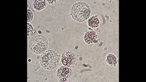
Squamous Epithelial Cells
The largest cellular constituents in urine sediment, containing abundant, flat, irregular cytoplasm and a small, distinct central nucleus, originating from the lower urethra and external genitalia.

Clue Cell
A squamous epithelial cell pathologically coated with numerous Gardnerella vaginalis coccobacilli, giving the cell borders a distinctively shaggy, granular appearance.

Transitional Epithelial Cells
Multiform epithelial cells lining the renal calyces, pelvis, ureters, and bladder, presenting as spherical, polyhedral, or caudate shapes with prominent, centrally located nuclei.

Renal Tubular Epithelial (RTE) Cells
The most clinically significant urine epithelial cells, originating from the nephron tubules, characterized by a coarsely granular cytoplasm and an eccentrically placed nucleus.
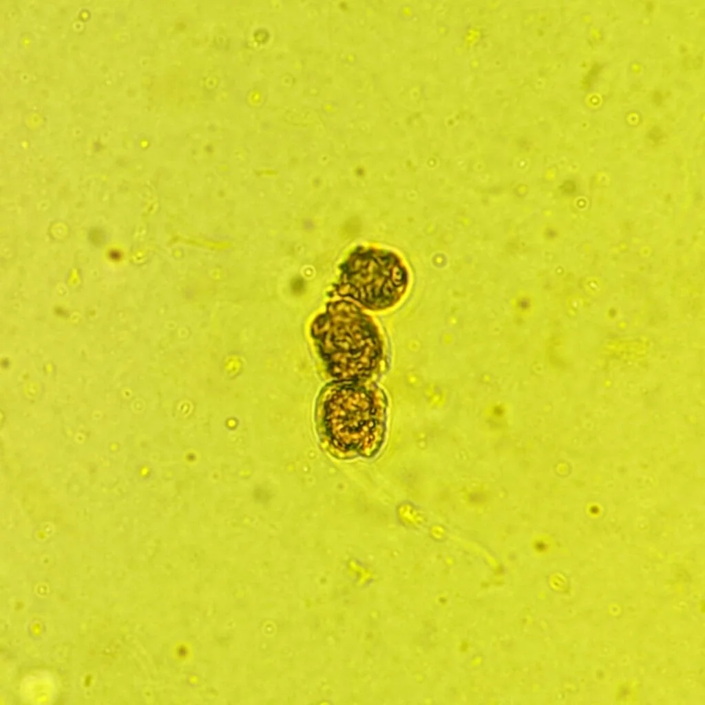
Oval Fat Bodies
Renal tubular epithelial cells that have engulfed filtered lipids and cholesterol, which display a characteristic Maltese cross appearance under polarized light.

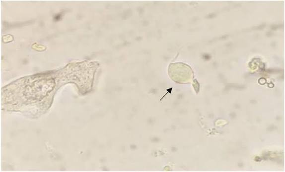
Trichomonas vaginalis
A flagellated protozoan parasite in urine sediment recognized on fresh wet mounts by its rapid darting motility, undulating membrane, and anterior flagella.
Cylindruria
The medical term describing the presence and excretion of urinary casts in the urine sediment.
Hyaline Cast
The most common and basic type of cast, composed entirely of uromodulin, displaying parallel sides, rounded ends, and a very low refractive index.

Red Blood Cell Cast
A cellular cast matrix containing incorporated red blood cells, diagnostic of intrinsic renal bleeding and active glomerular damage.

White Blood Cell Cast
A cast containing intact or degenerating polymorphonuclear leukocytes, serving as a primary hallmark to distinguish upper UTIs (pyelonephritis) from lower UTIs (cystitis).

Renal Tubular Epithelial Cell Cast
A cast embedded with sloughed renal tubular epithelial cells, indicating severe tubular destruction and acute tubular necrosis.

Fatty Cast

A highly refractile cast containing embedded fat droplets and oval fat bodies, associated with lipiduria and nephrotic syndrome.
Granular Cast
A urinary cast containing coarse or fine granules derived either from nonpathologic lysosomal excretion or pathologic disintegration of cellular casts and protein aggregates.
Waxy Cast
An advanced, brittle, highly refractile cast with fragmented edges and notched sides, representing prolonged urinary stasis and end-stage chronic renal failure.

Broad Casts
Markedly widened casts molded within dilated distal convoluted tubules or collecting ducts, commonly referred to as 'renal failure casts'.
Amorphous Urates
Normal acidic urine crystals appearing microscopically as yellow-brown amorphous granules and macroscopically as a distinct pink sediment that dissolves upon warming to 37∘C.
