Chapter 23: Aorta and Iliac Arteries PPT
 Call Kai
 Learn
 Practice Test
 Spaced Repetition
 Match
 Flashcards
 Knowt Play
1/71

There's no tags or description
Looks like no tags are added yet.
72 Terms
What is the incidence of an abdominal aortic aneurysm (AAA)?
60 per 1,000.
—12th leading cause of death; approximately 15,000 deaths annually from rupture.
Where do AAA’s occur more frequently?
In older men and are mostly located inferior to the renal arteries.
What are AAA’s commonly associated with?
Iliac, femoral, and popliteal aneurysms.
What are the indications for an aortoiliac duplex exam?
Pulsatile abdominal mass
Suspected or known aortic or iliac aneurysm
Claudication (hip or buttock) or ischemic rest pain
Decreased femoral pulses or abdominal bruit
Emboli in ischemic digits (blue toe syndrome)
Suspected inflow disease on physiologic studies
Follow-up after intervention
How long should patients fast overnight for an aortoiliac duplex exam?
8 to 12 hours.
—Minimizes scatter and attenuation from bowel gas.
—Patients can take morning medications with water.
—Gum chewing or smoking is discouraged.
What should also be done before the procedure/examination begins?
It should be explained to the patient and relative history should be obtained.
What kind of equipment is necessary for an aortoiliac duplex exam?
High-resolution equipment with good color flow and spectral Doppler.
—Must be able to penetrate to image deep vessels of abdomen.
What kind of transducers are mostly used?
Low-frequency transducers with a 2-5 MHz curved linear most commonly used as well as a 2-4 MHz sector phased array also helpful.
How does the AAA protocol begin?
At the level of the celiac axis and extends through bifurcation.
How should the aorta and iliac arteries be examined?
With B-mode in both transverse and sagittal planes.
How does the aorta appear normally?
Lies adjacent to the spine
Has smooth margins
No focal dilatations
Tapers distally

What kind of image is this representative of?
Normal tapering aorta in longitudinal view.
How are the transverse images with diameter measurements documented?
From the proximal aorta (near diaphragm), mid aorta (near renal arteries), and distal aorta (above bifurcation).
What kind of dimensions may the diameter measurements include?
Both anteroposterior (AP) and left-to-right lateral.
—Lateral dimensions are less reliable due to edge drop-off.
What measurements are also taken during the AAA protocol?
The proximal common iliac arteries.
What kind of measurements are also performed of the AAA protocol?
AP diameter measurements on longitudinal images.
—Measurements should be made from outer wall to outer wall.
—Care must be taken to be sure that measurements are taken perpendicular to the vessel.
—Important when aorta is angulated and elongated.

What kind of image is this representative of?
Longitudinal view of the aorta with calipers placed perpendicular to the long axis or the aorta.
What is important to note of an abdominal aortic aneurysm?
Length
Proximity to renal arteries
Presence and extent of intraluminal thrombus
Residual lumen
What should also be documented of an abdominal aortic aneurysm?
Dissection, intimal flaps, or other wall defects
Pseudoaneurysms
Stenosis and/or occlusion with characterization of any plaque visualized
What should also be performed during an AAA protocol?
Spectral doppler.
—Use 60 degrees or less
—Parallel to vessel wall
—Sample volume placed in center stream
Where should the PSV be recorded?
From proximal, mid, and distal aorta as well as from each common iliac artery.
What does the preintervention aortoiliac protocol include?
Common femoral (CFA), superficial femoral (SFA), and profunda femoral artery (PFA) origins.
—Diameter measurements and velocities are recorded from each segment.

What kind of image is this representative of?
Transverse view of common iliac stents with normal stent deployment.
What kind of good doppler techniques should be used for the postintervention aortoiliac protocol?
Angles between 45 and 60 degrees and parallel to vessel walls
Measure PSV and end-diastolic volume (EDV)
What does the typical postintervention exam include?
Limited images of aorta, common iliac, external iliac, and internal iliac arteries
May also include CFA, SFA, and PFA
Assess area of treatment
Must assess several centimeters above and below any area of treatment
What assists with locating and following vessels?
Color flow.
—Helps with placement of Doppler sample volume.
What is known about the location of iliac arteries?
They are deep and tortuous, so color flow helps follow.
—Lateral decubitus position may help evaluate.
What should be paid particular attention to when identifying increased velocities?
Scale for appropriate settings.
What are normal findings of an aorta/iliacs?
—An aortic diameter of less than 2 cm.
—An aorta that tapers as it courses distally.
—An aorta with smooth walls with well-defined margins.
—An aorta with a high-resistance doppler signal.
Hemodynamics in proximal aorta will have different waveform characteristics than distal aorta due to visceral organ branches.
Iliacs are high resistance, triphasic.
What kind of AAA has an aortic diameter greater than (>) 2 cm but less than (<) 3 cm or, irregular margins and nontapering profile?
Ectasia AAA.
What diameter measurement is consistent with an aneurysm?
Greater than (>) 3 cm, or focal diameter increases by more than 50% of normal segment.
What do true aneurysms involve?
All three layers of vessel wall.
Where in the aorta do AAA’s commonly occur?
Distal aorta (infrarenal).
What kind of AAA bulges, which involves the entire circumference of the aorta and is the most common type of aneurysm?
Fusiform AAA.
What kind of AAA has asymmetric outpouching dilations and is often caused by trauma or penetrating aortic ulcers?
Saccular AAA.
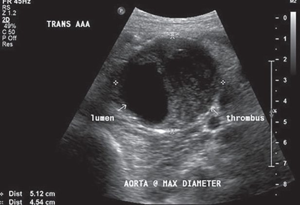
What kind of image is this representative of?
Transverse image of fusiform aneurysm.
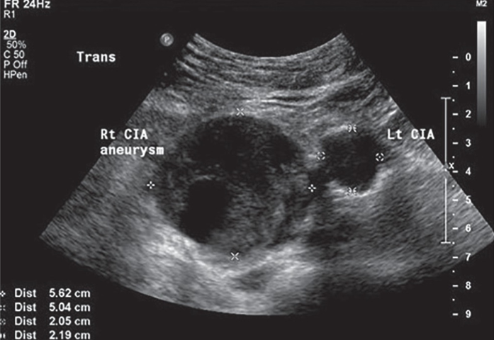
What kind of image is this representative of?
Transverse image of right common iliac artery aneurysm.

What kind of image is this representative of?
Longitudinal view of a saccular aneurysm.
What is it called for the iliac when the diameter increases by 50% when compared to normal segment?
Iliac artery aneurysm.
What diameter measurement is an iliac measurement considered an aneurysm?
With diameter of greater than (>) 1.5 cm.
—Usually associated with atherosclerotic disease.
—Often bilateral.
What may be present in a stenosis or occlusion?
Thrombus, plaque, and/or calcifications.
How may plaque appear on ultrasound?
Heterogeneous or homogeneous, with smooth or irregular borders.
How may calcifications appear on ultrasound?
Hyperechoic areas with shadowing.
How may thrombus appear on ultrasound?
Often homogeneous with smooth borders.
—Often found within aneurysm sac.
How should a stenosis or occlusion be carefully assessed with spectral doppler?
By walking through area of stenosis (proximal, in stenosis, and distal)
Documenting poststenotic turbulence
Documenting distal waveform changes (CFA, SFA, PFA)
—Distal waveforms may remain fairly normal with presence of extensive collateralization.
What is a greater than (>) 50% stenosis consistent with?
Increase in velocity of 100% (doubling)
Poststenotic turbulence
Distal waveform changes (dampened systolic component and loss of reversal component)
—Be careful to stay in aortoiliac vessels and not stay onto collaterals.
What is a less invasive alternative for an AAA repair?
EVAR (Endovascular Aortic Repair), because of the lower incidence of perioperative mortality and improved survival and recovery rates.
What does an aortoiliac duplex ultrasound following an EVAR involve?
It involves placement of stent graft device within aortic aneurysm sac and it is a catheter-based procedure.
—It excludes aneurysm from general circulation.
EVAR patients require close surveillance postprocedure, what complication is it called when flow is present within aneurysm sac after intervention?
Endoleak.
—Common complication of EVAR.
—Presence can increase pressure in aneurysm sac, continuing to make rupture a risk.
—Follow-up surveillance is done with either ultrasound or CT or both.
How is the patient positioned for an EVAR exam?
They are typically supine.
Where does an EVAR exam begin?
At the proximal aorta in a transverse plane.
During an EVAR exam, what is determined?
The proximal extent of the graft.
—It is usually below the renal arteries, but can be above.
—Has a hyperechoic signal along aortic walls.
—Distance from superior mesenteric artery (SMA) to proximal attachment site should be measured for detection of migration.
For an EVAR exam, where is an aortic diameter measured?
Above proximal attachment site to assess for dilatation of aneurysm neck.
For an EVAR exam, how are diameter measurements taken?
Throughout the length of the aorta, taking care to be perpendicular to walls.
—Residual sac diameters are important to follow.
—Vessel can be tortuous.
Why should residual sac characteristics be noted during an EVAR exam?
Areas of hyperechogenicity can be evidence of endoleak.
For an EVAR exam, transverse and sagittal views of distal attachment site are recorded. What evidence should be looked for?
Graft kinking or migration.
Where should color and spectral doppler of PSV be performed for an EVAR exam?
At the suprarenal aorta to confirm patency of renal arteries.
At the proximal attachment site, body of graft, graft limbs, and distal attachment site of stent graft.
Native vessels beyond distal attachment site.
With color and spectral doppler of an EVAR, what should the graft area be evaluating for?
Perigraft flow
Graft stenosis or thrombosis
Graft kinking
—Complications should be thoroughly documented.

What kind of image is this representative of?
Color duplex imaging of normal bifurcated endograft with a color box appropriately placed to include the excluded AAA sac.
How should a residual sac size appear over time?
It should be decreasing over time.
What is any increase in residual sac size, pulsatility of sac, or areas of echolucency in sac suggestive of?
An endoleak.
A residual sac that appears “spongy” (heterogeneous with hypoechoic areas) with an increase in sac size is suggestive of?
An endoleak.
How should flow patterns be of an residual sac?
Multiphasic (high resistance).
What type of endoleak is when there is an inadequate or ineffective seal at attachment site?***Know this!
Type 1.
What type of endoleak is when there is retrograde flow in branch vessel (lumbar, inferior mesenteric, accessory renal, and internal iliac arteries)?***Know this!
Type 2.
What type of endoleak is when there is a modular disconnection or defect in graft fabric?***Know this!
Type 3.
What type of endoleak is when there is porous graft or microleak in graft material?***Know this!
Type 4.

What kind of image is this representative of?
A diagram of potential sites of perigraft leaks (types 1 to 4).
What is a reliable tool for the diagnosis and surveillance of aortic pathology, both aneurysmal disease and occlusive disease?
Duplex ultrasound.
What is cost-effective and noninvasive and can be repeated without exposure to ionizing radiation?
Duplex ultrasound.
What is an accurate modality to detect complications following open surgery or endovascular treatment for aortic aneurysms or AIOD (Aortoiliac Occlusive Disease)?
Duplex ultrasound imaging with color and power Doppler.
What is very operator dependent and has optimal studies that require an experienced sonographer with knowledge of anatomy, endovascular and surgical techniques, and standard laboratory imaging protocols as well as good-quality equipment?
Duplex ultrasound.