Diagnostic Imaging 2- Exam 3- Benign Tumors- part 2
 Call Kai
 Learn
Learn Practice Test
 Spaced Repetition
 Match
Match Flashcards
 Knowt Play
1/131

There's no tags or description
Looks like no tags are added yet.
Name | Mastery | Learn | Test | Matching | Spaced | Call with Kai | Chat |
|---|
No analytics yet
Send a link to your students to track their progress
132 Terms
A benign tumor arising in membranous bone that is commonly found in adulthood
Osteoma
Osteoma is associated with what disease
Gardner`s syndrome (multiple osteoma colonic polyposis and soft tissue fibroma)
Most common location for osteoma
Para nasal sinuses (front and ethmoid) (MAINLY THIS)
Also inner/outer table of calvarium (skull)
Osteoma in the frontal sinus

Another osteoma in frontal sinus

A benign cartilaginous growth in the medullary cavity of bones preformed in cartilage
It arises as residual islands of cartilage left behind
Enchondroma
Age of occurrence for Enchondroma
10-30 years
What is the most common benign tumor of the hand
Enchondroma
Enchondroma radiographic features
Round/oval lucency with fine marginal line
Endosteal scalloping
Stippled or punctate calcifications
Slightly Expansile Enchondroma in the finger

Enchondroma with punctate calcifications

Enchondroma with pathological fracture

Age of occurrence for Enchondroma
10-30 years
What is the rule with an Enchondroma and the closer it forms to the axial skeleton
Greater chance for malignant degeneration
-echondroma becomes a malignant Chondrosarcoma
Treatment and prognosis for Enchondroma
Usually no treatment
-if symptomatic in long bones they they require surgical curettage and bone chips
What is multiple echondromas
Enchondromatosis (aka Ollier`s Disease)
Enchondromatosis (ollier`s disease) occurs at what age
Early childhood
What is it called when the young patient has Enchondromatosis with multiple soft tissue hemangiomas
Maffuccii syndrome
Another name for Enchondromatosis
Ollier`s disease
Example of Enchondromatosis
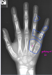
Calcified venous thrombi in the pelvis are called
Phelobliths
Another example of Enchondromatosis

Are phleboliths bad to have in the pelvis
No as long as they are blow the ischial spine and parallel to the pelvic brim

Multiple Enchondromas and multiple soft tissue hemangiomas (phleboliths) is called
Maffucci`s syndrome

Solitary vascular neoplasm which is slow growing and composed of newly formed capillary, CAVERNOUS or venous blood vessel
Hemangioma
Hemangioma age
4-5th decades
What is the most common benign tumor of the spine
Hemangioma
Most hemangiomas are
Asymptomatic
What causes symptoms of hemangioma
Expansion causing spinal stenosis and cord compression... need MRI if symptomatic
Locations of hemangioma
Vertebral body and lower Thoracics/upper lumbars
Calvarium with predilection for frontal bone
What do you see on X ray with with an hemangioma in the spine
Exaggerated vertical trabeculations

Example of hamangioma

What do you see on CT form a vertical view of a hemangioma in the spine
Polka-dot appearance

Another example of hemangioma of the spine

Radiographic skull features of hemangioma
Small round lytic lesion'
Sunburst or spoked wheel when viewed straight on
Occur in diploids space producing a palpable lump secondary to widening of the space
Hemangioma in the skull

Hemangioma of the frontal bone

Hemangioma on CT on a T1 weighted MRI

Treatment for hemangioma
No treatment
If symptomatic may undergo decompression surgery and need MRI
Symptomatic with skull then en bloc excision
What is a rare benign bone tumor that occurs between the ages of 5-25 and before the cessation of enchondral bone growth
Chondroblastoma
Clinical features and symptoms of Chondroblastoma
Often symptomatic for months to years
Mild joint pain, tenderness, joint swelling, and limitation of motion
Location of Chondroblastoma on the bone
Apophysis or epiphysis in a sub articular location
Difference between apophysis and epiphysis
Apophysis= growth center for bump on bone
Epiphysis= growth center for length of bone`
Most Chondroblastoma are on what bones
Proximal femur and distal femur
Radiographic features of Chondroblastoma
Round or oval eccentric lytic lesion of epiphysis or apophysis
Well defined sclerotic margin]
Lobulated
Treatment and prognosis of Chondroblastoma
Surgical curettage and packing with bone chips
Resection in expendable area
12 year old boy with Chondroblastoma

Example of Chondroblastoma in 17 yr old male

One more example of Chondroblastoma and explain why it isn't a giant cell tumor
Patient is too young and the giant cell likes metaphasis and epiphysis

Chondroblastoma in the proximal humerus

16 yr old with a Chondroblastoma in the proximal tibia, explain why its not a giant cell tumor
Giant cell tumors like age 20-40 so this patient is too young

Fibrous cortical defect (FCD) is most common in what ages
2-8 yrs with peak around 7-8 yrs
What is the term that encompasses FCD and NOF
Fibrous xanthoma
What is the only difference between an fibrous cortical defect and a NOF
Age
FCD is 2-8 and less than 2 cm
NOF is 8-20 and greater than 2 cm
Location of fibrous cortical defect
Posterior media surface of distal femur, tibia, fibula and proximal femur
Metaphyseal cortex of long bones of lower extremity
Round. Lyitc lesion that is less than 2 cm, well defined sclerotic margin
Eccentric and metaphyseal
Oval and extending parallel to the long axis which could produce cortical thinning
Lytic or bubbly with sclerotic border
Fibrous cortical defects
Treatment for fibrous cortical defect
No treatment needed, spontaneously regressed over a 2-5 year period
Fibrous cortical defect
Also could be a non ossifying fibroma

Fibrous cortical defect or non ossifying fibroma in distal femur

Should a non ossifying fibroma have pain
NO
Non ossifying fibroma and why is it not a fibrous cortical defect
It's too big to be a FCD

Fibrous cortical defect or a fibrous xanthoma

Fibrous cortical defect in the distal tibia

Age of occurrence for a nonossifying fibroma
8-20 with most occurring in 2nd demand
Do nonossifying fibroma (NOF) have symptoms
No they're asymptomatic
Favorite location of nonossifying fibroma
DISTAL TIBIA
Or distal femur, proximal tibia, fibula
Location of nonossifying fibroma on the bone
Eccentric and metadiaphyseal
Eccentric multi ocular ovoid lesion along the long axis of a bone which is about 2 cm with a dense sclerotic border
Endosteal scalloping with thinning and bulging of cortex and migrates towards the diaphysis
Nonossifying fibroma

Treatment and prognosis for nonossifying fibroma
No treatment, they should spontaneously regress with age
Large ones greater than 8 cm may require surgery
Nonossifying fibroma and why is it not an FCD
It's too big to be a fibrous cortical defect

Nonossifying fibroma in the distal tibia compared to an fibrous cortical defect in the distal tibia

One more non ossifying fibroma

What is a tumor like process that occurs in people typically older than 40 and effects extra in the northern latitude
Paget's disease
What is characterized by bone lysis followed by extensive repair attempts
Paget's disease
What are known as the great imitators of bone disease
Pagets and fibrous dysplasia
Age for Paget's disease
Most are older than 55
Etiology of Paget's
Mostly unknown but thought to have som genetic component as well
What is it when the bone undergoes increased resorption followed by increased bone formation, this new brown is disorganized and leads to bone softening
Paget's disease
What type of bone pathology is Paget's
Bone softening
Most Paget's disease cases are
Asymptomatic
What is given to treat Paget's disease
Bisphophonates or calcitonin
If there is sympomts with Paget's what is the most common symptom
Bone pain
What can occur if Paget's occurs around a joint
Secondary osteoarthritis
What type of bone deformity can occur with Paget's
Bowing of an extremity
What is something that you can feel with Paget's
Excessive warmth from hypervascularity
When it comes to Paget's disease ossous locations it is usually _________ and _______
Polyostotic (multiple locations) and asymmetric
What should you do if you see Paget's in one location
Bone scan to see if it has gone somewhere else
Common lcoations for Paget's
Pelvis
Lumbar spine
Thoracic spine
Proximal femur
Lab findings with Paget's
Increased hydroxyproline and alkaline phosphatase
Different stages of Paget's disease
1. Osteolytic/destructive
2. Mixed/combined
3. Sclerotic/ivory
4. Malignant degeneration (very rare)
Radiological feature of Paget's
Coarsened irregular trabecular pattern
Thickens cortex
Bone expansion
Osseous deformity
What is it when Paget's is in the skull
Osteoporosis circumscripta
-described as map like (geographic lysis) of the frontal and occipital regions
What does Paget's disease in the skull (osteoporosis curcumscripta) look like
Cotton wool appearance (fuzzy poorly defined)- blastic phase
What can happen when there is Paget's disease in the skull
Basilar invagination with foraminal encroachment
What two lines are used to help determine basilar invagination
McGreggors line and Chamberlins
Osteoporosis circumscripta

Another example of osteoporosis circumscripta

Enlarged cotton wool fluffy like densities in the skull from Paget's disease

Example of chamberlins line and McGregor`s line

Example of blastic phase of Paget's in the skull
Cotton wool appearance
