Nervous System

0.0(0)
Studied by 0 people
call kaiCall Kai
learnLearn
examPractice Test
spaced repetitionSpaced Repetition
heart puzzleMatch
flashcardsFlashcards
GameKnowt Play
Card Sorting

1/39

encourage image

There's no tags or description

Looks like no tags are added yet.

Last updated 8:02 PM on 10/7/26
Name
Mastery
Learn
Test
Matching
Spaced
Call with Kai
Chat

No analytics yet

Send a link to your students to track their progress

40 Terms

1
New cards

describe the map of the general Nervous system

CNS = spinal cord and brain: primary command center

PNS = 2 major pathways:

  • Sensory (Afferent): carries info towards CNS, sensory receptors, monitors internal and external environments

  • Motor (Efferent): carries info away CNS, splits into

    • somatic: skeletal muscle

    • visceral: involuntary effectors


<p>CNS = spinal cord and brain: primary command center</p><p>PNS = 2 major pathways: </p><ul><li><p>Sensory (Afferent): carries info towards CNS, sensory receptors, monitors internal and external environments</p></li><li><p>Motor (Efferent): carries info away CNS, splits into</p><ul><li><p>somatic: skeletal muscle</p></li><li><p>visceral: involuntary effectors</p></li></ul></li></ul><p></p>
2
New cards

who is the exception of the usual organization of nervous system?

tjeir nervous system is a nerve net

neurons not specialized in different divisions

neurons carry APs in both directions

<p>tjeir nervous system is a nerve net</p><p>neurons not specialized in different divisions</p><p>neurons carry APs in both directions</p>
3
New cards

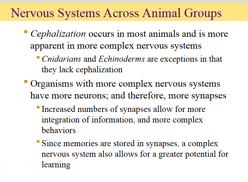
what is cephalization and where does it happen? who are the exceptions 2

an evolutionary trend where sensory organs, nervous tissue, and the mouth become concentrated at the front end (anterior) of an animal's body, effectively creating a head region.

it occurs in most animals

cnidarians and echinoderms are exceptions because they lack cephalization
<p><mark data-color="#fff1f1" style="background-color: rgb(255, 241, 241); color: inherit;">an evolutionary trend where </mark><strong><mark data-color="#fff1f1" style="background-color: rgb(255, 241, 241); color: inherit;">sensory organs, nervous tissue, and the mouth</mark></strong><mark data-color="#fff1f1" style="background-color: rgb(255, 241, 241); color: inherit;"> become concentrated at the front end (anterior) of an animal's body, effectively creating a </mark><strong><mark data-color="#fff1f1" style="background-color: rgb(255, 241, 241); color: inherit;">head region</mark></strong><mark data-color="#fff1f1" style="background-color: rgb(255, 241, 241); color: inherit;">.</mark></p><p>it occurs in most animals</p><p>cnidarians and echinoderms are exceptions because they lack cephalization</p>
4
New cards

which organisms have more synapses?

knowt flashcard image
5
New cards

What evolutionary trend is characterized by a high concentration of nervous tissue and sensory organs at the anterior (front) end of a vertebrate?

Cephalization

<p>Cephalization</p>
6
New cards

What unique structural feature do vertebrates possess that develops into the spinal cord?

hollow dorsal nerve cord

<p>hollow dorsal nerve cord</p>
7
New cards

Which protective structures encase the Central Nervous System (CNS) in vertebrates?

cartilage or bone

<p>cartilage or bone</p>
8
New cards

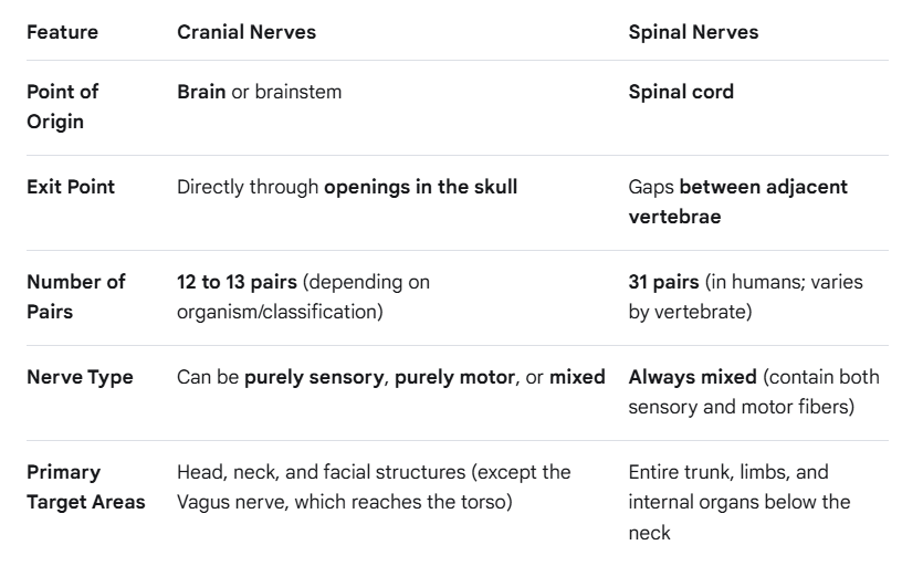
what are cranial nerves?

from where do cranial nerves physically emerge or exit?

how many pairs of cranial nerves are present, and how are they labeled?

They are specialized set of nerves that connect your brain directly to different parts of your head, neck and torso, travelling down the spinal cord first

They exit directly from the skull.

13 pairs, labeled with Roman numerals

<p>They are specialized set of nerves that connect your brain directly to different parts of your head, neck and torso, travelling down the spinal cord first</p><p>They <strong>exit directly from the skull</strong>.</p><p>13 pairs, labeled with Roman numerals</p>
9
New cards

what are spinal nerves?

where do they originate?

They branch from the spinal cord.

enter and exit between adjacent vertebrae


Spinal nerves are grouped and named based on the exact area of the vertebral column where they emerge:

  • Cervical: Neck region

  • Thoracic: Upper and mid-back (chest region)

  • Lumbar: Lower back

  • Sacral: Pelvis/hip region

  • Coccygeal: Tailbone region


<p>They <strong>branch from the spinal cord</strong>.</p><p>enter and exit between adjacent vertebrae</p><p></p><p>Spinal nerves are grouped and named based on the exact area of the vertebral column where they emerge:</p><ul><li><p><span><strong>Cervical:</strong> Neck region</span></p></li><li><p><span><strong>Thoracic:</strong> Upper and mid-back (chest region)</span></p></li><li><p><span><strong>Lumbar:</strong> Lower back</span></p></li><li><p><span><strong>Sacral:</strong> Pelvis/hip region</span></p></li><li><p><span><strong>Coccygeal:</strong> Tailbone region</span></p></li></ul><p></p>
10
New cards

whats the difference between spinal and cranial nerves?

are the 2 primary divisions of peripheral nervous system

<p>are the 2 primary divisions of peripheral nervous system</p><p></p>
11
New cards

whats gray and white matter

gray matter: neuronal cell bodies

white matter: tracts of axons and their myelin sheaths


spinal cord layout: gray matter is inside, and white matter is outside

brain layout: gray matter outside, white matter is inside


<p>gray matter: neuronal cell bodies </p><p>white matter: tracts of axons and their myelin sheaths </p><p></p><p>spinal cord layout: gray matter is inside, and white matter is outside</p><p>brain layout: gray matter outside, white matter is inside</p><p></p>
12
New cards

what are the 4 protective layers that keep the CNS safe from physical trauma? talk about all of them

  1. Skull and vertebral column protect the brain and spinal cord: skulls and backbone

  2. Meninges: protective sheets of connective tissue wrapped directly around the brain and spinal cord, number of layers varies by animal group

  3. Cerebral spinal fluid (CSF): its a clear fluid filling the spaces inside and around the CNS. Acts as a shock absorber, preventing brain from smashing against the hard skull

  4. Blood-brain barrier: A highly selective filter separating circulating blood from the brain tissue. Tight junctions block harmful things form coming in, brain ahs special transporters to let essential nutrients like glucose and amino acids pass through


<ol><li><p>Skull and vertebral column protect the brain and spinal cord: skulls and backbone</p></li><li><p>Meninges: protective sheets of connective tissue wrapped directly around the brain and spinal cord, number of layers varies by animal group</p></li><li><p>Cerebral spinal fluid (CSF): its a clear fluid filling the spaces inside and around the CNS. Acts as a shock absorber, preventing brain from smashing against the hard skull</p></li><li><p>Blood-brain barrier: A highly selective filter separating circulating blood from the brain tissue. Tight junctions block harmful things form coming in, brain ahs special transporters to let essential nutrients like glucose and amino acids pass through</p></li></ol><p></p>
13
New cards

talk a bit about the vertebrate brain

brain: extension of spinal cord

several cavities called ventricles that contian CSF

<p>brain: extension of spinal cord</p><p>several cavities called ventricles that contian CSF</p>
14
New cards

in the topic of the vertebrate brain, what are the primary and secondary brain vesicles

knowt flashcard image
15
New cards

what is the hindbrain and its parts?

3 regions:

  • Pons: located rostral to medulla

    • control alertness, initiate sleep and dreaming

  • Cerebellum: two hemispheres at back of brain

    • motor coordination

    • half neurons of brain

  • Medulla oblongata: located at top of spinal

    • breathing, heart rate


<p>3 regions: </p><ul><li><p>Pons: located rostral to medulla</p><ul><li><p>control alertness, initiate sleep and dreaming</p></li></ul></li><li><p>Cerebellum: two hemispheres at back of brain</p><ul><li><p>motor coordination</p></li><li><p>half neurons of brain</p></li></ul></li><li><p>Medulla oblongata: located at top of spinal</p><ul><li><p>breathing, heart rate</p></li></ul></li></ul><p></p>
16
New cards

what is the midbrain and its parts?

  • reflex responses coordination

  • size and function reduced in mammals

    • inferior colliculi

    • superior colliculi

    • Substantia nigra: controls and regulates motor functions and movement

    • VTA (Ventral tegmental area)= serves as a central component of the brain´s reward pathways


<ul><li><p>reflex responses coordination</p></li><li><p>size and function reduced in mammals</p><ul><li><p>inferior colliculi</p></li><li><p>superior colliculi</p></li><li><p>Substantia nigra: controls and regulates motor functions and movement</p></li><li><p>VTA (Ventral tegmental area)= serves as a central component of the brain´s reward pathways</p></li></ul></li></ul><p></p>
17
New cards

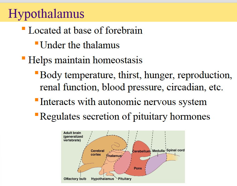
what is the forebrain and its parts?


<p></p>
18
New cards

what is the hypothalamus?

knowt flashcard image
19
New cards

what is the limbic system?

its a network of connected structures that lie between the cortex and the rets of the brain

  • influence emotions, motivation and memory

  • “emotional brain“

  • includes hypothalamus and other parts

    • amygdala: aggression and fear responses

    • hippocampus: converts short term memory to long term

    • olfactory bulbs: smell

    • Nucleus accumbens: reward, addiction

    • Cingulate cortex: executive function of brain, decision making and motivation and planning


<p>its a network of connected structures that lie between the cortex and the rets of the brain</p><ul><li><p>influence emotions, motivation and memory</p></li><li><p>“emotional brain“</p></li><li><p>includes hypothalamus and other parts</p><ul><li><p>amygdala: aggression and fear responses</p></li><li><p>hippocampus: converts short term memory to long term</p></li><li><p>olfactory bulbs: smell</p></li><li><p>Nucleus accumbens: reward, addiction</p></li><li><p>Cingulate cortex: executive function of brain, decision making and motivation and planning</p></li></ul></li></ul><p></p>
20
New cards

what does the thalamus do?

  • large grouping of gray matter above hypothalamus


<ul><li><p>large grouping of gray matter above hypothalamus</p></li></ul><p></p>
21
New cards

what does cerebrum do?

knowt flashcard image
22
New cards

what does the cortex do?

  • integrates and interprets sensory information and initiates voluntary movements

  • more folded in more advanced mammals

    • gyri: folds

    • sulci: grooves


<ul><li><p>integrates and interprets sensory information and initiates voluntary movements</p></li><li><p>more folded in more advanced mammals</p><ul><li><p>gyri: folds</p></li><li><p>sulci: grooves</p></li></ul></li></ul><p></p>
23
New cards

what are the cortical layers:

Cortical layers are the distinct horizontal layers of neurons that make up the cerebral cortex (the outer gray matter layer of the brain).

<p><span><strong>Cortical layers</strong></span> are the distinct horizontal layers of neurons that make up the <strong>cerebral cortex</strong> (the outer gray matter layer of the brain).</p>
24
New cards

what are cortical lobes?

cerebral cortex is divided into 4 major lobes


The 4 Major Cortical Lobes

  • Frontal Lobe (Pink): Located at the front; handles higher-level thinking, decision-making, planning, movement, and speech production.

  • Parietal Lobe (Blue): Located at the top-back; processes bodily sensations like touch, pressure, temperature, and spatial awareness.

  • Occipital Lobe (Green): Located at the very back; acts as the brain's primary visual processing center.

  • Temporal Lobe (Yellow): Located at the bottom/sides; handles hearing, language comprehension, memory, and smell.


<p>cerebral cortex is divided into 4 major lobes</p><p></p><p><strong>The 4 Major Cortical Lobes</strong></p><ul><li><p><span><strong>Frontal Lobe (Pink):</strong> Located at the front; handles higher-level thinking, decision-making, planning, movement, and speech production.</span></p></li><li><p><span><strong>Parietal Lobe (Blue):</strong> Located at the top-back; processes bodily sensations like touch, pressure, temperature, and spatial awareness.</span></p></li><li><p><span><strong>Occipital Lobe (Green):</strong> Located at the very back; acts as the brain's primary visual processing center.</span></p></li><li><p><span><strong>Temporal Lobe (Yellow):</strong> Located at the bottom/sides; handles hearing, language comprehension, memory, and smell.</span></p></li></ul><p></p>
25
New cards

what is the concept of cortical homunculus?

maps how much brain power (cortex space) is dedicated to processing sensations or movements for different parts of the body.

The mapping research was famously pioneered by neurosurgeons Wilder Penfield and Herbert Jasper.


1. The Normal Body vs. The Brain's Map

  • Row (a) Actual Proportions: Shows what humans and star-nosed moles look like in real life.

  • Row (b) Sensory Homunculus: Shows what the organisms would look like if their body parts were drawn proportional to the amount of brain tissue dedicated to handling their sensory inputs (touch, pressure, etc.).


This is because these areas are packed with high densities of sensory receptors, requiring a disproportionately large amount of space in the cerebral cortex to process detailed touch information.


<p>maps how much brain power (cortex space) is dedicated to processing sensations or movements for different parts of the body.</p><p>The mapping research was famously pioneered by neurosurgeons <strong>Wilder Penfield</strong> and <strong>Herbert Jasper</strong>.</p><p></p><p></p><p><strong>1. The Normal Body vs. The Brain's Map</strong></p><ul><li><p><span><strong>Row (a) Actual Proportions:</strong> Shows what humans and star-nosed moles look like in real life.</span></p></li><li><p><span><strong>Row (b) Sensory Homunculus:</strong> Shows what the organisms would look like if their body parts were drawn <strong>proportional to the amount of brain tissue</strong> dedicated to handling their sensory inputs (touch, pressure, etc.).</span></p></li></ul><p></p><p></p><p>This is because these areas are packed with high densities of sensory receptors, requiring a <strong>disproportionately large amount of space</strong> in the cerebral cortex to process detailed touch information.</p><p></p>
26
New cards

What is "somatotopic organization" in the cerebral cortex?

  • When specific regions of the cortex map directly to specific parts of the body for motor or sensory control.


Somatotopic organization means that specific areas of the brain map directly to specific parts of the body. Discovered by Wilder Penfield and Herbert Jasper, this map shows that brain space is divided by functional importance rather than actual body size. Highly sensitive or precise areas like the lips, tongue, and fingers take up the largest amounts of space on both the sensory and motor cortex strips. Meanwhile, large body parts like the back or legs get very little dedicated brain real estate.

<ul><li><p><span>When specific regions of the cortex map directly to <strong>specific parts of the body</strong> for motor or sensory control.</span></p></li></ul><p></p><p><span><strong>Somatotopic organization</strong></span> means that specific areas of the brain map directly to specific parts of the body. Discovered by <strong>Wilder Penfield and Herbert Jasper</strong>, this map shows that brain space is divided by functional importance rather than actual body size. Highly sensitive or precise areas like the <strong>lips, tongue, and fingers</strong> take up the largest amounts of space on both the sensory and motor cortex strips. Meanwhile, large body parts like the back or legs get very little dedicated brain real estate.</p>
27
New cards

what is the somatosensory system?

-mediates diverse range of sensations

3 subsystems:

  • cutaneous mechanoreceptors

  • proprioception

  • pain and temperature


<p>-mediates diverse range of sensations</p><p>3 subsystems:</p><ul><li><p>cutaneous mechanoreceptors</p></li><li><p>proprioception</p></li><li><p>pain and temperature</p></li></ul><p></p>
28
New cards

whats the somatosensory pathway?

your finger touches something, then the signal goes to dorsal ganglion cells in brain and then to the cranial nerve ganglia

the cell bodies of afferent fibers live outside the central nervous system

  • The dorsal root ganglia (DRG) harbor the sensory receptors and cell bodies for the body.

  • The trigeminal ganglia act as the equivalent sensory gateway specifically for the face and head.


Sensory information is organize din 2 different groups of nerve clusters before entering the CNS:

  • dorsal root ganglia: sensors from the body, outside of spinal cord and along spinal nerves

  • cranial nerve ganglia: sensors from head and face, along paths of sensory cranial nerves outside brain/skull


<p>your finger touches something, then the signal goes to dorsal ganglion cells in brain and then to the cranial nerve ganglia</p><p>the cell bodies of afferent fibers live outside the central nervous system</p><ul><li><p>The <strong>dorsal root ganglia</strong> (DRG) harbor the sensory receptors and cell bodies for the <strong>body</strong>.</p></li><li><p>The <strong>trigeminal ganglia</strong> act as the equivalent sensory gateway specifically for the <strong>face and head</strong>.</p></li></ul><p></p><p>Sensory information is organize din 2 different groups of nerve clusters before entering the CNS:</p><ul><li><p>dorsal root ganglia: sensors from the body, outside of spinal cord and along spinal nerves</p></li><li><p>cranial nerve ganglia: sensors from head and face, along paths of sensory cranial nerves outside brain/skull</p></li></ul><p></p>
29
New cards

what is the dermatome?

A dermatome is an area of skin that receives its sensory innervation primarily from a single spinal nerve root.

<p>A dermatome is an area of skin that receives its sensory innervation primarily from a single spinal nerve root.</p>
30
New cards

how does the transduction of mechanosensory afferent work?

this process is called mechanotransduction

an stimulus sensed by a mechanoreceptor changes the permeability of cation channels in afferent nerve endings.

then the Receptor Potential is generated

a receptor potential leads to an action potential, but only if it crosses a specific electrical threshold.

stronger physical stimulus will produce a larger electrical receptor potential

<p>this process is called mechanotransduction</p><p>an stimulus sensed by a mechanoreceptor changes the permeability of cation channels in afferent nerve endings.</p><p>then the Receptor Potential is generated</p><p>a receptor potential leads to an action potential, but only if it crosses a specific electrical threshold.</p><p>stronger physical stimulus will produce a larger electrical receptor potential</p>
31
New cards

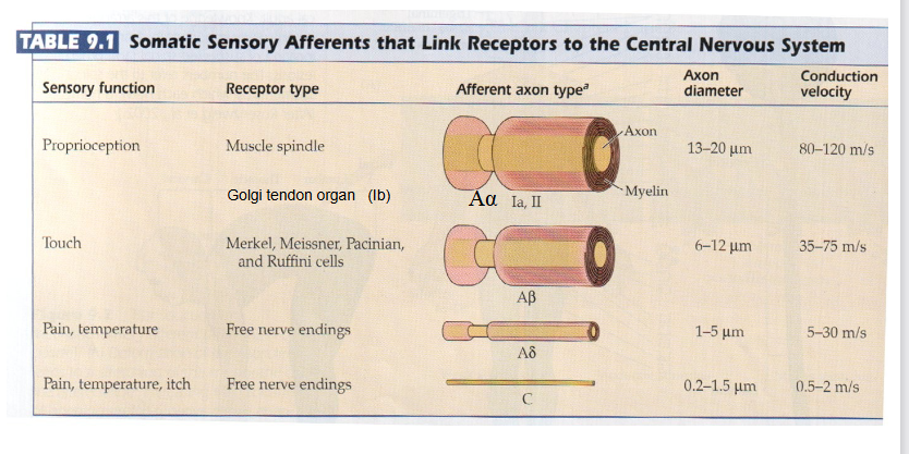
why is axon diameter and myelination importat to determine the speed of sensory information travellling to your brain?

  1. Thicker axon diameter = Faster signal speed.

  2. More myelin insulation = Faster signal speed.


pain temperature and itch are the slowest ones

<ol><li><p><span><strong>Thicker axon diameter</strong> = Faster signal speed.</span></p></li><li><p><strong>More myelin insulation</strong> = Faster signal speed.</p></li></ol><p></p><p>pain temperature and itch are the slowest ones</p><p></p>
32
New cards

what are afferents?

sensory pathways that carry electrical signals toward the central nervous system (the brain and spinal cord) from receptors in the rest of your body.

<p><span style="color: rgb(0, 0, 0);"><strong><mark data-color="#ffffff" style="background-color: rgb(255, 255, 255); color: inherit;">sensory pathways that carry electrical signals </mark><em><mark data-color="#ffffff" style="background-color: rgb(255, 255, 255); color: inherit;">toward</mark></em><mark data-color="#ffffff" style="background-color: rgb(255, 255, 255); color: inherit;"> the central nervous system (the brain and spinal cord)</mark></strong><mark data-color="#ffffff" style="background-color: rgb(255, 255, 255); color: inherit;"> from receptors in the rest of your body.</mark></span></p>
33
New cards

what are the three biological attributes that define how out somatonsensory afferents perceive and communicate different types of touch to the brain?

  1. Speed: thicker nerve fibers have more insulation and will send electrical signals faster to brain than thin ones.

  2. Size of Receptive field: whether or not you can feel fine details depends on how dense and localized the sensory nerves are in the specific body area. Fingers and lips have tiny and packed receptive fields, so they can feel microscopic textures. Meanwhile, your back have big and spread out fields, so you can know in which part you are being touched.

  3. Temporal dynamics: it is explaining how sensory nerves track time and change, whether they pay attention to sustained pressure or sudden movement.

    1. Rapidly adapting afferents: as soon as they detect a change in stimulus, they will fire electrical signals and if the stimulus stops, the nerve will go quiet.

      1. Think about putting a wristwatch or a t-shirt on in the morning. You feel it the exact moment it touches your skin (a change). After a few minutes, you completely forget it is there because the receptors have adapted and gone silent. If the watch slips down your wrist (another change), the receptors instantly fire again to alert you of the movement.

    2. Slowly adapting afferents: they never stop firing as long as the stimulus is present

      1. Size, weight, continuous shape

      2. hink about holding a coffee mug or a pen. Even if you hold it completely still with your eyes closed, your brain doesn't "forget" you are holding it. Because these nerves continuously fire the entire time, you maintain a constant awareness of the object's form, pressure, and edges so you don't accidentally drop it.


<ol><li><p><strong>Speed:</strong> thicker nerve fibers have more insulation and will send electrical signals faster to brain than thin ones. </p></li><li><p><strong>Size of Receptive field:</strong> whether or not you can feel fine details depends on how dense and localized the sensory nerves are in the specific body area. Fingers and lips have tiny and packed receptive fields, so they can feel microscopic textures. Meanwhile, your back have big and spread out fields, so you can know in which part you are being touched.</p></li><li><p><strong>Temporal dynamics: </strong>it is explaining how sensory nerves track time and change, whether they pay attention to sustained pressure or sudden movement. </p><ol><li><p><u>Rapidly adapting afferents</u>: as soon as they detect a change in stimulus, they will fire electrical signals and if the stimulus stops, the nerve will go quiet.</p><ol><li><p>Think about putting a wristwatch or a t-shirt on in the morning. You feel it the exact moment it touches your skin (a change). After a few minutes, you completely forget it is there because the receptors have adapted and gone silent. If the watch slips down your wrist (another change), the receptors instantly fire again to alert you of the movement.</p></li></ol></li><li><p><u>Slowly adapting afferents</u>: they never stop firing as long as the stimulus is present</p><ol><li><p>Size, weight, continuous shape</p></li><li><p><span>hink about holding a coffee mug or a pen. Even if you hold it completely still with your eyes closed, your brain doesn't "forget" you are holding it. Because these nerves continuously fire the entire time, you maintain a constant awareness of the object's form, pressure, and edges so you don't accidentally drop it.</span></p></li></ol></li></ol></li></ol><p></p>
34
New cards
<p>how can this graph help me understand the rapidly and slowly adapting afferents</p>

how can this graph help me understand the rapidly and slowly adapting afferents

Slowly adapting graph: fire a denser burst of spikes when you start holding something, and because stimulus is constant and static, it will keep firing those separated lines

Rapidly adapting graph: the stimulus is continous, you will feel the watch at first, but then you will not, so it goes on and off

<p>Slowly adapting graph: fire a denser burst of spikes when you start holding something, and because stimulus is constant and static, it will keep firing those separated lines</p><p>Rapidly adapting graph: the stimulus is continous, you will feel the watch at first, but then you will not, so it goes on and off</p>
35
New cards

what is the anatomical structure of glabrous skin?

there r 3 structural layers:

  1. Epidermis: has free nerve endings and merkel cell-neurite complexes

  2. Dermis:has meissner corpuscles and ruffini corpuscles

  3. Subcutaenous: houses pacinian corspuscles


<p>there r 3 structural layers:</p><ol><li><p>Epidermis: has free nerve endings and merkel cell-neurite complexes</p></li><li><p>Dermis:has meissner corpuscles  and ruffini corpuscles</p></li><li><p>Subcutaenous: houses pacinian corspuscles </p></li></ol><p></p>
36
New cards

what are merkel cell afferent and meissner afferent?

2 types of encapsulated cutaneous mechanoreceptors that convey tactile information through myelinated AB sensory axons


merkel: epidermis

meissner: beneath epidermis

<p>2 types of encapsulated cutaneous mechanoreceptors that convey tactile information through myelinated AB sensory axons</p><p></p><p>merkel: epidermis</p><p>meissner: beneath epidermis</p>
37
New cards

what are pacinian afferent and ruffini afferent?

they are deeply situated in skin

pacinian: deep in dermis and subcutaneous layer, most sensititve mechanoreceptors

ruffini: deep in dermis

<p>they are deeply situated in skin</p><p>pacinian: deep in dermis and subcutaneous layer, most sensititve mechanoreceptors</p><p>ruffini: deep in dermis</p>
38
New cards

what are proprioceptors? their 2 types?

sensory receptors located in muscles, tendons, and joints that deliver continuous information about limb position, other body parts in space, and the load on tendons and joints


trasnmits info to cerebellum and cerebral cortex


  1. muscle spindles: in skeletal muscles, consisting of specialized intrafusal muscle fibers, consists of group Ia and II afferents. Ia manages movement of limbs and group II manages static limb positions, encodes info on muscle length changes

  2. golgi tendon organs: has a group Ib afferent, distributed among collagen fibers of the tendons, provides info on muscle


39
New cards

what is the primary somatosensory cortex?

is the primary cortical area of the brain responsible for processing mechanoreceptive and proprioceptive sensations

it comprises Brodmannss areas 3a, 3b, 1 and 2
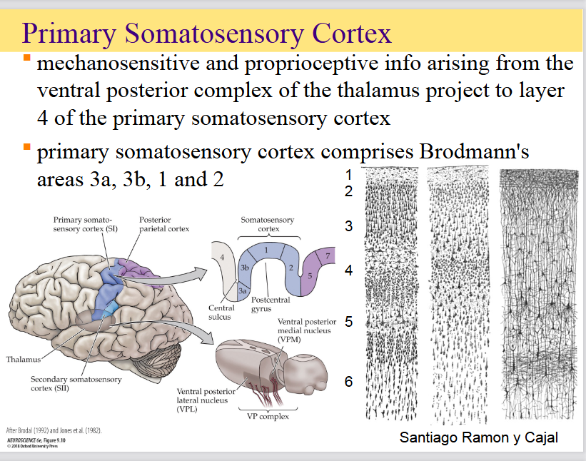
<p><span>is the primary cortical area of the brain responsible for processing mechanoreceptive and proprioceptive sensations</span></p><p><span>it comprises Brodmannss areas 3a, 3b, 1 and 2</span></p>
40
New cards

what is the somototopic organization of cerebral cortex?

Each specific region of the cortex corresponds directly to a distinct part of the body—either receiving sensory input from it or controlling it through motor output

Body regions requiring high sensory precision—such as the fingers, hands, face, lips, and tongue—occupy disproportionately large territories of the cortical map compared to areas like the trunk, arm, or leg

<p><span>Each specific region of the cortex corresponds directly to a distinct part of the body—either receiving sensory input from it or controlling it through motor output</span><br></p><p><span>Body regions requiring high sensory precision—such as the </span><strong>fingers, hands, face, lips, and tongue</strong><span>—occupy disproportionately large territories of the cortical map compared to areas like the trunk, arm, or leg</span></p>