Intro to Imaging Medical Terms
 Call Kai
 Learn
Learn Practice Test
 Spaced Repetition
 Match
Match Flashcards
 Knowt Play
1/104

There's no tags or description
Looks like no tags are added yet.
Name | Mastery | Learn | Test | Matching | Spaced | Call with Kai | Chat |
|---|
No analytics yet
Send a link to your students to track their progress
105 Terms
Anterior (Ventral)
Forward or front part of body or forward part of an organ

Posterior (Dorsal)
Back part of the body or organ (top of foot is considered dorsal)

Superior
nearer of the head or situated above

Inferior
nearer to the feet or situated below

Lateral
part away from the median plane of body or away from the middle of another body part to the right or left

Medial
parts toward median plan of body or towards the middle of another body part

Proximal
parts nearer point of attachment, point of reference, origin, or beginning, toward center of body
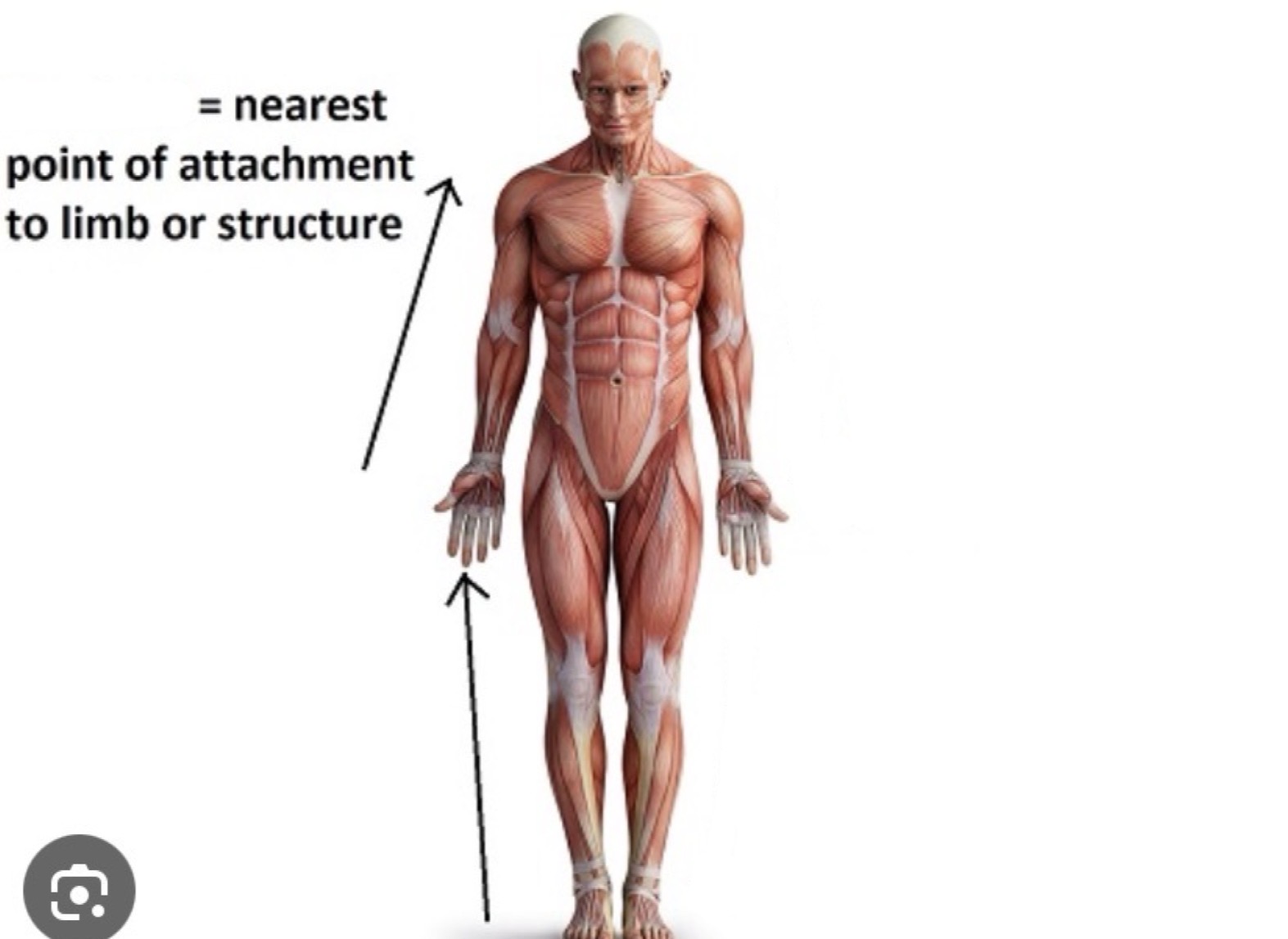
Distal
parts farthest from point of attachment, point of reference, origin or beginning; away from the center of body

Supine
lying on the back

Prone
lying face down

Sagittal
divides the entire body or body part into right and left segments
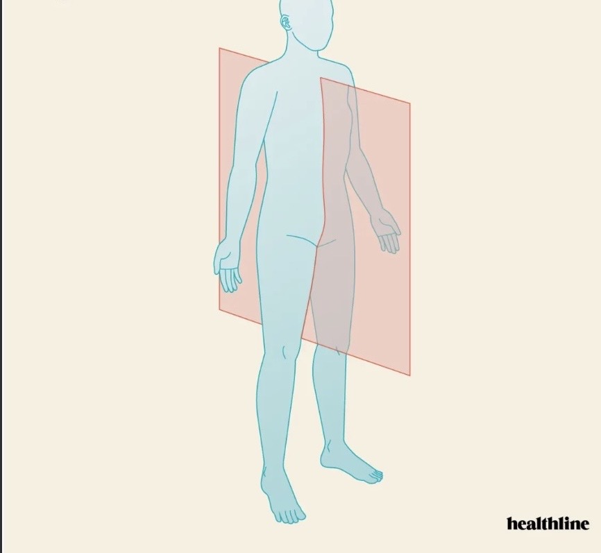
Coronal
divides the entire body or body part into anterior or posterior segments

Transverse
divides the body or body part into superior and inferior portions (also known as axial or horizontal)

Oblique
a plane that passes through a body part at any angle among the sagittal, coronal, or transverse

Contrast
ability to distinguish between different structures or tissues on an image
Artifact
any error or distortion in the image that does not accurately represent the underlying anatomy or tissue
Lesion
injury or other damaging change to an organ or tissue
Pathology
a disease process
Anomaly
variation from the normal pattern
Protocol
inclusive order of necessary images acquired during an examination
Modality
specific types of technology or technique used to create an image of the body for diagnostic purposes (i.e. CT, MRI, Interventional, Rad Tx, Ultrasound, Mammo)
Patient history
process of talking to the patient to obtain pertinent information for more accurate reason for exam (AIDET)
Diagnosis
process of identifying or determining the nature and cause of a disease, injury, or medical condition
Procedure
specific steps and techniques used to obtain medical images of the body for diagnostic purposes
Report
document that summarizes the findings of a diagnostic imaging examination
Contraindicated
any reason for a patient to not receive an exam or treatment
CPR
Cardiopulmonary Resuscitation
Emphysema
abnormal, permanent enlargement of airspaces distal to the terminal bronchioles
Renal Function Test
BUN, Creatinine, eGFR
Anaphylaxis
severe, potentially life-threatening allergic reactions that can occur rapidly
DNR
Do Not Resuscitate
COPD
Chronic Obstructive Pulmonary Disease
CHF
Congestive Heart Failure
c
with
s
without
h/o
history of
STAT
Immediately
tid
three times daily
qd
every day
qid
four times a day
sob
short of breath
Peripheral
located away from the center of near the surface
artho-
joint
cardi-
heart
cost-
rib
ile-
ileum
contra-
against
hyper-
above, a greater concentration
hypo-
below, a lesser concentration
-ectomy
surgical removal
-itis
inflammation
Radiographer
skilled imaging professional trained to obtain x-ray images and assist the radiologist in x-ray procedures
Positioning
used in 2 ways in radiography. 1.) overall posture of the patient or general body position. 2.) specific placement of the bod part in relation to the table or Image Receptor (IR)
X-ray tube
evacuated glass bulbs with positive (anode) and negative (cathode) electrodes
kVp
measure of potential difference across the x-ray tube (quality)
mAs
when mA is multiplied by the exposure time. Indicates the amount of radiation in the exposure (quantity)
mA
measure of the current flow rate in the x-ray tube circuit
Grid
devices used to prevent scatter radiation from reaching the IR
Collimator
allows the radiographer to vary the size of the radiation field and to indicate with a light beam the size, location, and center of the field
Bucky
located beneath the table surface; it is a moving grid that incorporates a tray to hold an IR
Radiolucent
transparent to x-rays
Radiopaque
block radiation from reaching the IR
Exposure index
determines the underexposure or overexposure of an image (also known as s-number, digital index number (DI)
Fluoroscopy
x-ray machine designed for direct viewing of the x-ray image
Digital Radiography (DR)
has radiation receptors within the table, bucky, or stand-alone DR receptors that transmit digital signals directly to the computer system
Scatter radiation
Patient is the principal source of occupational exposure to radiographers. Ionizing radiation that occurs as a result of their interaction between the primary beam and matter.
Source to Image Detector (SID)
distance between the x-ray type target and the image receptor. Measured along the central ray
Object to Image Detector (OID)
distance between the object and the image receptor
Image receptor
device that captures a radiographic image
Contrast media
Gas or radiopaque substance that is injected into the body
Anode
electrode at which oxidation occurs; electrically positive, target end of the x-ray tube
Cathode
negative electrode; the negative side of the x-ray tube. Contains the focusing cup and filament
Automatic Exposure Control (AEC)
automatic exposure control - ionization chamber located between the patient and the image receptor
Lead apron
coverings worn by radiographers who are in a radiographic or fluoroscopic room with the x-ray beam turned on. Worn for radiation protection when performing portable radiography. Absorbs most of the scatter radiation that strikes the apron

Dosimeter
monitoring device worn by radiographer that measures the amount of radiation received
In-Profile
an outline or silhouette view of an anatomic structure that has a distinctive shape
Central ray
imaginary photon in the center of the x-ray beam that is directed toward the center of the object being radiographed
Echogenic
structure that procedures echoes
Hyperechoic
brighter than surrounding tissue
Hypoechoic
darker than surrounding tissue
Isoechoic
have the same echogenicity
Homogeneous
uniform composition
Heterogeneous
differing composition
Doppler
recognized the frequency shift resulting from moving objects
Transducer
instrument used to obtain an image of the human body using ultrasound. Converts one form of energy to another
Sonographer
skilled medical professional trained to utilize ultrasound to image the human body
Scan
The act of performing a sonography examination
Ultrasound
sound range above the normal hearing range of humans
Gain
amplification of the electrical signal
Depth
varies by the depth of the echoes that are displayed on the monitor (depth range)
Frequency
number of cycles per unit of time
Time-gain compensation
control that alters the brightness of an image at a specific depth
A-mode
amplitude mode - ultrasound display in which the depth of the returning echo was represented on the x-axis, while the strength (amplitude) of the reflector was represented along the y-axis
B-mode
brightness mode - ultrasound display in which a dot with varying degrees of brightness is used on the screen. The stronger the returning echo, the brighter the dot (grayscale)
M-mode
motion mode - demonstrates the motion of structures along a single scan line
Color Doppler
doppler shift information presented as a color or hue superimposed over a grayscale image
Power Doppler
amplitude mode of doppler when it is not the shift itself that prevents signal but rather the strength (amplitude) of the shift
Spectral Doppler
type of ultrasound technique that graphically displays blood flow velocities over time
Suprapubic
anatomical region located above the pubic bone
Insonation
process of exposing tissue or an object to ultrasound waves