horse state part C - internal diseases
 Call Kai
 Learn
Learn Practice Test
 Spaced Repetition
 Match
Match Flashcards
 Knowt Play
1/24

There's no tags or description
Looks like no tags are added yet.
Name | Mastery | Learn | Test | Matching | Spaced | Call with Kai | Chat |
|---|
No analytics yet
Send a link to your students to track their progress
25 Terms
Swallowing and Oesophageal Disorders
Main disorders:
1⃣ Dysphagia = abnormal swallowing
2⃣ Esophageal obstruction / choke
3⃣ Megaesophagus
4⃣ Esophageal stricture / stenosis
5⃣ Other disorders:
Rupture / perforation / laceration
Esophagitis
Esophageal diverticulum
Esophageal cysts
1⃣ DYSPHAGIA
Dysphagia = abnormality/difficulty in swallowing.
⚠ It is a clinical sign, NOT a specific disease.
The causes can be divided according to where the problem occurs:
Oral, Pharyngeal, Esophageal, Systemic
1. ORAL CAUSES
Problem occurs before food is properly transferred into the pharynx.
Causes:
Facial paralysis, Lip lesions, Temporomandibular joint disorders, Hyoid disorders, OA → pain
Lingual abnormalities
Inability to move ingesta
Dental disorders
Congenital/acquired palatal defects
Foreign bodies, Neoplasia
🧠 Think: MOUTH cannot prepare/move food → ORAL dysphagia
2. PHARYNGEAL CAUSES
Pharyngeal paralysis
Can result from:
Glossopharyngeal nerve damage, Guttural pouch mycosis, Botulism, Heavy-metal poisoning
Other causes:
Pharyngeal compression
e.g. strangles
Pharyngeal cysts, Epiglottic lesions, Laryngeal abnormalities
3. ESOPHAGEAL CAUSES
Megaesophagus, Choke, Stenosis/stricture, Cysts, Rupture, Neoplasia
4. SYSTEMIC CAUSES
Nutritional myodegeneration
Equine grass sickness
Tetanus
🩺 CLINICAL SIGNS OF DYSPHAGIA
Slow and Messy eating
Halitosis (bad breath)
Quidding
Productive cough
Nasal reflux of: Saliva, Ingesta, Fluids
Weight loss
🌾 What is QUIDDING?
Horse chews food → Forms a partially chewed bolus → Cannot process/swallow it properly → Drops/spits it from mouth
➡ Quidding = rejection/dropping of semi-masticated food
Often associated with:
➡ Dental/oral disease.
⭐ ORAL vs NASAL REFLUX
👄 Food/reflux from MOUTH
➡ Think oral cavity
👃 Food/fluid from NOSE
➡ Think pharyngeal or esophageal problem
🔎 DIAGNOSIS OF DYSPHAGIA
History, Clinical signs
Physical examination, Oral examination
Nasal endoscopy
X-ray
Fluoroscopy
Treatment ➡ Treat the underlying cause
2⃣ 🚨 ESOPHAGEAL OBSTRUCTION – CHOKE
Choke = acute obstruction of the esophagus.
⚠ Important in horses: CHOKE ≠ tracheal obstruction
→ The horse can usually still breathe because the obstruction is in esophagus, not trachea
🔍 COMMON CAUSE
Typically caused by:
🌾 Dry fibrous material
→ Dry food enters esophagus → Absorbs saliva → Swells → Bolus expands → Blocks esophageal lumen → CHOKE!!🚨
📍 PREDISPOSED SITES
Three important narrow regions:
1⃣ Cranial cervical esophagus
2⃣ Apertura thoracica cranialis
= thoracic inlet
3⃣ Diaphragmatic esophageal hiatus
🧠 NECK → CHEST ENTRANCE → DIAPHRAGM
🩺 CLINICAL SIGNS – CHOKE
Dysphagia, Distress, Reflux, Saliva from nose and mouth, Ingesta from nostrils, Extended neck, Dehydration
Typical picture:
Horse eats → Suddenly distressed → Stretches neck → Tries repeatedly to swallow → SALIVA + FOOD FROM NOSE (green frothy nasal discharge)


🚨 MAJOR COMPLICATION
Aspiration pneumonia
Refluxed material → Enters respiratory tract → Aspiration → Pneumonia
Clinical sign: Cough
🔎 DIAGNOSIS – CHOKE
Nasogastric tube ⭐
Try to pass tube. Tube reaches obstruction → Cannot pass into stomach
→ Supports diagnosis of esophageal obstruction
Also:
➡ Distension of cervical esophagus may be visible/palpable!
💊 TREATMENT – CHOKE ⭐⭐⭐
1. Sedation
➡ Alpha-2 agonist + butorphanol
Sedation also helps lower the head, reducing aspiration risk.
2. Spasmoanalgesia (Buscopan)
Purpose:
➡ Relax esophageal muscle
➡ Reduce pain/inflammation
Can include NSAIDs
3. Massage
If obstruction is accessible in cervical esophagus:
➡ Gentle external massage may help.
4. LAVAGE THROUGH NASOGASTRIC TUBE ⭐
Use: Saline/water
Repeated gentle lavage helps soften and remove obstruction.
🚫 DO NOT use oil for lavage
Why?
Oil → Can be aspirated → Enters lungs → Cannot be cleared effectively → Severe lipoid/granulomatous pneumonia
5. IV FLUIDS
Choke can cause:
Dehydration
Electrolyte abnormalities
Therefore:
➡ IV rehydration
➡ Electrolyte supplementation
Also supports tissue perfusion.
6. SURGERY
If conservative treatment fails:
➡ Esophagotomy
Used only when necessary because esophageal surgery has a relatively high complication risk.
3⃣ MEGAESOPHAGUS
Megaesophagus = dilation/distension of the esophagus associated with loss/reduction of normal motility.
Esophageal motility ↓ → Food cannot move efficiently toward stomach → Food + fluid accumulate → Esophagus stretches → MEGAESOPHAGUS
🔍 ETIOLOGY
Congenital:
Especially described in Friesian horses, associated with developmental/connective-tissue abnormalities
Acquired
Can be associated with:
Vascular ring abnormalities/strictures
Herpesvirus
Myeloencephalitis
Nerve damage
Chronic esophageal obstruction
🩺 CLINICAL SIGNS
Coughing, Nasal reflux of ingesta, Distension of cervical esophagus
Potential consequence:
🚨 Aspiration pneumonia
🔎 DIAGNOSIS
⭐ Contrast radiography
Contrast enters esophagus → X-ray → Dilated esophagus becomes visible
💊 TREATMENT
Feeding management is important:
Feed food and water from an elevated position
Example: Step-up/elevated feeding station
Goal: Use gravity to assist passage of food toward stomach.
4⃣ ESOPHAGEAL STRICTURE / STENOSIS
Stricture/stenosis = narrowing of the esophageal lumen.
Frequently occurs around: Thoracic inlet
3 types
adventitia and muscularis
mucosa and submucosa
annular stenosis.
🔍 ETIOLOGY
Most commonly:
➡ Sequel to choke/esophageal obstruction
Why?
Choke → Pressure/inflammation damages esophageal wall → Healing → Fibrosis/scar tissue → Scar contracts → LUMEN NARROWS
Can also be:
Congenital, Acquired, Parasitic
🪱 GASTEROPHILUS INERMIS
Migrating bot-fly larvae can cause:
➡ Esophageal inflammation → Stenosis → Secondary dilation of esophagus cranial to obstruction
Diagnosis: Endoscopy
Treatment: Ivermectin
🔎 DIAGNOSIS – STRICTURE
⭐ Contrast radiography
Can demonstrate:
➡ Narrowed segment
➡ Dilation proximal to narrowing
💊 TREATMENT
Conservative:
Antibiotics if indicated, NSAIDs
Feeding management
Balloon Dilatation: Balloon placed into narrowed segment → Inflated → Stretches stenotic area → Lumen becomes wider
Surgical
Possible procedures:
Esophagomyotomy, Esophagopexy, Partial resection
5⃣ ESOPHAGEAL RUPTURE / PERFORATION / LACERATION 🚨
Esophageal wall is damaged.
Causes:
External trauma, Kicks, Stick wounds
Misuse/trauma from nasogastric tube
⚠ Why is it dangerous?
Esophagus ruptures → Saliva + ingesta leak into neck tissues → Massive bacterial contamination → Severe inflammation/infection → PHLEGMON + subcut emphysema
Phlegmon = diffuse spreading infection/inflammation of soft tissues.
6⃣ ESOPHAGITIS
= Inflammation of the esophagus
Causes:
Choke, Reflux, Pyloric stenosis / delayed gastric emptying
💊 Treatment
Feeding management
Antibiotics if bacterial infection is involved
NSAIDs
Treatment of delayed gastric emptying/underlying disease
7⃣ ESOPHAGEAL DIVERTICULUM
= pouch/outpouching of esophageal wall. (almost always cercical region)
TRUE ➡ Involves all layers
FALSE ➡ Does not involve all layers; involvement of the muscular layer.
Traction arises from fibrotic contraction of all tissue layers, whereas pulsion involves mucosal protrusion through a muscular defect.
8⃣ ESOPHAGEAL CYSTS
Two types:
Intramural Inclusion cyst ➡ Squamous epithelial lining, youngs and yearlings, congenital
Esophageal Duplication cyst
🩺 Clinical Signs
Dysphagia, Regurgitation/reflux
🔎 Diagnosis
Endoscopy, USG, X-ray
Therapy
surgical technique recommended for managing congenital intramural esophageal cysts → MARSUPIALIZATION (The cyst wall is incised, and its edges are sutured to the horse's neck skin, creating an open pouch (stoma). Complete resection is HIGH complication risk.
Equine grass sickness → toxicoinfection with clostridium botulinum. affecting the enteric nervous stsrem - neuropathy. acute, subacute, chronic. Clostridium tetani causes spastic paralysis, while Clostridium botulinum causes flaccid paralysis.
🧠 QUICK COMPARISON ⭐⭐⭐
Disorder | Main problem | Key clue |
|---|---|---|
Dysphagia | Abnormal swallowing | Clinical sign, many causes |
Choke | Acute obstruction | 👃 Food/saliva from nose + NG tube won't pass |
Megaesophagus | ↓ Motility + dilation | Dilated esophagus + reflux |
Stricture | Narrowed lumen | Often after previous choke |
Rupture | Hole/tear | Saliva + food enter tissues → phlegmon |
Esophagitis | Inflammation | Often after choke/reflux |
Diverticulum | Esophageal pouch | Food can accumulate |
Cyst | Mass/cyst | Dysphagia + regurgitation |
What is choke? → Acute obstruction of the esophagus, usually by impacted feed material.
What is the typical sign the owner notices? → ⭐ Feed, saliva and/or fluid coming from the nostrils shortly after attempting to eat or drink.
Which horses were specifically mentioned as predisposed to choke? → Friesian horses.
How can you diagnose choke in the field? → Pass a nasogastric tube; it cannot be advanced beyond the obstruction. Cervical esophageal distension may also be palpable.
What spasmolytic drug can you use? → Buscopan (hyoscine butylbromide).
What reproductive drug did Zert say can also help relax the esophagus? → Oxytocin, particularly for certain esophageal obstructions.
How do you treat choke conservatively? → Sedate → keep the head low → carefully lavage through a nasogastric tube → massage a palpable cervical obstruction → repeat attempts if necessary.
Why should the horse's head be kept low during lavage? → To allow lavage fluid/feed material to drain from the nose and reduce aspiration into the lungs.
If lavage is unsuccessful, should you immediately operate? → No. Continue conservative treatment and repeated careful lavage when appropriate; surgery is generally a last resort.
If the horse needs fluids, where should they be given? → ⭐ IV, not orally/nasogastrically while the esophagus is obstructed.
What important complication can occur during choke/treatment? → ⭐ Aspiration pneumonia.
How do you diagnose aspiration pneumonia? → Clinical respiratory examination plus thoracic ultrasound; pulmonary consolidation/fluid-related changes may be detected, with B-lines/comet-tail artifacts depending on pulmonary involvement.
How do you treat aspiration pneumonia? → Broad-spectrum antimicrobial therapy + NSAIDs/supportive care, with additional respiratory support depending on severity.
Stomach Diseases and Verminous Chronic Gastritis
Main conditions:
1⃣ Equine grass sickness
2⃣ Equine Gastric Ulcer Syndrome – EGUS
3⃣ Gastric impaction
4⃣ Verminous chronic gastritis
Gasterophilosis
Habronemiasis
Trichostrongylosis
5⃣ Gastric neoplasia – SCC
1⃣ 🌱 EQUINE GRASS SICKNESS
Equine Dysautonomia/Equine grass sickness = highly fatal neuropathy of grazing horses linked to neurotoxic enzymes
Main lesion: ➡ Degeneration of neurons of the autonomic nervous system
Especially:
⭐ Enteric nervous system
Therefore:
Autonomic/enteric neurons degenerate → GIT cannot function normally → Severe ↓ gastrointestinal motility → Mainly GIT signs
🔍 Etiology
The exact cause is not completely established.
Your notes suggest:
➡ Possible toxicoinfection associated with Clostridium botulinum
Intestinal infection → Toxin production → Damage to autonomic neurons → Loss of GIT motility
🩺 THREE CLINICAL FORMS ⭐⭐⭐
🔴 ACUTE
Severe disease.
Colic, Severe ↓ gut motility, Dysphagia, Gastric distension, Gastric reflux, Muscle tremors
Outcome: ☠ Death usually in <4 days
🟠 SUBACUTE
Similar to acute form, but less severe/slower progression
🟡 CHRONIC
More prolonged disease.
Dullness, ↓ GIT motility, Severe/progressive weight loss, Hard, dry feces, Tachycardia, Crusty nasal discharge

🔎 Diagnosis
Presumptive:
based on: Clinical signs, History, Physical examination
Definitive:
⭐ Histopathology / biopsy → Looking for characteristic neuronal degeneration.
Post-Mortem:
May find:
Gastric distension, Colon containing hard + dry feces
Why?
↓ Enteric nervous function → decreased Motility → GI contents stagnate → Water absorbed → Hard/dry feces
💊 Treatment
No specific curative treatment.
➡ Supportive treatment only
Due to poor prognosis:
➡ Euthanasia often recommended
2⃣ 🔥 EQUINE GASTRIC ULCER SYNDROME – EGUS
Gastric ulcer = an open sore/defect in the stomach mucosa
🧪 NORMAL STOMACH
Glandular epithelium continuously secretes:HCl – hydrochloric acid
The stomach therefore needs protection against its own acid.
Protective mechanisms include:
🛡 Mucus/bicarbonate barrier
💧 Alkaline saliva
🩸 Normal mucosal blood flow and epithelial defenses
If protection fails → Acid damages mucosa → ULCERATION
📍 TWO REGIONS
Squamous mucosa: Most commonly affected, Approximately 80%
Glandular mucosa: Approximately 20%

🧠 WHY IS SQUAMOUS MUCOSA VULNERABLE?
Glandular stomach is designed to secrete and resist acid, while the squamous mucosa has much less acid protection.
Especially during exercise → Acidic gastric contents → splash upward → Contact squamous mucosa → Damage
🔍 ETIOLOGY / RISK FACTORS
Diet
High concentrate and Low roughage
Why? Less roughage → Less chewing → Less alkaline saliva → Less buffering of acid → Increased ulcer risk
Other factors: Stress, Other GIT disease, Systemic illness, Anorexia, withholding food, NSAID therapy
🏇 INTENSE EXERCISE ⭐
Very common in:
Racehorses, Performance horses >90%
Exercise → Increased abdominal pressure / acid movement → Acid contacts squamous mucosa → Ulceration
💊 WHY NSAIDs CAN CAUSE ULCERS?
NSAIDs → ↓ Prostaglandins → ↓ Mucosal protection/blood flow/bicarbonate → Glandular defenses weaken → Ulceration
🩺 Clinical Signs
Can range from no clinical signs to Anorexia, Colic, Depression, Weight loss, Diarrhea
🔎 Diagnosis
⭐ Gastroscopy / endoscopy
➡ Direct visualization of gastric mucosa, and the response to treatment
💊 TREATMENT – EGUS ⭐⭐⭐
Management
Decrease/stop ulcerogenic NSAIDs where possible
↓ Concentrate feeding, ↑ Roughage
Avoid prolonged fasting
GASTRIC ACID SUPPRESSION
⭐ Omeprazole (Proton-pump inhibitor – PPI). Blocks gastric proton pumps → ↓ HCl secretion → ↑ Gastric pH → Allows ulcer healing
Histamine H₂ receptor antagonists. Example Cimetidine → ↓ Histamine stimulation of acid secretion → ↓ HCl
🛡 OTHER TREATMENT
Antacids, Mucosal protectants
3⃣ 🥕 GASTRIC IMPACTION
= Persistent and progressive accumulation of ingesta in the stomach.
Food accumulates → Cannot leave normally → Stomach becomes progressively full → Risk of gastric rupture

🔍 Etiology
Dry feed that swells/forms a mass, Example: Beet pulp
Other causes:
Dental disease → poor mastication, Inadequate water supply, Rapid food intake, Pyloric outflow obstruction, Bezoars
Liver Disease → may cause secondary gastric impaction. Example: Ragwort poisoning
🩺 Clinical Signs
Colic, Abdominal distension, Anorexia, Lethargy, Recumbency
🔎 Diagnosis
⚠ Definitive diagnosis can be difficult.
May only be diagnosed during Exploratory celiotomy
Rectal Examination: May detect displacement of spleen
Ultrasound: May show abnormally large/distended stomach
💊 TREATMENT
🚨 Main concern = GASTRIC RUPTURE
Therefore: Withhold feed and Limit/control water intake
Gastric Lavage via Nasogastric tube → Soften/remove accumulated ingesta.
💧 IV Therapy → Correct dehydration/electrolytes
💊Analgesia for Pain/colic
Surgery ➡ Gastrotomy: Incision into stomach → Remove impaction (Often unsuccessful / poor prognosis)
4⃣ 🪱 VERMINOUS CHRONIC GASTRITIS
Three important parasites:
🪰 Gasterophilus = BOT FLY
🪱 Habronema = STOMACH WORM + SUMMER SORES
🪱 Trichostrongylus axei = STOMACH HAIR WORM
A. 🪰 GASTEROPHILOSIS
Very common gastric parasite of horses.
Caused by:
➡ Bot-fly larvae
Gasterophilus haemorrhoidalis, G. intestinalis, G. nasalis

🔄 LIFE CYCLE ⭐⭐⭐
Adult bot fly → Eggs deposited on horse's hair (Face, Lips, Intermandibular region, Forelegs) → Horse licks itself → Eggs enter mouth → Hatch to L1 → L1 burrows into oral mucosa → Molt → L2 → Migrate toward pharynx → Enter stomach → Molt → L3 → Attach/remain in GIT for approximately 10–12 months⭐ → L3 passed in feces
→ Pupate in soil → Adult fly emerges after approximately 3–9 weeks⭐
🔄 Cycle repeats
⚙ Pathogenesis
Larvae attach to gastric mucosa → Local irritation/inflammation → Mucosal damage → Bleeding → Ulceration → Possible secondary infection
➡ possible release of Hemolytic toxin
🩺 Clinical Signs
Oral larval migration may cause:
Pus pockets in gums, Salivation, Head shaking, Loss of appetite, Chewing problems
Gastric infection can contribute to → Gastritis/ulceration
🔎 Diagnosis
Coprology
Larvae may be found seasonally, especially: April / beginning of May
⭐ Best: Gastroscopy/endoscopy: Directly visualize larvae attached to stomach.
💊 Treatment:⭐ Ivermectin
B. 🪱 HABRONEMIASIS
Important because it has 3 forms:
1⃣ Gastric
2⃣ Cutaneous
3⃣ Conjunctival
🦠 Etiology
Nematodes:
Habronema muscae, Habronema microstoma
➡ Adults live in gastric mucosa.
Draschia megastoma
➡ Produces large nodules in stomach wall.
🐴 FINAL HOST
➡ Horse and other equids
Adult worms normally live in Stomach
🪰 INTERMEDIATE HOST
⭐ Muscid flies – Musca
🔄 LIFE CYCLE ⭐⭐⭐
Adults in stomach → Eggs/L1 passed in feces → Fly larvae ingest eggs/L1 → Develop to infective L3 → Adult fly feeds on moist areas of horse (Eyes, Genitalia, Nostrils, Lips/wounds) → L3 leave fly → Horse swallows L3 → L3 reach stomach → Develop into adults → GASTRIC HABRONEMIASIS
🫃 GASTRIC FORM ⭐Most common
Causes:
➡ Gastritis and Sometimes granuloma formation
H. muscae + H. microstoma
➡ Gastritis, Usually without large granulomas
Draschia megastoma ⭐ → Causes: Large (up to 10cm) granulomatous nodules in gastric mucosa. Contain Worms and Necrotic material.
🧠 DRASCHIA = DRAMATIC BIG NODULE
🪰 ABERRANT HABRONEMIASIS
What if larvae deposited by flies do NOT reach the stomach?
L3 deposited in:
👁 Eye
🩹 Skin/wound
👃 Nostrils
🍆 Genitalia
⬇
Cannot complete normal life cycle → Remain in tissue → Strong local hypersensitivity → Granulomatous inflammation ➡ ABERRANT FORM
👁 CONJUNCTIVAL FORM
Can cause:
Conjunctivitis, Eyelid thickening, Granulomatous lesions
🩹 CUTANEOUS FORM
Causes:
Non-healing wounds, Ulceration, Severe itching, Granulomatous skin lesions, Granular dermatitis
⭐ Called: 🌞 SUMMER SORES
Why summer? 🪰 More flies → More larvae deposited in wounds → Habronema lesions

🔎 Diagnosis
Endoscopy
ELISA
Identify non-healing reddish granulomas
Larvae may be identified in lesions
💩 FECAL EXAM
⚠ Eggs are difficult to detect!!
➡ Eggs are very dense and may not float in standard flotation solutions
💊 Treatment
⭐ Ivermectin
➡ Especially against adult worms/systemic parasitic burden.
Local aberrant lesions may additionally require:
➡ Local wound/inflammation management.
C. 🪱 TRICHOSTRONGYLOSIS
Etiology:
⭐ Trichostrongylus axei
Also called: Stomach hair worm
Common in horses grazing on pasture shared or previously used by infected ruminants!
🔄 Life Cycle
Infective L3 → Horse ingests L3 while grazing→ Larvae penetrate gastric mucosa → Molt/develop → Adults emerge into gastric lumen
⚙ Pathogenesis
Causes: Catarrhal gastritis
🔎 Diagnosis
⭐ Coproculture
💊 Treatment
⭐ Ivermectin
🧠 THREE GASTRIC PARASITES ⭐⭐⭐
Parasite | What is it? | Key feature | Diagnosis | Treatment |
|---|---|---|---|---|
Gasterophilus | 🪰 Bot-fly larvae | Larvae attached to stomach | Endoscopy | Ivermectin |
Habronema/Draschia | 🪱 Nematodes | Gastritis + summer sores; Draschia → huge nodules | Endoscopy/ELISA | Ivermectin |
Trichostrongylus axei | 🪱 Stomach hair worm | Catarrhal gastritis | Coproculture | Ivermectin |
5⃣ 🧬 GASTRIC NEOPLASIA
Squamous Cell Carcinoma – SCC
Important gastric neoplasia in horses.
🩺 Clinical Sign
Most characteristic:
➡ Chronic progressive weight loss
Can also eventually cause nonspecific GI signs.
🔎 Diagnosis
⭐ Biopsy
💊 Treatment
Generally:
❌ Not attempted
➡ Usually advanced when diagnosed + Poor prognosis
⭐ EXAM ESSENTIALS
🌱 GRASS SICKNESS
➡ Highly fatal autonomic neuropathy
➡ Especially enteric nervous system
➡ Severe ↓ GIT motility
➡ Possible C. botulinum toxicoinfection association
➡ Acute: colic + dysphagia + reflux + tremors → death <4d
➡ Chronic: dull + thin + hard/dry feces
➡ Definitive Dx = histopathology/biopsy
➡ Supportive only / euthanasia often recommended
🔥 EGUS
➡ Acid damages insufficiently protected gastric mucosa
➡ Squamous mucosa especially affected
➡ High concentrate + low roughage + fasting + stress + NSAIDs + intense exercise
➡ Gastroscopy ⭐
➡ Omeprazole
➡ More roughage / less concentrate
🥕 GASTRIC IMPACTION
➡ Progressive accumulation of ingesta
➡ Dry/swelling feed + poor mastication/water + outflow obstruction
➡ Risk = gastric rupture 🚨
➡ Difficult diagnosis
➡ Withhold feed + NG lavage + IV fluids
➡ Gastrotomy possible but difficult/poor success
🪱 VERMINOUS CHRONIC GASTRITIS
GASTEROPHILUS
BOT FLY
➡ Eggs on HAIR
➡ Horse LICKS
➡ Mouth → stomach
➡ Larvae remain 10–12 months
➡ Endoscopy
➡ Ivermectin
HABRONEMA
STOMACH WORM + SUMMER SORES
➡ Musca = intermediate host
➡ Swallowed L3 → gastric form
➡ Skin/eye L3 → aberrant form
➡ Draschia → 10 cm gastric granulomas
➡ Ivermectin
TRICHOSTRONGYLUS AXEI
STOMACH HAIR WORM
➡ L3 ingested
➡ Penetrates gastric mucosa
➡ Catarrhal gastritis
➡ Coproculture
➡ Ivermectin
Small Intestine Diseases and Verminous Enteritis
Main groups:
1⃣ Chronic inflammatory bowel disease – CIBD
2⃣ Idiopathic chronic diarrhea
3⃣ Intestinal obstruction
4⃣ Ulcerative duodenitis
5⃣ Small intestinal volvulus
6⃣ Intussusception
7⃣ Verminous/parasitic enteritis
8⃣ Alimentary lymphoma
9⃣ Bacterial diseases
🔟 Viral diseases
1⃣ CHRONIC INFLAMMATORY BOWEL DISEASE – CIBD
CIBD = collective term for a group of chronic enteropathies with similar clinical signs.
➡ Chronic inflammation/infiltration of intestinal wall → Poor intestinal function → Malabsorption → Weight loss ± diarrhea
Etiology: Often uncertain.
Possible causes: Immune-mediated disease or Infectious agents (Parasites, Bacteria)
🩺 Clinical Signs
⭐ Weight loss, Diarrhea, Colic, Fever
🔎 Diagnosis
⭐ Definitive diagnosis ➡ Histopathology
USG, Blood samples
💊 Treatment
First: ➡ Corticosteroids, Examples: Dexamethasone, Prednisolone
Purpose: → Suppress intestinal inflammation / immune reaction
If ineffective: ➡ Combine with azathioprine (immunosuppressive drug)
2⃣ 💩 IDIOPATHIC CHRONIC DIARRHEA
Idiopathic = cause unknown
In horses, the exact cause of chronic diarrhea often remains undiagnosed.
Possible Causes/Associations:
Previous intestinal infection, Dietary change
Especially seen in Foals and Yearlings
🩺 Clinical Signs
Chronic diarrhea and Weight loss
🔎 Diagnosis
⭐ Diagnosis of exclusion
Meaning:
Rule out parasites → Rule out infection → Rule out inflammatory disease → Rule out other causes
⬇
No cause identified
⬇
➡ Idiopathic chronic diarrhea
💊 Treatment
Symptomatic:
Antidiarrheal therapy (Codeine phosphatase), Probiotics, Activated charcoal
3⃣ 🚧 INTESTINAL OBSTRUCTION
Two major types:
A. SIMPLE OBSTRUCTION ➡ Blood supply is maintained
B. STRANGULATING OBSTRUCTION ➡ Blood supply is compromised ⭐ This difference is extremely important.
🟡 SIMPLE OBSTRUCTION
The lumen becomes blocked, but the vascular supply remains intact initially.
Causes: Feed material, Parasites, Extraluminal mass
🔴 STRANGULATING OBSTRUCTION 🚨
Obstruction + vascular compromise → Intestine strangulated →↓ Venous drainage → ↓ Arterial perfusion → Ischemia → Necrosis → Loss of intestinal barrier →
🔥 ENDOTOXEMIA
🩺 Clinical Signs – Strangulation
Usually much more severe: Severe pain, ↑ Heart rate, Abnormal mucous membrane color, Progressive systemic deterioration
🚨 Surgical emergency.
4⃣ 🔥 ULCERATIVE DUODENITIS
More common in foals than adults.
🔍 Etiology
Excess HCl + pepsin reaching the duodenum
🩺 Clinical Signs
Nonspecific: Fever, Mild–moderate colic, Dullness, Diarrhea
🔎 Diagnosis
⭐ Duodenoscopy = most specific.
Long endoscope: Approximately 2 meters, can be performed in: Foals up to approximately 6 months
Also: X-ray, Bloodwork
💊 Treatment
Goal: Reduce acid + protect mucosa
H₂ antagonist (Cimetidine) or Proton-pump inhibitor (Pantoprazole) + Mucosal protector (Sucralfate)
5⃣ 🔄 SMALL INTESTINAL VOLVULUS
= A segment of small intestine twists: ➡ >180° around the axis of its mesentery
⚙ What Happens?
SI twists → Mesenteric vessels twist → Blood flow compromised → Ischemia → Necrosis → Severe acute colic
Therefore:
➡ Strangulating obstruction
🔍 Cause:
Can be associated with altered/local abnormal peristalsis
More commonly affects foals
🩺 Clinical Sign
⭐ Acute, severe colic
💊 Treatment / Prognosis
Requires rapid surgical assessment/treatment.
Prognosis depends strongly on Duration + Degree of ischemia
The longer it is twisted → the more bowel dies → worse prognosis.
6⃣ INTUSSUSCEPTION
= One segment of intestine invaginates into the adjacent segment
One segment slides inside another → Obstruction + Possible vascular compromise

7⃣ 🪱 VERMINOUS / PARASITIC ENTERITIS
Important parasites:
A. Anoplocephala
B. Parascaris equorum
C. Strongyloides westeri
D. Eimeria leuckarti
A. 🪱 ANOPLOCEPHALA
Equine Tapeworms ➡ Cestodes
All live in different intestinal locations and:
➡ Can occur simultaneouslyThey attach to: Intestinal mucosa
⭐ THREE SPECIES
Species | Location | Size | Importance |
|---|---|---|---|
A. perfoliata | ⭐ Ileocecal junction | ~20 cm | Most pathogenic |
A. magna | Jejunum | ~80 cm | Largest |
Paranoplocephala mamillana | Duodenum | ~5 cm | Smallest |
⭐ A. PERFOLIATA
Most important/pathogenic:
Ileocecal junction is narrow → Many tapeworms accumulate → Inflammation + obstruction → Colic → Potential intestinal damage/rupture → Peritonitis
🔄 LIFE CYCLE – ANOPLOCEPHALA ⭐⭐⭐
Final host: Horse
Intermediate host: Oribatid pasture mite
Infective stage: ⭐ Cysticercoid
Adult tapeworm in horse → Eggs passed in feces → Pasture mite eats eggs → Egg develops into cysticercoid → Horse grazes → Accidentally eats infected mite → Cysticercoid released → Develops into adult → Attaches to intestinal wall

⚙ Pathogenesis
Can cause:
Catarrhal and Hemorrhagic enteritis, Mucosal ulceration, Intestinal obstruction, Intestinal rupture, Peritonitis
81% of constipations and 22% of spastic colic’s are caused by tapeworms!!
🩺 Clinical Signs
Constipation, Colic, Diarrhea, Weight loss, Anemia, Dull coat
🔎 Diagnosis
⭐ ELISA / PCR. ELISA can help assess: Exposure/infection burden
Coprology is difficult as egg shedding is low and intermittent/discontinuous. Therefore: Negative fecal examination does not reliably exclude infection.
💊 Treatment
⭐ Praziquantel, can be used alone or in combination with: Moxidectin, Ivermectin
B. 🪱 PARASCARIOSIS
Etiology:⭐ Parascaris equorum
Nematode / ascarid, ca. 40 cm, in Small intestine
Especially common in: ⭐Foals and young horses
🔄 LIFE CYCLE – PARASCARIS ⭐⭐⭐
Direct life cycle ➡ NO intermediate host
Eggs passed in feces → Develop to infective stage in environment → Horse ingests infective eggs → larva hatch in stomach → intestine → penetrate intestinal wall → LIVER → parenchyma → bloodstream → LUNGS → molting to L4 → Trachea → cough → Swallows → stomach → small intestine → Develop into adults

⚙ Pathogenesis
Migration/adults can cause:
Pneumonia, Chronic intestinal inflammation, Colic, Intestinal obstruction with heavy burdens
Toxin release with affinity for nervous system → Possible neurological signs such as convulsions
🩺 Clinical Signs
Inappetence, Fever, Diarrhea, Coughing, Colic, Poor condition. GREEN nasal discharge (Alicia said 90% of foals with this has parascariosis)
🔎 Diagnosis
➡ Coprology (flotation method)
💊 Treatment: ⚠ Resistance is an important problem for this parasite. it is one of the hardest to kill!
Resistant to: Benzimidazoles, Ivermectin, Pyrantel (require higher doses)
Can use: tiabendazol, mebendazol, fenbendazol
C. 🪱 STRONGYLOIDES WESTERI
Threadworm present in Duodenum, mainly in Foals
Important: One of the first nematodes to infect young foals!
👩 Adult Worms in Horse
Inside horse: Parasitic females
They reproduce: Parthenogenetically/asexually
Males and free-living females occur: In environment
🔄 LIFE CYCLE ⭐⭐⭐
Embryonated eggs/L1 passed in feces.
In environment there are 2 pathways:
A. HOMOGONIC DEVELOPMENT:
L1 → Molts → Infective L3 → Enters horse by Skin penetration OR Ingestion
B. HETEROGONIC DEVELOPMENT:
L1 → Free-living male + female adults → Sexual reproduction → Larvae → Develop into infective L3 → Enter horse
🐴 MIGRATION IN HORSE
🫁 Tracheal Route
L3 penetrates skin → Subcutaneous tissues → Lymphatic/blood circulation → Lungs → Pharynx → Cough → Swallow → Duodenum → Adult female
Peroral infection may also ultimately lead to intestinal establishment.
🐴 MARE → FOAL TRANSMISSION ⭐
Lactogenic/transmammary transmission: Dormant larvae in mare → Activated around parturition/lactation → Larvae passed in milk → Foal infected
This explains why very young foals can become infected early!

🩺 Clinical Signs
Especially: Enteritis in young foals
Can cause → Severe watery diarrhea
🔎 Diagnosis ➡ Coprology? (Alicia said sedimentation of the milk from the mother, and observe the parasite)
💊 Treatment
Tiabendazole, Fenbendazole
NEVER use ivermectin in foals (→ intoxication and death), only in adult horses!
D. 🦠 EIMERIA LEUCKARTI
Protozoa/coccidia, in the small intestine
Affected: Young horses/foals, particularly up to approximately 1½ years
⚙ Pathogenesis
Parasite enters → Intestinal epithelial cells → Reproduces intracellularly → Numbers increase →💥 Cell ruptures → Intestinal epithelial damage
Can cause: Catarrhal enteritis and Hemorrhagic inflammation
🩺 Clinical Signs
Mild/light watery diarrhea, Jaundice, Anorexia, Fever, Weight loss, Emaciation
🔎 Diagnosis: Coprology
💊 Treatment
⭐ Symptomatic/supportive therapy only (atb, hydration)
Recovery:
➡ Approximately 2–3 weeks
🧠 PARASITE COMPARISON ⭐⭐⭐
Parasite | Type | Location | Key clue |
|---|---|---|---|
Anoplocephala perfoliata | Cestode | Ileocecal junction | 🕷 Mite IH + colic |
Parascaris equorum | Nematode | SI | 🫀 Liver → 🫁 lung migration |
Strongyloides westeri | Nematode | Duodenum | Foal + 🥛 transmammary + diarrhea |
Eimeria leuckarti | Protozoa | SI | Young horse + epithelial cell destruction |
8⃣ 🧬 ALIMENTARY LYMPHOMA
Neoplastic disease involving intestinal/GI lymphoid tissue.
🩺 Clinical Signs
Weight loss ⭐, Malabsorption, Colic, Diarrhea
🔎 Diagnosis: ⭐ Histopathology = definitive
💊 Treatment / Prognosis:
➡ Extremely poor prognosis, Euthanasia generally recommended
9⃣ 🦠 BACTERIAL DISEASES OF SMALL INTESTINE
A. ANTERIOR ENTERITIS
Proximal enteritis / duodenitis-proximal jejunitis
Possible agents: Salmonella Typhimurium, Clostridium perfringens
🩺 Clinical Signs
Abdominal pain, Ileus, Large-volume gastric reflux may occur, Hypovolemia, Endotoxemia, Shock
🔎 Diagnosis
Cultivation/culture
Clinical diagnosis also relies heavily on examination and GI findings.
💊 Treatment
🚨 Aggressive supportive therapy
Nasogastric decompression, IV fluids, Antibiotics when indicated
Manage endotoxemia/pain
B. 🌊 POTOMAC HORSE FEVER
Acute enterocolitis syndrome.
Etiology: Neorickettsia risticii
Geographically important especially: USA / Canada
Affects: Small + large intestine
⚙ Pathogenesis
Enterocolitis → Severe intestinal inflammation → Endotoxemia → Systemic disease
🩺 Clinical Signs ⭐
Acute fever, Profuse watery diarrhea, Edema, Abortion, 🚨 Acute laminitis, Death
🔎 Diagnosis
⭐ PCR, blood testing
💊 Treatment:
Tetracyclines
Vaccination is available in endemic regions.
C. 🦠 EQUINE PROLIFERATIVE ENTEROPATHY – EPE
Etiology: ⭐ Lawsonia intracellularis
Mainly affects:
➡ Foals / weanlings
⚙ What Happens?
Lawsonia infects intestinal cells → Causes proliferation/thickening of intestinal mucosa → Poor absorption → Protein loss → Weight loss + edema
🩺 Clinical Signs
Diarrhea, Lethargy, Fever, Peripheral edema ⭐, Colic, Weight loss
🔎 Diagnosis
ELISA, IFAT, USG (USG may demonstrate: Thickened intestinal wall)
💊 Treatment
Supportive therapy + Antibiotics such as:
Erythromycin, Rifampin, Doxycycline
🔟 🦠 VIRAL DISEASES
A. ROTAVIRUS
Family: Reoviridae
Important cause of Diarrhea in young foals, Especially: <2 months, but can be a few days to several months.
⚙ Pathogenesis ⭐
Rotavirus damages INTESTINAL VILLI → ↓ Absorptive surface → Malabsorption → Maldigestion → Watery diarrhea
🩺 Clinical Signs
Fever, Depression, Watery diarrhea, Dehydration
📊 Epidemiology
⭐ High morbidity: Many foals affected
⭐ Low mortality: Most survive with appropriate supportive care
🔎 Diagnosis
Rapid antigen tests, ELISA
🛡 Prevention
Vaccination strategies, including vaccination of pregnant mares where appropriate
Hygiene, Reduce stress, Support colostral immunity
B. CORONAVIRUS
➡ Can cause intestinal disease
⚠ The line “high mortality in youngs” should not be your main exam definition for equine coronavirus without additional course context.
🧠 BIG PICTURE – HOW TO ORGANIZE QUESTION 3
               SMALL INTESTINE DISEASE
                         │
       ┌─────────────────┼──────────────────┐
       │                 │                  │
 INFLAMMATORY        MECHANICAL         INFECTIOUS
       │                 │                  │
     CIBD            Obstruction         Parasites
 Duodenitis           Volvulus           Bacteria
                      Intussusception      Viruses
       │                                    │
       │                             ┌──────┴──────┐
       │                             │             │
 MALABSORPTION                     FOALS        ADULTS
       │
 WEIGHT LOSS
⭐ PARASITES – EXAM ESSENTIALS
🪱 ANOPLOCEPHALA
➡️ Tapeworm
➡️ A. perfoliata = ileocecal junction = most pathogenic
➡️ Intermediate host = mite
➡️ Infective stage = cysticercoid
➡️ Colic/obstruction/intestinal damage
➡️ ELISA/PCR
➡️ Praziquantel
🪱 PARASCARIS
➡️ Young horses
➡️ Small intestine
➡️ Direct life cycle
➡️ GUT → LIVER → LUNG → TRACHEA → SWALLOW → GUT
➡ Cough + colic ± obstruction
➡ Coprology
🪱 STRONGYLOIDES WESTERI
➡ Young foals
➡ Duodenum
➡ Parasitic females in horse
➡ Free-living generation possible
➡ Important transmammary/lactogenic transmission
➡ Severe watery diarrhea
➡ Benzimidazole / ivermectin
🦠 EIMERIA LEUCKARTI
➡ Coccidia
➡ Young horses
➡ Reproduces in intestinal epithelial cells
➡ Cell rupture → enteritis
➡ Coprology
➡ Supportive treatment
⭐ OTHER EXAM ESSENTIALS
CIBD
Chronic weight loss + diarrhea → histopathology → corticosteroids
Simple obstruction
Lumen blocked, blood flow intact
Strangulation
Lumen + blood flow blocked → ischemia/endotoxemia
Volvulus
SI twists >180° around mesentery → severe acute colic
Intussusception
One intestine telescopes into another
Ulcerative duodenitis
Foal + HCl/pepsin → ulcer → acid suppression + sucralfate
Anterior enteritis
Proximal SI inflammation → ileus/reflux → hypovolemia/endotoxemia
Potomac horse fever
Neorickettsia → fever + watery diarrhea + laminitis → tetracycline
EPE
Lawsonia → foal + intestinal thickening + weight loss + peripheral edema
Rotavirus
Foal → villous damage → malabsorption → watery diarrhea
4. Large Intestine Diseases, Equine Strongylidosis & Cyathostomosis
Main topics:
1⃣ Large-intestine anatomy
2⃣ Colic
3⃣ Intestinal obstruction
4⃣ Pelvic flexure impaction
5⃣ Left dorsal displacement
6⃣ Right dorsal displacement
7⃣ Acute colitis
8⃣ Cecal tympany
9⃣ Large strongyles
🔟 Small strongyles – cyathostomosis
1⃣1⃣ Pinworms – oxyurosis
LARGE-INTESTINE ANATOMY
The large intestine begins at the: CECUM, and ends with: DESCENDING/SMALL COLON
CECUM: Located mainly on: Right side of abdominal cavity
Parts:
Base/basis, Body/corpus, Apex, Taeniae, Haustra
LARGE COLON: ⭐⭐⭐
Learn the pathway:
RIGHT VENTRAL COLON → STERNAL FLEXURE → LEFT VENTRAL COLON → PELVIC FLEXURE⭐ → LEFT DORSAL COLON → DIAPHRAGMATIC FLEXURE —> RIGHT DORSAL COLON → TRANSVERSE COLON → SMALL / DESCENDING COLON
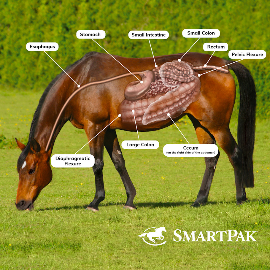
🧠 Think of it as a double layered horse shoe!
1. 🐴 COLIC
Colic = acute abdominal pain.
⚠ Colic is NOT a disease.
It is a: CLINICAL SIGN / SYNDROME indicating abdominal pain
🔍 Causes
Usually associated with disruption of normal gastrointestinal function/motility.
Predisposing factors include:
Diet, Management, Parasites
Pain may result from:
Distension, with accumulation of Gas, Fluid, Ingesta
Obstruction
Mesenteric tension (Torsion, Hernias, Intussusception)
Ischemia (Vascular occlusion / strangulation)
Inflammation (Mucosal inflammation, Irritation, Ulceration)
🩺 Clinical Signs
Horse may: Paw the ground, look toward flanks, Kick abdomen, lie down more than normal, Roll, Sweat
⚠ FALSE COLIC:
Not every horse showing colic behavior has primary GI disease.
Other painful diseases can mimic colic: → Uterine torsion, Rhabdomyolysis (monday morning disease), Severe bladder distension, Laminitis
2. LARGE-INTESTINE OBSTRUCTION
Important sites according to your notes:
⭐ Pelvic flexure
⭐ Left dorsal colon
⭐ Right dorsal colon
🟢 SIMPLE OBSTRUCTION
Blood supply remains normal initially.
Causes:
Impaction, Enteroliths, Trichobezoars, Foreign bodies (Sand, Nylon)
Strictures, Adhesions/fibrous bands, Large-colon displacement
🟡 INCOMPLETE OBSTRUCTION
Some ingesta and gas can still pass.
🔴 COMPLETE OBSTRUCTION
Nothing passes → Ingesta + gas accumulate rapidly → Severe intestinal distension → Abdominal distension → Pressure on Diaphragm and Vena cava → Respiratory + cardiovascular compromise can develop.
🚨 STRANGULATING OBSTRUCTION
Most dangerous. = Obstruction + compromised vascular supply
Causes: Volvulus, Intussusception, Strangulating lipoma
⚙ Pathogenesis
Strangulation → Blood supply compromised → Intestinal ischemia → Necrosis → Loss of mucosal barrier → Bacteria + endotoxins cross intestinal wall → ENDOTOXEMIA
Also: Fluid sequestration → HYPOVOLEMIA
🩺 Clinical Signs
Severe colic, Hypovolemia
Pale → congested/cyanotic abnormal mucous membranes
Tachycardia, Rapid deterioration, Endotoxemia
🚨 EMERGENCY
3. 🌾 PELVIC FLEXURE IMPACTION
One of the classic sites of large-colon impaction.
📍 Anatomy
Pelvic flexure connects: LEFT VENTRAL COLON → PELVIC FLEXURE → LEFT DORSAL COLON
Important: There is a marked reduction in lumen diameter here → Natural site for obstruction.
🔍 Etiology
Dry / inadequately digested feed → Moves through large colon → Reaches narrow pelvic flexure → Fails to pass → IMPACTION (forstoppelse)
🩺 Clinical Signs
Usually relatively mild:
Mild abdominal pain, Slight ↑ heart rate, Usually no severe toxemia initially
🔎 Diagnosis
Clinical signs, Auscultation of intestinal sounds, Rectal examination is also important clinically
💊 Treatment
Oral fluids, IV fluids, Mild analgesics, Laxatives, Withhold feed until resolved
If medical treatment fails: ➡ Surgery
4. 🔄 LEFT DORSAL DISPLACEMENT
Nephrosplenic / Renosplenic Entrapment
The large colon moves dorsally on the LEFT side, and becomes trapped around/between Spleen, Nephrosplenic/renosplenic ligament, Left kidney region
⚙ What Happens?
Colon moves dorsally → Passes over/around spleen → Becomes entrapped in nephrosplenic region → Colon may rotate → Ventral colon becomes positioned dorsally
+Dorsal colon becomes positioned ventrally
The spleen may be:
➡ Displaced ventrally, Congested/engorged

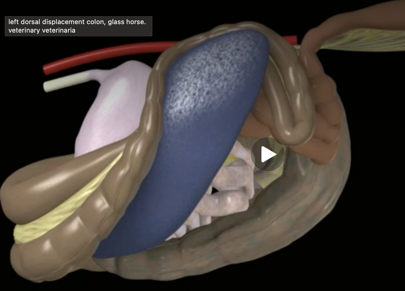
🩺 Clinical Signs
➡ Abdominal pain / colic. Severity varies.
🔎 Diagnosis
Rectal examination ⭐ ➡ Abnormal colon position
USG ➡ Useful for assessing spleen/kidney/colon relationship
Abdominocentesis may yield splenic blood if the displaced spleen is inadvertently entered.
💊 Treatment
Conservative techniques may include:
Feed restriction, Short-term anesthesia
Positioning/rolling ➡ Right lateral recumbency and Roll over the back
If unsuccessful/severe: Surgery
5. 🔄 RIGHT DORSAL DISPLACEMENT
Colon is displaced: Between cecum and right body wall
⚙ Development:
Pelvic flexure impaction → Pelvic flexure moves cranially toward diaphragm → Gas distends sternal + diaphragmatic flexures → Colon migrates caudally along ventral abdominal wall → Colon may twist along long axis around caecocolic attachment → Colon becomes edematous
Usually: Not initially severely ischemic
🩺 Clinical Signs
Abdominal pain, Gas distension
🔎 Diagnosis
Rectal examination
Important clue: ❌ Cannot identify normal: Cecum, Pelvic flexure, because anatomy has been displaced.
💊 Treatment
Medical
Analgesics, Fluids
Surgical
Remove/decompress intestinal contents, Reposition colon
good prognosis and low reoccurrence
6. ACUTE COLITIS
= Acute inflammation of the large intestine/colon.
Etiology:
Salmonellosis, Clostridial infection, Antibiotics, NSAIDs, Contaminated feed
⚙ Pathogenesis ⭐
Inflammation damages mucosa → Intestinal barrier is lost → Luminal bacteria + toxins penetrate mucosa → Systemic inflammation → ENDOTOXEMIA
Also: Severe fluid loss into intestine → Diarrhea and Dehydration
🩺 Clinical Signs
Fever, Inappetence, Colic, Diarrhea
Bloody diarrhea can occur especially with: Salmonella, Clostridial disease
Signs of: Endotoxemia
🔎 Diagnosis
Clinical signs, Dehydration
Blood: ↑ PCV ➡ Hemoconcentration from dehydration
Metabolic acidosis
Leukopenia
💊 Treatment
Aggressive IV fluids, Flunixin meglumine
Intestinal adsorbents/supportive therapy
Antibiotics when specifically indicated, including selected bacterial infections such as clostridial disease
7. 🎈 CECAL TYMPANY
= Gaseous distension of the cecum
🔍 Etiology
Rapid fermentation, Colonic displacement, Obstruction
🩺 Clinical Signs
Distended abdomen, Abdominal pain
🔎 Diagnosis
Auscultation + percussion
→ ⭐ Characteristic: High-pitched “PING”
Also: Rectal examination
💊 Treatment
➡ Remove/decompress gas, for example by catheter/trocarization when clinically indicated AND Correct underlying cause
9⃣ 🪱 EQUINE STRONGYLIDOSIS
LARGE STRONGYLES
Location of adults: Cecum + colon (Proximal ventral colon)
🪱 TWO GROUPS
Genus STRONGYLUS
⭐ Larvae migrate through organs
S. vulgaris ➡ Cranial/anterior mesenteric artery
S. edentatus ➡ Liver
S. equinus ➡ Liver + pancreas
Genus TRIodontophorus
Examples:
T. serratus
T. tenuicollis
T. brevicauda
T. minor
➡ No major extraintestinal larval migration like Strongylus spp.
🪱 LARGE STRONGYLES – STRONGYLUS spp. ⭐⭐⭐
Large strongyles are located as adults in the:
➡ Cecum and colon
They have a direct life cycle:
Eggs in feces → Develop in environment → Infective L3 → Horse ingests L3 while grazing → Larvae penetrate intestinal mucosa→ Larval migration differs between species → Larvae eventually return to large intestine → Adults attach to intestinal mucosa using their large buccal capsule → Suck tissue/blood → many small bleeding ulcers
1⃣ 🩸 STRONGYLUS VULGARIS ➡ Arteritis parasitaria ⭐⭐⭐
🔄 MIGRATION
Infective L3 → Enters large intestine → Remains there for maximum approximately 10 days → Penetrates intestinal mucosa → develops/migrates as L4
→ Migrates through arteries toward → A. mesenterica cranialis, Abdominal aorta and Thoracic aorta → Larvae form small nodules in arterial walls → PARASITIC ARTERITIS → Damage to vessel wall → Aneurysm + thrombus formation → nodules/lesions release larvae → Larvae migrate back toward large intestine → Develop into adults
🚨 WHY DOES S. VULGARIS CAUSE COLIC?
The important lesion is:
Arteritis → Thrombus → Thromboembolism / impaired intestinal blood supply → ISCHEMIA → INFARCTION → NECROSIS of intestinal wall → Severe colic
🩸 ADULT WORMS
Adults return to large intestine, they have a Large buccal capsule, in which attaches strongly to the intestinal mucosa and draws mucosal tissue into the buccal capsule → Bleeding mucosal ulcers
2⃣ 🫀 STRONGYLUS EQUINUS
Larvae penetrate intestine → Migrate across the abdominal cavity → LIVER → Migrate to PANCREAS → Eventually return to large intestine → Develop into adults
Migration period according to Alica: ⭐ 8–9 months
⚙ Consequences
Larval migration through soft tissues causes:
Tissue injury, Inflammation, Nodules, Fibrosis
Even after deworming: Parasite dies/is eliminated, But damaged tissue heals with FIBROTIC TISSUE → Can cause later problems
3⃣ 🫀 STRONGYLUS EDENTATUS ⭐⭐⭐
⚠ You MUST explain the migration and how it can lead to PERITONITIS.
🔄 MIGRATION
Larvae penetrate intestinal wall → Migrate to LIVER → Form small nodules in liver → Migrate from liver toward FLANK / abdominal wall region → Larvae migrate through abdominal tissues/peritoneal region → Cause tissue damage + inflammation → PERITONITIS
Migration period: 9–11 months
🧠 THREE SPECIES – DO NOT MIX THEM UP ⭐⭐⭐
Species | Main migration | Major consequence | Time from lecture |
|---|---|---|---|
S. vulgaris | 🩸 Cranial mesenteric artery → aorta | Arteritis parasitaria → thrombus → ischemia/infarction | L3 initially SI/LI mucosa max ~10 d before migration |
S. equinus | 🫀 Liver → pancreas | Tissue damage + fibrosis | 8–9 months |
S. edentatus | 🫀 Liver → flank/peritoneal region | ⭐ Severe peritonitis + hepatic fibrosis | 9–11 months |
🩺 Clinical Signs – Strongylidosis
Anemia, Colic, Poor condition, Diarrhea, Fever, Anorexia, Weight loss
🔎 Diagnosis
Coprology, Necropsy
⭐ Larval culture for species differentiation. Strongyle eggs look very similar on routine fecal examination.
💊 Treatment
Fenbendazole (kills migrating larva, small strongyle is resistant)
Moxidectin (kills both adults and encysted larva)
🔟 🪱 SMALL STRONGYLES - CYATHOSTOMOSIS
Also called: Cyathostomins
Genus Trichonema
Cyathostomes
More than 50 species
Location of adults:
➡ Cecum + colon
Small strongyles are very common in horses.
⭐ BIG DIFFERENCE FROM LARGE STRONGYLESLARGE STRONGYLES
➡ Larvae MIGRATE to organs/vessels
SMALL STRONGYLES
➡ Larvae DO NOT perform major extraintestinal migration
➡ Instead, they ENCYST in the mucosa of the cecum and colon
🧠 LARGE = LEAVE, SMALL = STAY
🔄 LIFE CYCLE – CYATHOSTOMINS ⭐⭐⭐
Direct life cycle
➡ No intermediate host
Eggs shed in feces → Develop in environment → Infective L3 → Horse ingests L3 while grazing → L3 reach cecum + colon → Invade the large-intestinal mucosa → Form encysted/nodular stages → Develop toward L4 → Larvae emerge from mucosa back into intestinal lumen → Develop into adult worms → Adults produce eggs
→ Eggs passed in feces 🔄
😴 HYPOBIOSIS ⭐
The encysted larvae can:
➡ ARREST THEIR DEVELOPMENT
They remain dormant in the intestinal mucosa.
➡ They may remain encysted for up to ~3 years
⚙ PATHOGENESIS
Small strongyles are generally considered less individually pathogenic than the large Strongylus spp., but:
⚠ Heavy infections can cause severe disease.
1⃣ ENCYSTMENT
Larvae invade intestinal mucosa → Form nodules → Damage mucosa →Cause:
Inflammatory reaction, Edema, Mucosal bleeding, Thickening/damage of intestinal wall
2⃣ MASS EMERGENCE ⭐⭐⭐
Large numbers of encysted larvae can reactivate and emerge from the mucosa at approximately the same time.
Thousands of larvae emerge → Large areas of mucosa are damaged → Severe inflammation → Loss of intestinal barrier/function → Protein/fluid loss → Severe watery diarrhea
This clinical syndrome is:
⭐ LARVAL CYATHOSTOMINOSIS
So remember:
The dangerous part is not only having encysted larvae — it is especially when MANY emerge simultaneously.
🩺 CLINICAL SIGNS
⭐ Weight loss, Rough/dull coat
⭐ Watery diarrhea
Colic, Fever, Anemia, Poor condition
With severe larval cyathostominosis:
➡ Severe enteritis/colitis
➡ Protein loss may occur
➡ Rapid deterioration can occur
🔎 DIAGNOSIS
Coprology / fecal egg count
Necropsy
Larval culture for species identification
⚠ IMPORTANT LIMITATION
Encysted larvae may not be detected by fecal egg counts.
❗ Low fecal egg count does NOT necessarily mean there are few encysted cyathostomin larvae.
💊 TREATMENT
⭐ MOXIDECTIN
Important because it has activity against:
Adult cyathostomins
Larval stages
⭐ Encysted larvae
⭐ LARGE vs SMALL STRONGYLES
LARGE STRONGYLES | SMALL STRONGYLES | |
|---|---|---|
Adults | Cecum + colon | Cecum + colon |
Infective stage | L3 | L3 |
Intermediate host | None | None |
Life cycle | Direct | Direct |
Larval behavior | ⭐ Migrate outside intestine | ⭐ Encyst in intestinal mucosa |
Main danger | Organ/vessel damage | Mucosal damage |
Important syndrome | S. vulgaris arteritis | Larval cyathostominosis |
Hypobiosis | Not key feature | ⭐ Very important |
Severe disease mechanism | Migration | ⭐ Mass larval emergence |
Key treatment in notes | Moxidectin | ⭐ Moxidectin |
1⃣1⃣ 🪱 PINWORMS – OXYUROSIS
Etiology: ⭐ Oxyuris equi
Type:
➡ Nematode
Location:
➡ Cecum + colon
🔄 LIFE CYCLE
Adult females in colon → Pregnant female migrates toward anus → Deposits eggs in sticky fluid around anus → Severe irritation/pruritus → Horse rubs tail/perineum against objects → Eggs enter environment → Develop to eggs with infective stage L3 → Horse ingests infective eggs → small intestine → large intestine → L4 with large buccal capsule ingests on wall → destroy wall → L5 → adult in colon
🩺 Clinical Signs ⭐
Main sign:
🍑 INTENSE PERIANAL ITCHING
Horse rubs rear end → Hair breaks/falls out → Focal alopecia around tail/perianal area → Skin trauma → Possible secondary infection
🔎 Diagnosis
⭐ Perianal tape/swab test
Why not rely on ordinary fecal exam?
Because:
➡ Female deposits eggs around anus, not primarily into feces.
💊 Treatment
1⃣ Anthelmintic:
Moxidectin
Ivermectin orally
2⃣ Very important:
➡ Clean eggs from perianal skin/environment
🧠 WHOLE QUESTION IN ONE MAP
              LARGE INTESTINE
                    │
       ┌────────────┴────────────┐
       │                         │
    DISEASES                 PARASITES
       │                         │
       ├─ Colic                  ├─ LARGE STRONGYLES
       ├─ Obstruction            │      ↓
       ├─ Pelvic impaction       │    MIGRATE
       ├─ Left displacement      │      ↓
       ├─ Right displacement     │  S. vulgaris → artery
       ├─ Acute colitis          │  S. edentatus → liver
       └─ Cecal tympany          │  S. equinus → liver/pancreas
                                 │
                                 ├─ SMALL STRONGYLES
                                 │      ↓
                                 │   ENCYST
                                 │      ↓
                                 │  Cyathostomosis
                                 │
                                 └─ OXYURIS
                                        ↓
PERIANAL ITCHPrinciples of Treatment and Prevention of Parasitic Infections in Horses
MAIN PRINCIPLE
Treatment and prevention of equine parasitic infections have TWO main components:
1⃣ MANAGEMENT
➡ Decrease the number of infective parasite stages in the environment
2⃣ THERAPEUTIC CONTROL
➡ Deworming with anthelmintics at appropriate intervals / according to parasite burden
⭐ GOAL = BREAK THE PARASITE LIFE CYCLE
Horse has parasites → Eggs/larvae contaminate environment → Develop into infective stages → Horse becomes reinfected
⬇
🔄 Cycle continues
Therefore:
MANAGEMENT + ANTHELMINTICS → Reduce environmental contamination → Reduce reinfection → BREAK LIFE CYCLE
⚠ ANTHELMINTIC RESISTANCE
Traditionally, parasite control relied heavily on repeated anthelmintic treatment.
Anthelmintics → Kill intestinal worms → ↓ Egg production → ↓ Environmental contamination
BUT:
Frequent/inappropriate deworming → Selection pressure → Resistant parasites survive → Reproduce
⬇
💊 ANTHELMINTIC RESISTANCE
Therefore:
❌ Deworming alone is NOT sustainable
We need:
⭐ MANAGEMENT + MONITORING + TARGETED/APPROPRIATE TREATMENT
1⃣ 🌱 MANAGEMENT
1.💩 MANURE REMOVAL ⭐
Remove manure regularly from:
Pasture, Paddocks, Stables, Every 24–72 hours!
Strongyle eggs are passed in feces → Eggs hatch → Larvae develop → Infective L3 develop in environment within approximately 5–7 days
Therefore → 💩 Remove feces BEFORE larvae become infective
2.💧 DECREASE HUMIDITY
Many free-living parasite stages survive/develop better in moist conditions
3.♨ COMPOST MANURE ⭐
Many parasite eggs have resistant shells and survive adverse environmental conditions
Proper composting → Heat generated → Eggs/larvae devitalized → ↓ Environmental contamination
4.🚜 PASTURE MANAGEMENT
Can include:
Deep plowing, Reseeding, Manure removal, Pasture rotation
5.🍽 FEEDING MANAGEMENT
❌ Do not feed directly from contaminated ground. Instead: Use elevated feeders
6.🆕 QUARANTINE NEW HORSES ⭐
New arrivals may introduce resistant or unfamiliar parasite populations.
Therefore:
New horse → QUARANTINE → Fecal examination → Appropriate parasite treatment if indicated → Only then introduce to herd/pasture
→ Larvicidal treatment before turnout of new arrivals.
7.🌱 PASTURE ROTATION
Move horses from Contaminated pasture → Pasture with lower parasite contamination
➡ Deworming appropriately before moving animals!
Most important: Cleanest pasture → foals + young horses
Young horses:
➡ Less developed immunity, and more susceptible to important parasites such as Parascaris
8.🪰 INSECT CONTROL
Important because flies act as ➡ Intermediate hosts/vectors for some parasites.
Habronema:🪰 Muscid flies transmit L3
Gasterophilus: Adult bot flies deposit eggs on horse
Therefore:
➡ Implement fly-control programs.
FOALS
Foals are especially susceptible to:
Parascaris, Strongyloides westeri, Other nematodes
Tapeworms during the first year
Recommendations:
➡ Regular deworming approximately every 60 days/2months
with drugs:
➡ Safe and effective against ascarids
Protocols during first year should account for:
➡ Nematodes and Cestodes
⚠ For your exam, remember this as the course protocol; modern parasite-control programs may adjust treatment based on local resistance and monitoring.
2⃣ 🔬 MONITORING ENDOPARASITES
Why monitor?
➡ Identify parasite burden
➡ Decide whether treatment is needed
➡ Assess effectiveness
➡ Reduce unnecessary drug use
➡ Slow resistance
💩 BASIC FECAL FLOTATION
Detects: Parasite eggs
Problem: ⚠ Some parasites shed eggs intermittently
Therefore: A negative fecal sample does not always exclude infection!
Also, encysted/larval stages may not produce eggs.
🔬 DIRECT MICROSCOPY
Feces can be examined directly for: Eggs/larvae/other parasite stages.
🔢 McMASTER METHOD ⭐
A quantitative fecal egg-count technique.
→ Eggs per gram (EPG)
Therefore helps estimate the intensity of egg shedding/infection
If 200 EPG → start deworming
If 600 EPG → HEAVY infestation
🧫 LARVAL CULTURE
Useful especially because: Strongyle eggs can look very similar
Culture larvae → Examine larvae → Help differentiate strongyle groups/species
☠ POST-MORTEM EXAMINATION
Can identify:
Adult worms, Larvae, tissue migration, Nodules, Intestinal lesions
3⃣ 💊 ANTHELMINTICS
Four major chemical families of equine anthelmintics:
Chemical family | Important drug/example | Main association |
|---|---|---|
Macrocyclic lactones | Ivermectin, moxidectin | Strongyles, bots, several nematodes |
Acylated quinoline pyrazines | Praziquantel | Tapeworms/Anoplocephala |
Tetrahydropyrimidines | Pyrantel | Luminal nematodes ± tapeworm activity depending dose |
Benzimidazoles | Fenbendazole, mebendazole, oxibendazole, thiabendazole | Broad nematode activity |
A. 💊 MACROCYCLIC LACTONES ⭐⭐⭐
Ivermectin, Moxidectin
Very important broad-spectrum equine anthelmintics.
⭐ MOXIDECTIN:
Important against:
Adult AND Larval strongyles
⭐ Encysted cyathostomin larvae
Migrating large strongyle larvae
⭐ IVERMECTIN:
Important against:
Large strongyles
Luminal nematodes
Strongyloides
Habronema
⭐ Gasterophilus larvae
Some ectoparasites such as mites
B. 💊 PRAZIQUANTEL (Acylated quinoline pyrazines)⭐⭐⭐
Main thing to remember:
🪱 PRAZIQUANTEL = TAPEWORMS
Especially:
Anoplocephala perfoliata, A. magna
Paranoplocephala mamillana
Often available combined with Ivermectin or Moxidectin
Why important?
If horse repeatedly receives only nematocidal drugs without effective cestocidal treatment:
➡ Tapeworm infection can persist.
C. 💊 PYRANTEL (Tetrahydropyrimidine)
Effective mainly against luminal stages including:
Adult large strongyles
Small strongyles – resistance can occur
Ascarids
Pinworms
Also: Anoplocephala perfoliata at appropriate tapeworm dosing.
D. 💊 BENZIMIDAZOLES – BZs
Examples:
Fenbendazole, Mebendazole, Oxibendazole, Thiabendazole
Broad-spectrum activity.
Can target:
Large strongyles
Cyathostomins ⚠ resistance common
Parascaris
Oxyuris
Other:
💊 PIPERAZINE
Parascaris, Pinworms, Some small strongyles
❌ Poor/not effective for large strongyles.
4⃣ 🪰 PYRETHROIDS
External / environmental parasite control
Examples:
Cypermethrin
Permethrin
Pyrethrum
Used particularly for:
➡ Insect/ectoparasite control: lice, ticks, insects (dipteran, fleas, lice)
⚙ Mechanism
Synthetic pyrethroids such as cypermethrin/permethrin: ➡ Affect/modulate sodium channels in parasite/insect nerves → Prolonged neuronal excitation → Paralysis/death
Pyrethrum: → Contact insecticidal action
⭐ WHICH DRUG FOR WHICH PARASITE? ⭐⭐⭐
Parasite | Main treatment |
|---|---|
🪱 Small strongyles / cyathostomins | ⭐ Moxidectin; fenbendazole depending resistance |
🩸 Large/migratory strongyles | Ivermectin / moxidectin⭐ |
🪱 Parascaris | Depends strongly on resistance; BZ/pyrantel/ML according to susceptibility |
🪱 Tapeworm – Anoplocephala | ⭐ Praziquantel |
🪰 Gasterophilus / bots | ⭐ Ivermectin / ML |
🪱 Strongyloides westeri | Ivermectin or fenbendazole |
🍑 Oxyuris equi | ML or BZ + environmental/perianal cleaning |
🪰 Habronema | Ivermectin + local lesion/fly management |
1⃣ SMALL STRONGYLES
Cyathostomins ➡ Moxidectin or Fenbendazole where effective
Why moxidectin important?
⭐ Activity against encysted larvae
2⃣ LARGE STRONGYLES
Treatment:
➡ Ivermectin
➡ Moxidectin
Important because:
➡ Activity against adults + important migrating larval stages.
3⃣ 🪱 PARASCARIS
Especially important in:
➡ Foals/young horses
⚠ Anthelmintic resistance is a major consideration.
Your notes mention resistance to:
Macrocyclic lactones
Benzimidazoles
Pyrantel
Therefore:
➡ Drug choice should depend on known/local efficacy
Possible classes:
BZs: tiabendazol, mebendazol, fenbendazol
Pyrantel
MLs where still effective
⚠ Heavy Parascaris infections in foals need particular care because rapid killing of a very large worm burden can contribute to intestinal obstruction.
4⃣ 🪱 TAPEWORMS
Anoplocephala perfoliata
A. magna
Paranoplocephala mamillana
⭐ Drug of choice:
PRAZIQUANTEL
Often combined with:
➡ Ivermectin/moxidectin
5⃣ 🪰 BOT FLIES
Gasterophilus spp.
Larval stages are susceptible to:
➡ Macrocyclic lactones
Especially:
⭐ Ivermectin
Yellowish eggs may be visible with naked eye on horse hair.
6⃣ 🪱 STRONGYLOIDES WESTERI
Threadworm
➡ Ivermectin or Fenbendazole
Fenbendazole:
➡ 50 mg/kg
compared with standard:
➡ 7.5 mg/kg
⭐ those numbers are lecturer-specific.
7⃣ 🍑 OXYURIS EQUI
Pinworm
Treatment:
Macrocyclic lactones
Benzimidazoles
Pyrantel has variable efficacy according to notes
BUT:
Remember management too:
Female lays sticky eggs around anus → Environment/perineum contaminated → Reinfection
Therefore:
⭐ Clean perianal area + environment
⚠ ANTHELMINTIC RESISTANCE ⭐⭐⭐
This is a major part of modern parasite control.
Repeated unnecessary treatment → Susceptible worms die → Resistant worms survive → Resistant worms reproduce → Population becomes increasingly resistant
Therefore:
❌ Do not rely only on frequent blind deworming.
Instead:
➡ Fecal monitoring
➡ Appropriate drug choice
➡ Treat strategically/appropriately
➡ Environmental management
➡ Avoid unnecessary treatment
➡ Monitor treatment efficacy
🧪 HOW CAN WE CHECK IF A DEWORMER WORKS?
Conceptually:
Fecal egg count before treatment → Deworm → Repeat fecal egg count after treatment → Compare egg counts
If expected reduction does not occur:
➡ Suspect anthelmintic resistance / treatment failure
This is the principle of the:
⭐ Fecal Egg Count Reduction Test – FECRT
🧠 WHOLE QUESTION IN ONE MAP
         PARASITE CONTROL
                │
      ┌─────────┴─────────┐
      │                   │
 MANAGEMENT           THERAPY
      │                   │
 manure removal       anthelmintics
 composting               │
 pasture rotation         ├─ ML
 clean feeding            ├─ BZ
 quarantine               ├─ pyrantel
 fly control              └─ praziquantel
      │
      └─────────┬─────────┘
                │
           MONITORING
                │
         FECAL EGG COUNT
                │
       TARGETED TREATMENT
                │
      ↓ DRUG RESISTANCE
⭐ EXAM ESSENTIALS
If you need to answer this quickly:
“The goal of parasite control is to break the parasite life cycle by combining environmental management with appropriate anthelmintic treatment. Management includes frequent manure removal, composting, pasture rotation, reducing humidity, avoiding feeding directly from contaminated ground, quarantine and fecal examination of new horses, use of the cleanest pasture for young horses, and insect control. Parasite burden is monitored by fecal examination, especially quantitative methods such as McMaster. Anthelmintic treatment should be selected according to the parasite and resistance situation rather than relying only on frequent routine treatment. Important drugs include ivermectin and moxidectin for strongyles, moxidectin especially for encysted cyathostomins, praziquantel for tapeworms, and ivermectin for Gasterophilus and Habronema. Anthelmintic resistance is an important problem, so management, monitoring and appropriate treatment must be combined.”
🧠 FINAL MEMORY
M-M-M
🌱 MANAGEMENT
Reduce parasites in environment
🔬 MONITORING
Know what/how much is present
💊 MEDICATION
Treat appropriately
⬇
🔨 BREAK THE LIFE CYCLE
And remember the three strongest drug associations:
PRAZIQUANTEL → TAPEWORM 🪱
MOXIDECTIN → ENCYSTED CYATHOSTOMINS 😴
IVERMECTIN → BOTS 🪰
PERITONITIS
Peritonitis = inflammation of the peritoneum → tissue lining the abdominal cavity and organs.
Normal peritoneum:
Secretes fluid → lubricates abdominal cavity
Minimizes adhesion formation
Has antibacterial properties
ETIOLOGY
May occur primary or secondary.
🦠 Infectious / septic:
Surgical complications, Intestinal perforation, Uterine perforation, Metritis, Post-castration, Enteritis, Septicemia
⚪ Non-septic:
Ruptured bladder/ureter / kidney, Bile, gastric or pancreatic juice, Foreign body, Neoplasia, Uroliths, Hepatitis, Gastric rupture
🪱 Parasitic:
Larval migration
🩸 Traumatic:
Uterine artery hemorrhage, Penetrating abdominal wound, Blunt abdominal wound, Ruptured diaphragm
PATHOGENESIS
1⃣ Fibrin + adhesions
Peritonitis → ↓ fibrinolytic activity → precipitation/deposition of fibrin → adhesions + pain
2⃣ Fluid loss + shock
Inflammatory response → activation of leukocytes + immunoglobulins
→ proteins + fluid + electrolytes move from plasma into abdominal cavity → hypovolemia + hypoproteinemia → cardiovascular collapse / shock
🧠 Remember:
FIBRIN → ADHESIONS
FLUID/PROTEIN LOSS → HYPOVOLEMIA → SHOCK
CLINICAL SIGNS
🚨 Peracute peritonitis
E.g. gastric rupture, May be found dead, Profound toxemia, Rapid circulatory failure, Death within 4–12 h
🔥 Acute peritonitis
Tachycardia, Tachypnoea
Congested → cyanotic mucous membranes
Cold extremities, Dehydration, Depression, Sweating, Immobility, Sensitivity to pressure / abdominal pain
⏳ Subacute / chronic peritonitis
Dullness, ↓ Appetite, Progressive weight loss
Abdominal pain → low-grade, intermittent or absent
Intermittent fever, Variable bowel sounds, Chronic diarrhea in some cases
DIAGNOSIS
⭐ Peritonitis is characterized by:
↑ amount of peritoneal fluid
↑ cellular content
↑ protein content
💉 Abdominocentesis → examine peritoneal fluid
Cytology of peritoneal fluid = definitive diagnosis
Biochemistry:
↑ Lactate
↑ WBC
↑ Albumin/globulin ratio
Appearance of peritoneal fluid:
Turbid + off-white → haemoperitoneum / intestinal infarction
Turbid + brown/green → contamination with intestinal contents
Other diagnostics:
USG, X-ray, Laparoscopy
🔹 TREATMENT
1. Treat the underlying cause
2. Medical treatment
💧 IV fluid therapy
💊 NSAIDs
💉 Antibiotics → Penicillin = first choice
3. Correct
Cardiovascular shock
Endotoxic shock
4. Fibrinolytic medications
Heparin
Fragmented heparin
→ ↓ fibrin/adhesion formation
5. Abdominal drainage + lavage
Drain peritoneal fluid
Lavage with sterile solution
Via cannula or Foley catheter
🪱 PARASITIC PERITONITIS ⭐
1⃣ Strongylus edentatus
Larval migration → can cause peritonitis
2⃣ Setariosis
Caused by:
Setaria equina
Setaria labiato-papillosa
Setaria cervi
Transmission: 🦟 Mosquitoes
Pathogenesis:
Mosquito infection
→ adult worms 10–15 cm long in abdominal cavity
→ produce microfilariae in blood
→ larvae may migrate to eyes + other organs
Clinical consequences:
Peritonitis, CNS signs, Visual impairment
Diagnosis:
🔬 Microfilarial larvae in blood
ELISA → antibodies
Treatment:
💉 Ivermectin
🧠 EXAM SEQUENCE
PERITONITIS = inflammation of peritoneum
E:
Septic / non-septic / parasitic / traumatic
⬇
P:
↓ fibrinolysis → fibrin → adhesions + pain
Fluid + protein + electrolytes leave plasma → hypovolemia + hypoproteinemia → shock
⬇
CS:
Peracute: toxemia → circulatory failure → death
Acute: tachycardia + tachypnoea + dehydration + abdominal pain
Chronic: dullness + ↓ appetite + weight loss + intermittent pain/fever
⬇
Dx:
⭐ Abdominocentesis + cytology
↑ fluid + ↑ cells + ↑ protein
⬇
Tx:
⭐ Underlying cause + IV fluids + NSAIDs + ATB + shock treatment + heparin ± abdominal drainage/lavage
Parasitic peritonitis = Strongylus edentatus migration + Setaria.
SETARIA:
🦟 Mosquito → abdominal worms → microfilariae in blood → eyes/CNS/peritoneum → Dx blood/ELISA → Tx ivermectin
Asthma
EQUINE ASTHMA / RECURRENT AIRWAY OBSTRUCTION (RAO)
= common condition characterized by laboured breathing due to obstruction of the bronchioles.
Pulmonary hypersensitivity associated with stabling + feeding hay
Usually reversible with environmental dust control
🧠 Think: Dust/hay → hypersensitivity → small airway obstruction
ETIOLOGY
Hypersensitivity to inhaled dust → inflammation of small airways.
Important inhaled triggers:
Dust, Mould spores, Fungi, Actinomycetes, Gases
⭐ Associated with stabling and hay feeding
⭐ Tends to increase with age → commonly >7 years
PATHOGENESIS ⭐
Allergen exposure
⬇
Hypersensitivity + airway inflammation
⬇
3 important effects:
1. Bronchospasm → smooth muscle contraction of bronchi
2. Mucus hypersecretion
3. Inflammatory bronchitis
⬇
Reduced airway lumen
⬇
↓ Expiratory airflow → laboured expiration / dyspnoea
Chronic cases:
Persistent inflammation → structural changes:
Metaplasia, Hyperplasia, Emphysema
🧠 Remember the 3:
SPASM + MUCUS + INFLAMMATION → narrowed airway → ↓ expiration
CLINICAL SIGNS
Severity ranges from subclinical → mild → severe.
🟢 Subclinical
Mainly performance horses
Exercise intolerance
🟡 Mild
Occasional/sporadic cough
During feeding and During exercise
Slight nasal discharge
🔴 Severe
Chronic cough, Dyspnoea, Bilateral nasal discharge, ↑ Respiratory rate
Exercise intolerance, Increased expiratory effort
⭐ Classic severe sign = double expiratory effort
DIAGNOSIS
1⃣ History + clinical signs ⭐
Typical:
Horse >7 years, Chronic cough >3 months, Double expiratory effort, Dilated nostrils
2⃣ Cough reflex + auscultation
↑ Susceptibility to cough reflex
Crackles, Wheezing
3⃣ Endoscopy
May show:
Mucopurulent discharge in trachea + bronchi
Congestion of airway mucosa
Thickening of airway mucosa
Severe cases:
Collapse of trachea + bronchi during expiration/coughing
4⃣ Additional tests
Tracheal aspirate
BAL / cytology
X-ray
5⃣ Inhalation challenge test
Expose horse to mouldy hay/straw
→ worsening of clinical signs
and/or
→ changes in tracheal aspirate/BAL cytology
6⃣ Response to treatment
Improvement following:
Corticosteroids
Bronchodilators
⚠ Corticosteroids when respiratory infection is unlikely.
TREATMENT⭐⭐⭐
1⃣ ENVIRONMENTAL MANAGEMENT — MOST IMPORTANT
Reduce allergen/dust exposure:
“Dust-free” environment
Pasture + fresh grass
Wet hay before feeding
Avoid straw bedding
Good ventilation
🧠 Without environmental control → drugs alone are not enough.
2⃣ Corticosteroids
→ ↓ pulmonary inflammation
Systemic: Dexamethasone
Inhaled: Beclomethasone
3⃣ Bronchodilators
→ rapid relief of bronchospasm
Rapid, relatively short-lived action:
Albuterol, Clenbuterol
4⃣ Mucolytics
→ ↓ viscosity of respiratory secretions
Acetylcysteine
5⃣ Mucokinetic agents
→ aid clearance of respiratory secretions
PROGNOSIS
Changes associated with RAO are mostly reversible with appropriate environmental management and treatment.
⚠ Once structural damage / emphysema develops → changes may become irreversible!
➡ Generally favourable prognosis if managed before permanent structural damage.
CIZEK'S LIST — RESPIRATORY PATHOGENS
🦠 VIRAL
Equine rhinopneumonitis — EHV-1
Equine herpesvirus 2 — EHV-2
Equine herpesvirus 5 — EHV-5
→ Interstitial pneumonia
Adenovirus
Equine influenza virus
African horse sickness virus
Morbillivirus
Reovirus
Equine rhinovirus
🦠 BACTERIAL
Clostridium tetani → lockjaw / tetanus
Streptococcus equi → strangles
Burkholderia mallei → glanders
Rhodococcus equi → pneumonia in foals ⭐
For Rhodococcus equi, I would say pyogranulomatous/abscessing bronchopneumonia in foals rather than simply “interstitial pneumonia.”
🧠 EXAM SEQUENCE
EQUINE ASTHMA / RAO
E:
Dust/hay/mould hypersensitivity, especially stabled horses >7 years
⬇
P:
Allergen exposure
→ bronchospasm + mucus hypersecretion + inflammatory bronchitis
→ ↓ airway lumen
→ ↓ expiratory airflow
Chronic → metaplasia + hyperplasia + emphysema
⬇
CS:
Exercise intolerance → cough → nasal discharge → dyspnoea + double expiratory effort
⬇
Dx:
History + CS → auscultation → endoscopy → tracheal aspirate/BAL ± X-ray ± challenge test
⬇
Tx:
⭐ ENVIRONMENT FIRST
Dust control + pasture/wet hay + ventilation
corticosteroids
bronchodilators
mucolytics/mucokinetics
⬇
Prognosis:
Favourable and mostly reversible → until structural damage/emphysema occurs.
Pleuropneumonia and Verminous pneumonia
A. PLEUROPNEUMONIA
Pleuropneumonia = infection/inflammation of the lung AND pleural space.
⭐ You know the pleura is involved when there is fluid around the lungs → PLEURAL EFFUSION.
Pleural effusion = excess fluid around the lungs.
ETIOLOGY
Usually secondary to factors that lower respiratory defence → secondary bacterial infection.
1⃣ Stress / long-distance transport ⭐⭐⭐
“Transit fever”
Long-distance travel
→ prolonged head elevation → prevents normal drainage of respiratory secretions → secretions + bacteria descend into lower airways → pleuropneumonia⭐ Very common cause!
2⃣ Viral respiratory disease
Predisposes to secondary bacterial infection:
EHV-1, EHV-2, EHV-4, EHV-5
Equine influenza
3⃣ Bacterial infection
Streptococcus equi → strangles
Rhodococcus equi
Burkholderia mallei → glanders
Pseudomonas
Bordetella bronchiseptica
4⃣ Immunosuppression
Corticosteroid therapy
Cushing's disease
5⃣ Aspiration / inhalation pneumonia
Due to:
Dysphagia, Esophageal obstruction, Pharyngeal paralysis, Choke, Inhaled foreign bodies
6⃣ Trauma
Direct thoracic trauma, Penetrating chest wounds
7⃣ Pulmonary emboli/infarction
E.g. secondary to: jugular phlebitis + thrombosis
PATHOGENESIS ⭐⭐⭐
1⃣ EXUDATIVE PHASE
Initial bacterial colonization of lung tissue
→ inflammatory response
→ protein-rich inflammatory exudate
⬇
2⃣ FIBRINOPURULENT PHASE
Bacterial infection progresses
→ large amount of septic fibrinopurulent exudate
→ accumulates in pleural cavity
⬇
3⃣ ORGANIZATION PHASE
Fibrin becomes organized
→ fibrous tissue
→ thick “pleural peel”
CLINICAL SIGNS
Fever, Reluctance to move
Abduction of elbows, Tachypnoea
Dyspnoea → inspiratory + expiratory
Cough, Nasal discharge
Sternal edema, Stilted gait, Reluctance to lie down
🧠 Think: A very painful horse that doesn't want to move or lie down + respiratory disease.
DIAGNOSIS
1⃣ History + clinical signs
⭐ Ask: Has the horse recently travelled a long distance?
2⃣ Thoracic auscultation ⭐
Ventral lung field:
→ muffled / absent sounds due to pleural fluid
Dorsal lung field:
→ abnormal lung sounds
Heart sounds audible over larger-than-normal area
→ suggests pleural effusion
🧠 Fluid sinks → ventral sounds disappear.
3⃣ Percussion
Reduced/dull ventrally
→ pleural effusion
4⃣ X-ray
Fluid line → pleural effusion
Consolidated lung
5⃣ Ultrasound ⭐
Shows:
Pleural effusion, Consolidated lung, Abscesses, Fibrin, Adhesions
⭐ USG gives more detail than X-ray.
6⃣ Blood
Early/severe bacterial sepsis or toxemia:
Leukopenia, Neutropenia + left shift, Hemoconcentration, Azotemia
Later stage:
Leukocytosis, Mature neutrophilia, Hyperfibrinogenemia, Hyperglobulinemia, Hypoalbuminemia
7⃣ Thoracocentesis ⭐
Collect pleural fluid for:
Cytology, Bacterial culture
⚠ Include anaerobic culture
8⃣ Respiratory sampling
TTW = transtracheal wash
BAL = bronchoalveolar lavage
🔹 TREATMENT
💉 Systemic antibiotics → treat bacterial infection
💊 NSAIDs → analgesic + anti-inflammatory
Supportive care: Pleural drainage + lavage ⭐ → remove infected pleural fluid. If large quantities of thick, organized fibrinopurulent material cannot be adequately drained → Thoracotomy
🪱 B. VERMINOUS PNEUMONIA
Important parasites:
⭐ 1. Dictyocaulus arnfieldi
⭐ 2. Migrating larvae of Parascaris equorum
⭐ 3. Echinococcus equinus → equine cystic echinococcosis
🪱 1. DICTYOCAULUS ARNFIELDI ⭐⭐
Nematode / lungworm = Dictyocaulus arnfieldi
Predilection site:
→ Bronchi + bronchioles
Adults develop/live in the lungs.
Life cycle — DIRECT ⭐
Infective larvae are ingested during grazing → Migrate from intestine → Reach lungs → Adults develop in bronchi/bronchioles → Eggs + larvae carried up to trachea
→ Coughed up + swallowed → Released with feces onto pasture → Infect new host

🔹 Pathogenesis
Larvae/adults irritate respiratory mucosa
→ inflammation of bronchi, chronic bronchitis, pneumonia
Severe cases:
→ airway damage/obstruction
→ compensatory emphysema
🔹 Clinical signs ⭐
Severe paroxysmal/productive cough
Dyspnoea
Mucopurulent nasal discharge, Fever
🔹 Diagnosis
Larvoscopy / detection of larvae in feces
20 larvae / 1 g feces → high-intensity infection
🔹 Treatment ⭐
Ivermectin, Moxidectin, Macrocyclic lactones
Pasture management, Change pasture during season, Reduce reinfection.
🪱 2. PARASCARIS EQUORUM
Nematode = Parascaris equorum
Large worm → about 40 cm, adults located in small intestine

Very common in older foals
⭐ Verminous pneumonia occurs because of MIGRATING LARVAE.
Life cycle — DIRECT ⭐
Eggs passed in feces → Develop to infective stage in environment → Eggs ingested → Larvae penetrate intestinal wall → Liver
→ Lungs → Trachea → Irritation → horse coughs → Larvae swallowed → Return to small intestine → Become adults
Pathogenesis
Migrating larvae cause:
Focal eosinophilic inflammation, Bleeding, Calcified subpleural nodules, Pneumonia
In liver:
“Milk spots” = scar tissue
Intestinal infection:
Chronic intestinal inflammation, Colic
Release of toxins with affinity for the nervous system
🔹 Clinical signs
Cough, Fever, Inappetence, Pneumonia, ± Colic / GI signs
🔹 Diagnosis
Coprology / fecal examination

🔹 Treatment
⚠ Parascaris equorum has important anthelmintic resistance.
Resistance can occur to:
Benzimidazoles, Ivermectin / macrocyclic lactones, Pyrantel
→ treatment choice should account for local resistance and efficacy.
🪱 3. ECHINOCOCCUS EQUINUS — EQUINE CYSTIC ECHINOCOCCOSIS
🔹 Etiology
Cestode / tapeworm
Echinococcus equinus
→ causes equine cystic echinococcosis
→ produces fertile hydatid cysts

🔹 Hosts ⭐
Definitive hosts (DH):
Dog, Red fox, Arctic fox, cats
Intermediate hosts (IH):
⭐ Horse / donkey (also sheep, goat, cattle, pig)
🧠 DOG = definitive host → HORSE = intermediate host
🔹 Life cycle + Pathogenesis
Horse ingests Echinococcus eggs from contaminated environment/grass → Larvae migrate into tissues → Formation of hydatid cysts → Cysts + surrounding inflammation
→ If pulmonary involvement is extensive → respiratory impairment / pneumonia-like signs
🔹 Clinical signs
Usually related to the number and size of cysts.
If many cysts:
Shortness of breath, Cough, Respiratory disease
🔹 Diagnosis
Imaging, including X-ray when pulmonary cysts are suspected
🔹 Treatment / prognosis
Single cyst → may cause little clinical problem
Many cysts → no practical treatment for the cyst burden → poor prognosis / euthanasia may be considered
🧠 VERMINOUS PNEUMONIA — EXAM MEMORY
3 PARASITES:
1⃣ Dictyocaulus arnfieldi ⭐⭐⭐
→ LUNGWORM
→ bronchi/bronchioles
→ chronic bronchitis
→ severe paroxysmal cough + dyspnoea
→ severe = compensatory emphysema
→ Dx larvae in feces
→ Tx IVERMECTIN
2⃣ Parascaris equorum ⭐⭐
→ mainly foals
→ larvae migrate INTESTINE → LIVER → LUNGS → TRACHEA → swallowed → INTESTINE
→ coughing + pneumonia
→ Dx coprology
3⃣ Echinococcus equinus ⭐
→ cystic echinococcosis / hydatid cysts
→ dog = DH, horse = IH
→ multiple pulmonary cysts → cough + dyspnoea
→ imaging/X-ray.
Exercise intolerance associated with upper respiratory tract diseases and nasopharyngeal myiasis
General concept
Upper respiratory tract diseases can cause partial airway obstruction → reduced airflow during exercise → respiratory noise + exercise intolerance / poor performance.
🐴 1. HYPERTROPHY OF ALAR FOLDS
Vibrating sound during inspiration
Characteristic “high blowing” noise
Dx
Place sutures from the skin to the dorsal opening of the false nostrils
Compare respiratory noise before vs after
Tx
Resection of the alar folds
🩸 2. PROGRESSIVE ETHMOIDAL HEMATOMA (PEH) ⭐
Etiology
Unknown
Expanding submucosal hemorrhages on the surface of the ethmoidal turbinate labyrinth
Mucosal capsule may split → bloody discharge
More common in horses >4 years
CS
⭐ Low-grade recurrent hemorrhage/epistaxis from nostril
Dirty nasal discharge, Nasal obstruction, Facial swelling
Rarely: Nervous signs, Blindness
Dx
History + physical examination
⭐ Endoscopy
X-ray, CT
Tx
Chemical ablation → 10% formalin
🦠 3. PRIMARY & SECONDARY SINUSITIS / EMPYEMA
Empyema = collection of pus.
Primary sinusitis
Usually starts with URT viral infection → impaired mucociliary clearance → stagnation of mucus → opportunistic bacterial infection → hyperplasia of sinus lining → narrowing of ostia → inspissation/thickening of pus
Secondary sinusitis ⭐
Usually associated with dental periapical suppuration.
Especially roots of 4th–6th maxillary cheek teeth
→ fracture/necrosis/devitalization → infection spreads into sinus.
CS
Initially mucoid unilateral nasal discharge → Later purulent + malodorous
Facial swelling, Nasal obstruction
Dx
History + CS, Physical examination
Percussion
⭐ Oral/dental examination
Endoscopy
X-ray
Tx
Conservative:
Systemic antibiotics, Steam inhalation, Light exercise
Surgical:
Catheter placement/drainage, Radical surgery if required
🟡 4. SINUS CYSTS
Etiology
Unknown
Some features similar to PEH
Arise around drainage ostium
Contain yellow fluid → blood pigment degradation
CS
Nasal obstruction, Facial swelling, Rarely ocular proptosis
Mucoid nasal discharge
Dx
CS + physical examination
Endoscopy, X-ray
Tx
⭐ Fronto-nasal flap surgery
🍄 5. MYCOTIC RHINITIS & SINUSITIS
Etiology
Opportunistic fungal infection secondary to other predisposing/supportive conditions.
CS
Low-grade unilateral purulent nasal discharge
± Epistaxis
Dx
⭐ Endoscopy
Tx
Topical antifungal treatment
Benzimidazole
Foley balloon catheter
🔴 6. NEOPLASIA & POLYPS
Etiology
True tumors are uncommon:
Squamous cell carcinoma
Adenocarcinoma
Osteoma
Polyps:
Pedunculated inflammatory proliferations
Covered by mucous membrane
May be associated with dental periapical disease
CS
Putrid nasal discharge mixed with blood
Ocular proptosis
Dx
Physical examination, Endoscopy, X-ray, CT
Tx
Fronto-nasal flap surgery
🟢 7. CHRONIC GUTTURAL POUCH EMPYEMA + CHONDROIDS
⭐Pathogenesis
Failure of drainage
→ accumulation of pus/mucus in guttural pouch → pus becomes inspissated → solid concretions form → CHONDROIDS
⭐ Important association: Streptococcus equi → strangles
CS
Bilateral purulent nasal discharge
Swelling of parotid region
Dx
Clinical signs, X-ray, Endoscopy is also useful
Tx
Drainage, Foley balloon catheter, Lavage
🐴 8. LARYNGEAL HEMIPLEGIA / RECURRENT LARYNGEAL NEUROPATHY
⭐⭐⭐Pathogenesis
Progressive functional loss of recurrent laryngeal nerve → permanent dysfunction of intrinsic laryngeal muscles → inadequate arytenoid movement → partial airway obstruction → exercise intolerance.
CS
⭐ Exercise intolerance
⭐ Inspiratory noise
Often asymptomatic at rest
May have an unusual whinny
Dx
Palpation of larynx:
Atrophy of intrinsic laryngeal musculature
Arytenoid depression
“Grunt-to-the-stick” test
⭐ Endoscopy
Tx
⭐ Prosthetic laryngoplasty (“tie-back”)
Mimics function of the CAD muscle
Tracheotomy/intubation when indicated
Permanent tracheostomy in selected severe cases
🧠 RLN → inspiratory noise + poor performance → endoscopy → tie-back
🪰 9. NASOPHARYNGEAL MYIASIS ⭐⭐⭐
Definition
Myiasis = parasitic infestation of a living mammal by larval stages (maggots) of dipterous flies.
Etiology
⭐ Rhinoestrus purpureus “Horse nose bot”
Botfly
Larvae/maggots affect nasal/nasopharyngeal region
Other parasites causing rhinitis:
Habronema muscae
Draschia megastoma
Life cycle ⭐
Adult fly deposits larvae in horse's nostrils → Larvae migrate through nasal mucosa + sinuses → Feed/develop within host → Develop to L3 → L3 migrate back toward nostrils → Fall onto ground → Pupate
⬇
After approximately 15–30 days → adult flies emerge
Pathogenesis
Larvae migrate and feed within nasal tissues
→ irritation + mucosal damage → rhinitis/sinusitis → may predispose to secondary bacterial infection
Severe disease may involve deeper respiratory structures.
Clinical signs⭐
Initially:
Serous nasal discharge
As mucosa is damaged:
Bloody nasal discharge
Also:
Rubbing nose, Head shaking, Sneezing, Wheezing, Snorting
Mild fever
Secondary bacterial infection
Diagnosis
⭐ Endoscopy → visualize larvae
Treatment
Few larvae (e.g. 1–10): → may be self-limiting/self-healing
Many larvae (e.g. 20–50): → ⭐ Ivermectin (Macrocyclic lactones)
Prevention:
→ preventive treatment in August
🧠 RHINOESTRUS → nose/sinuses → bloody discharge + head shaking → endoscopy → IVERMECTIN
⚠ OTHER IMPORTANT CONDITIONS previously asked on state:
Glanders (Burkholderia mallei), 3 forms: Nasal, Pulmonary, Cutaneous
Equine rhinitis → deformed face (?)
🧠 CAUSES OF POOR PERFORMANCE / EXERCISE INTOLERANCE ⭐⭐⭐
Know this list:
Laryngeal hemiplegia / recurrent laryngeal neuropathy
Epiglottic entrapment (EE)
Arytenoid chondritis
Guttural pouch infection
Aryepiglottic fold entrapment
Tracheal stenosis / stricture / collapse
Arytenoid chondroma
Ethmoidal hematoma
Nasal polyps
Pharyngeal and subepiglottic cysts
Rhinitis
Nasal granuloma
🧠 EXAM MEMORY — QUESTION 9
If you draw this question, start with:
“Upper respiratory tract diseases can cause airway obstruction, abnormal respiratory noise and exercise intolerance.”
Then remember the major diseases:
ALAR FOLD
→ high blowing → resection
ETHMOID HEMATOMA
→ recurrent epistaxis → endoscopy → 10% formalin
SINUSITIS
→ primary = respiratory infection
→ secondary = dental disease
SINUS CYST
→ facial swelling → fronto-nasal flap
MYCOTIC
→ unilateral purulent discharge → topical antifungal
NEOPLASIA/POLYPS
→ bloody/putrid discharge → surgery
GUTTURAL POUCH EMPYEMA
→ S. equi → chondroids → lavage
LARYNGEAL HEMIPLEGIA ⭐
→ inspiratory noise + exercise intolerance → endoscopy → tie-back
NASOPHARYNGEAL MYIASIS ⭐
→ Rhinoestrus purpureus
→ larvae in nose/sinuses
→ serous → bloody discharge + head shaking
→ endoscopy → ivermectin
Infectious diseases of the respiratory tract
Viral
Equine rhinotracheitis / rhinopneumonitis — EHV-1 + EHV-4
EHV-2 + EHV-5
Equine influenza
African horse sickness
Equine viral arteritis
Equine rhinovirus
Equine adenovirus
Reovirus
Equine morbillivirus / Hendra virus
Bacterial
Strangles — Streptococcus equi
Glanders — Burkholderia mallei
Rhodococcus equi
Lockjaw / tetanus — Clostridium tetani
Parasitic
Dictyocaulus arnfieldi, Parascaris equorum, Cystic echinococcosis, Rhinoestrus purpureus
🦠 1. EQUINE RHINOTRACHEITIS / RHINOPNEUMONITIS ⭐⭐⭐
Etiology
Alphaherpesviridae
EHV-1, EHV-4
EHV-1 ⭐
Associated with:
Respiratory disease, Abortion, Neonatal disease, CNS/neurological disease
Transmission
Direct or indirect contact with:
Nasal secretions
Aborted fetus, Fetal membranes, Fetal fluids
CS
Fever, Serous nasal discharge, Cough, Submandibular lymphadenopathy
Dx
Nasopharyngeal swab
PCR
Virus isolation, ELISA, IFA
Tx
Supportive treatment
Antibiotics → secondary bacterial infection
Respiratory disease often self-limiting
Prevention
💉 Vaccination
According to your lecture:
Adults → once/year
Foals → 3 doses starting at 4–6 months
Breeding animals → before breeding season
Pregnant mares → 5th + 7th + 9th month
🧠 EHV-1 = RESPIRATORY + ABORTION + NEONATAL + CNS
🦠 2. EHV-2 & EHV-5
EHV-2
→ respiratory infection:
Fever, Watery nasal discharge, Enlarged mandibular LN, Cough
EHV-5 ⭐
→ associated with equine multinodular pulmonary fibrosis (EMPF)
🦟 3. AFRICAN HORSE SICKNESS ⭐⭐
Etiology
Reoviridae → Orbivirus
Severe disease with very high mortality.
Transmission ⭐
🦟 Culicoides midges
❌ No direct horse-to-horse transmission
Clinical forms
🫁 Pulmonary form
Pulmonary edema, Cough, Lung congestion, Severe respiratory disease
❤ Cardiac form
Pyrexia, Edema of head + neck
Dx
PCR, ELISA, VNT
Prevention
💉 Vaccination in endemic areas
🧠 AHS = CULICOIDES → LUNG EDEMA or HEAD/NECK EDEMA

🦠 4. EQUINE VIRAL ARTERITIS (EVA) ⭐⭐
Etiology
Arteriviridae → Equine arteritis virus
Transmission
Aerosol, In utero, ⭐ Venereal,
⭐ Long-term carrier state in stallions
Pathogenesis
Virus causes vasculitis of small vessels
CS
Fever, Cough, Respiratory distress, Abortion, including late gestation
Dx
Nasopharyngeal swab, PCR
ELISA
Tx
Supportive
Prevention
Vaccination
🧠 EVA = VASCULITIS + VENEREAL + CARRIER STALLION + ABORTION
🦠 5. EQUINE INFLUENZA ⭐⭐⭐
Etiology
Orthomyxoviridae → Influenza A
H7N7, H3N8
CS
Classic:
Pyrexia, Nasal discharge
⭐ Harsh, dry cough
Weakness
Dx
PCR, ELISA, Virus isolation
Tx
Supportive
Prevention
💉 Vaccination, 2×/year
🧠 INFLUENZA = FEVER + HARSH DRY COUGH
🦠 6. EQUINE RHINOVIRUS
Etiology
Picornaviridae → equine rhinovirus
Transmission
Respiratory route
venereal transmission
CS
Pneumonia in young foals
Often subclinical in adults
Dx
ELISA, VNT
🦠 7. EQUINE ADENOVIRUS
Can cause:
Acute upper respiratory disease
Conjunctivitis
Bronchopneumonia
GIT infection
🦠 8. MORBILLIVIRUS / HENDRA VIRUS ⭐
Important: ⚠ ZOONOTIC
CS
Bloody nasal discharge
Pyrexia, Pneumonia, Severe respiratory disease
High mortality! Death may occur within 1–3 days
🧠 HENDRA = ZOONOTIC + BLOODY NOSE + PNEUMONIA
🧫 9. STRANGLES / EQUINE DISTEMPER ⭐⭐⭐
Etiology
Streptococcus equi
Common, especially in young horses.
Pathogenesis
S.equi → swelling + abscessation of submandibular/retropharyngeal lymph nodes → may compress upper respiratory structures → respiratory obstruction → “strangles”
Immune-mediated complication: ⭐ → After infection, some horses can develop excessive immune-complex formation → deposition in blood vessel walls → vasculitis → purpura hemorrhagica
CS
Fever, Anorexia, nasal discharge, Productive cough, Dyspnoea, Difficulty swallowing
⭐ Enlarged + painful lymph nodes, Abscess formation
Edema may occur with purpura hemorrhagica

Dx
Culture
PCR
Nasopharyngeal sample
Guttural pouch lavage
ELISA/serology where indicated
Tx
Depending on disease stage:
Penicillin, NSAIDs
Soft food
Hot packs on superficial LN/abscesses
Drain mature abscesses
Tracheotomy if severe airway obstruction
Guttural pouch lavage → empyema/chondroids
Surgical removal when necessary
Prevention
💉 Vaccination
🧠 STRANGLES = S. EQUI → LN ABSCESSES → AIRWAY COMPRESSION
☠ 10. GLANDERS ⭐⭐⭐
Etiology
Burkholderia mallei
⚠ ZOONOTIC
Pathogenesis
Causes ulcerating nodules in: Upper respiratory tract, Lungs, Skin
Acute: may rapidly be fatal
Chronic: recurrent abscessation/nodular disease
3 FORMS ⭐⭐⭐
👃 1. Nasal form
Catarrhal → purulent nasal discharge
Nodules → Nodules ulcerate → Heal with characteristic stellate scars
🫁 2. Pulmonary form
Small tubercle-like nodules in lungs
Nodules break down → Contents enter bronchioles
Infection may extend into URT
Lung consolidation
Pneumonia
🦵 3. Cutaneous form = FARCY
Nodules along lymphatic vessels
Especially extremities
Ulceration
Sticky, highly infectious pus

Dx
CFT = complement fixation test
Tx
Doxycycline, Trimethoprim, Streptomycin
⚠ But for the exam, emphasize that in eradication programs such as in Europe, infected horses are generally not treated → euthanasia/culling according to official disease-control measures.
Prevention
❌ No vaccine
🧠 GLANDERS = B. MALLEI + ZOONOTIC + 3 FORMS:
NASAL – PULMONARY – FARCY
🫁 11. RHODOCOCCUS EQUI ⭐⭐⭐
Etiology
Rhodococcus equi
⭐ Major cause of severe pneumonia in foals, especially around 1–4 months
Can have significant mortality.
Pathogenesis
⭐ Pyogranulomatous pneumonia / lung abscesses
May also cause polysynovitis
CS
Lethargy, Fever, Cough, Nasal discharge, Tachypnoea, Respiratory distress, Dyspnoea
Dx
⭐ Transtracheal wash + culture/PCR
Plus:
USG, X-ray
Tx
Azithromycin, Erythromycin
Long treatment → around 2 months
Penicillin + streptomycin as another listed protocol
Supportive respiratory treatment:
Expectorants
Bronchodilators → e.g. salbutamol
Mucolytics
NSAIDs
🧠 RHODOCOCCUS = FOAL 1–4 MONTHS + LUNG ABSCESSES + TTW
🧫 12. LOCKJAW / TETANUS
Etiology
Clostridium tetani → produces neurotoxins
Respiratory relevance:
Neurotoxin → spastic paralysis → respiratory muscle involvement → hypoxia/hypoxemia → death from:
Respiratory failure/hypoxemia
± aspiration pneumonia
Prevention ⭐
💉 Vaccination
🪱 PARASITIC RESPIRATORY DISEASES (Alica said no parasites on this question)
From your previous questions:
1. Dictyocaulus arnfieldi
→ lungworm
→ bronchi/bronchioles
→ chronic bronchitis + severe cough + dyspnoea
→ ivermectin
2. Migrating Parascaris equorum larvae
→ liver → lungs → trachea → swallowed
3. Cystic echinococcosis
→ hydatid cysts, potentially involving lungs
4. Rhinoestrus purpureus
→ nasopharyngeal myiasis
→ larvae in nose/sinuses
→ nasal discharge + head shaking
→ ivermectin
🧠 EXAM MEMORY — QUESTION 10
If you need to quickly structure the answer:
VIRAL
EHV-1/4 → respiratory + abortion/CNS with EHV-1
EHV-5 → multinodular pulmonary fibrosis
Influenza → harsh dry cough
AHS → Culicoides + pulmonary edema/head edema
EVA → vasculitis + carrier stallion + abortion
Hendra → zoonotic + severe pneumonia
BACTERIAL
Strangles → S. equi + LN abscesses
Glanders → B. mallei + zoonotic + nasal/pulmonary/farcy
Rhodococcus → foals 1–4 months + lung abscesses
Tetanus → spastic paralysis → respiratory failure
PARASITIC
Dictyocaulus* + Parascaris + Echinococcus + *Rhinoestrus
⭐ The 6 I would know in the most detail
EHV-1/4 → Influenza → African horse sickness → Strangles → Glanders → Rhodococcus equi
Those give you enough material to build a substantial oral answer even if you don't remember every detail of the smaller diseases.
Heart rhythm disorders
Arrhythmia = disturbance of the rate, regularity, or site of formation of cardiac electrical impulses.
Two main groups:
🐢 Bradyarrhythmia → <24 BPM
🏃 Tachyarrhythmia → >50 BPM
❤ NORMAL CARDIAC CONDUCTION SYSTEM
1⃣ SA node → starts the heartbeat
Primary pacemaker
Determines heart rate
Produces electrical impulse
→ both atria contract → blood moves into ventricles
2⃣ AV node → delays the impulse
Receives impulse from SA node
⭐ Delays conduction
Allows atria to finish emptying and ventricles to fill
3⃣ Bundle of His
Continuation of AV node
Conducts impulse down interventricular septum
Divides into:
Right and Left bundle branch
4⃣ Purkinje fibers
Spread impulse through ventricular myocardium
→ ventricular contraction
→ blood ejected into pulmonary artery + aorta
🧠 SA → AV → BUNDLE OF HIS → PURKINJE → VENTRICULAR CONTRACTION
🐢 BRADYARRHYTHMIAS <24 BPM
1. THIRD-DEGREE AV BLOCK ⭐⭐⭐
= Complete AV block
⚠ Your notes say “slow conduction,” but the important concept is:
⭐ NO communication/conduction between atria and ventricles.
Etiology
Inflammation, Degenerative changes
Pathogenesis
SA node continues to depolarize atria, BUT → impulses do not reach ventricles
Atria + ventricles beat independently!
Ventricles are activated by a subsidiary pacemaker (Junctional, Ventricular)
CS
Severe exercise intolerance
Frequent syncope (fainting)
Dx
Auscultation:
Very slow ventricular rate, often <20 BPM
ECG:
⭐ P waves and QRS complexes have NO relationship
Tx:
Improve conduction/rate:
Vagolytic → atropine, glycopyrrolate
Sympathomimetic → isoproterenol
Corticosteroid → dexamethasone
Definitive option:
⭐ Cardiac pacemaker
🧠 3rd DEGREE = P AND QRS DIVORCED → SYNCOPE → PACEMAKER
2. SECOND-DEGREE AV BLOCK ⭐⭐⭐
Not all SA/sinus impulses are conducted through the AV node to the ventricles.
→ some atrial contractions occur without ventricular contraction
⭐ This is important in horses because some forms of second-degree AV block can occur physiologically at rest due to high vagal tone and disappear with exercise.
Etiology
Your notes include:
Electrolyte imbalance, e.g. hypercalcemia
AV node disease
High vagal tone can cause physiological block
CS
Physiological → often no clinical signs
Pathological/severe:
Exercise intolerance, Collapse
Dx
Auscultation:
Slow/irregular heart rate
Periodically dropped beat
ECG:
⭐ P wave NOT followed by QRS complex
Can range from:
Occasional blocked P wave to Most P waves being blocked
Tx
Physiological → no treatment required (Exercise should abolish vagally mediated block)
Persistent/pathological cases → investigate underlying cause
🧠 2nd DEGREE = SOME P WAVES HAVE NO QRS
3. SINUS BRADYCARDIA / SINUS ARRHYTHMIA / SA BLOCK
Sinus bradycardia
→ SA node fires normally but too slowly
Sinus arrhythmia
→ variation in intervals between sinus impulses
SA block
→ impulse from SA node fails to reach atrial myocardium
Dx
SA block — auscultation:
Regular systolic/diastolic intervals
Then a pause
Sinus bradycardia/arrhythmia:
Rhythmic variation in diastolic intervals
Tx
Exercise
Vagolytic → atropine
Sympathomimetic → isoproterenol
4. SINUS ARREST
SA node fails to fire
→ pause lasting ≥2 normal P–P intervals
Uncommon in horses!
Etiology
Inflammatory changes, Degenerative changes, Sinus node disease
Conditions causing high vagal tone
Dx
Auscultation:
Prolonged pause
ECG:
⭐ Irregular/prolonged P–P interval
Tx
High-dose dexamethasone
🧠 SINUS ARREST = SA NODE DOESN'T FIRE → LONG PAUSE
🏃 TACHYARRHYTHMIAS >50 BPM
5. ATRIAL FIBRILLATION ⭐⭐⭐
Very important equine arrhythmia.
Pathogenesis
Chaotic electrical activity within atria
→ atria do not contract normally
→ irregular impulses reach AV node
→ irregular ventricular rhythm
Can be associated with:
Cardiac disease/heart failure, Systemic illness, Electrolyte abnormalities, Colic and other conditions
CS
⭐ Exercise intolerance, May have poor performance.
Dx
Auscultation:
⭐ Irregularly irregular rhythm
ECG:
❌ No P waves
⭐ Fibrillation (f) waves
Irregular R–R intervals
Usually normal QRS morphology
Tx
⭐ Quinidine → oral or IV according to protocol
Your lecture also lists:
Digitoxin/digoxin, Furosemide, Vasodilators such as acepromazine in selected cardiac cases
🧠 ATRIAL FIBRILLATION = NO P WAVES + f WAVES + IRREGULARLY IRREGULAR
6. SUPRAVENTRICULAR TACHYCARDIA
= Rapid rhythm originating above the ventricles.
Uncommon in horses!
Important:
If HR becomes extremely high, e.g. >150 BPM → ↓ ventricular filling → ↓ cardiac output → cardiovascular collapse
Dx
Auscultation:
⭐ Rapid, regular rhythm
ECG:
Increased HR
Usually narrow/normal-looking QRS complexes
Tx
Drugs that slow AV nodal conduction:
Calcium-channel blockers
β-blockers
Digoxin in selected cases
🧠 SVT = FAST + REGULAR + SUPRAVENTRICULAR
7. VENTRICULAR TACHYCARDIA ⭐⭐⭐
Etiology
Congenital/cardiac disease, Electrolyte imbalance, Acid-base imbalance, Hypoxia, Ischemia, Toxins, Drugs, CNS disease
Pathogenesis
Rapid impulses originate in ventricular myocardium
Ventricles contract rapidly/inefficiently → ↓ cardiac output → potentially life-threatening arrhythmia
CS
Can cause signs of cardiovascular compromise/CHF:
Weakness, Syncope, Jugular pulsations/distension, Peripheral/ventral edema, Pleural effusion, Pericardial effusion, Pulmonary edema, Ascites
Dx
Auscultation:
Rapid regular rhythm
ECG:
⭐ >60 beats/min
P waves may be normal
P waves may occur before, during or after the QRS complex
⭐ Bizarre/abnormal QRS complexes
Treatment
Treatment is indicated when:
Horse shows clinical signs at rest
Ventricular rate is excessively high
Antiarrhythmic drugs:
⭐ Lidocaine WITHOUT epinephrine
Quinidine, Procainamide, MgSO₄, Amiodarone
🧠 VENTRICULAR TACHYCARDIA = >60 BPM + BIZARRE QRS + P WAVES UNRELATED → LIDOCAINE WITHOUT EPINEPHRINE
🩺 CARDIAC AUSCULTATION — FIRDA
Remember:
F – Frequency → heart rate
I – Intensity → loudness
R – Rhythm → regular/irregular
D – Demarcation → distinction between heart sounds
A – Adventitious sounds → murmurs/additional abnormal sounds
🫀 VALVE AUSCULTATION
LEFT side
Pulmonary valve
Aortic valve
Mitral valve
RIGHT side
Tricuspid valve
🧠 LEFT = PAM
Pulmonary – Aortic – Mitral
RIGHT = Tricuspid
💊 GENERAL THERAPY OF ARRHYTHMIAS ⭐⭐⭐
FIRST:
⭐ Rule out extracardiac causes before giving anti-arrhythmic drugs.
For example:
Electrolyte disturbances, Acid-base abnormalities, Hypoxia, Systemic disease, Drugs/toxins
Goals is to Prevent:
Weakness, Syncope, Seizures, CHF
Progression to lethal arrhythmia
Bradycardia:
⭐ Atropine trial
→ determines whether high vagal tone is responsible.
Lecture also lists:
Clenbuterol, Terbutaline, Propantheline bromide
Tachycardia:
Acute:
Diltiazem, ⭐ Lidocaine
Long-term:
Digoxin, Atenolol, Propranolol
🧠 EXAM MEMORY
The easiest way to separate the important ECG findings:
Arrhythmia | ⭐ ECG clue |
|---|---|
2nd-degree AV block | P wave → NO QRS sometimes |
3rd-degree AV block | P and QRS completely unrelated |
Sinus arrest | Long P–P pause |
Atrial fibrillation | NO P waves + f waves + irregular rhythm |
SVT | Very fast, usually narrow QRS |
Ventricular tachycardia | Fast + abnormal/wide QRS |
🔥 Five sentences to know perfectly
2nd-degree AV block: Some P waves are not followed by QRS complexes.
3rd-degree AV block: There is complete AV dissociation, so P waves and QRS complexes have no relationship.
Sinus arrest: The SA node fails to fire, producing a prolonged pause.
Atrial fibrillation: There are no P waves, fibrillation waves are present, and the rhythm is irregularly irregular.
Ventricular tachycardia: Rapid ventricular impulses produce a fast rhythm with abnormal/wide QRS complexes.
Valvular and septal heart disorders
Two main groups:
Congenital
Ventricular septal defect (VSD), Patent ductus arteriosus (PDA), Tetralogy of Fallot, Patent foramen ovale (PFO)
Acquired
Mitral regurgitation, Aortic regurgitation, Tricuspid regurgitation, Pulmonic valve insufficiency, Endocarditis
🫀 CONGENITAL DISORDERS
1. VENTRICULAR SEPTAL DEFECT — VSD ⭐⭐⭐
⭐ Most common congenital heart defect in foals
= hole in the interventricular septum
Pathogenesis
Because pressure is higher in the left ventricle: LEFT → RIGHT SHUNT
Blood moves from LV → RV → increased blood flow/volume overload → cardiac dilation + hypertrophy
CS
Failure to thrive. Exercise intolerance, Pale mucous membranes, Heart murmur
Dx
Auscultation → murmur
ECG
⭐ Echocardiography/Doppler → visualize defect + blood shunting
Tx
Depends on severity:
Small/restrictive VSD → may have normal life
Exercise restriction when indicated
vasodilators → acepromazine
🧠 VSD = HOLE BETWEEN VENTRICLES → HIGH PRESSURE LEFT → LOW PRESSURE RIGHT

2. PATENT DUCTUS ARTERIOSUS — PDA ⭐⭐
Normal fetal circulation:
Ductus arteriosus connects Pulmonary artery ↔ aorta → allows fetal blood to bypass the non-functioning lungs
After birth:
normally closes within the first days → becomes ligamentum arteriosum
PDA
Patent = open, → ductus arteriosus fails to close
After birth, systemic pressure is higher:
AORTA → PULMONARY ARTERY → excessive pulmonary blood flow → blood returns to left heart → ⭐ left-sided volume overload
CS
Cough, Exercise intolerance
Pale MM, Dyspnea
⭐ Prominent continuous “machinery-like” murmur
Dx
⭐ Echocardiography
Left ventricular enlargement
ECG
Doppler → abnormal blood flow
Tx
Furosemide
ACE inhibitors
🧠 PDA = DUCTUS STAYS OPEN → CONTINUOUS MACHINERY MURMUR → LEFT-SIDED VOLUME OVERLOAD

3. TETRALOGY OF FALLOT ⭐⭐⭐
Congenital disorder consisting of 4 abnormalities:
P-V-R-A
Pulmonic stenosis
Ventricular septal defect
Right ventricular hypertrophy
Aorta displaced/overriding (The displaced aorta receives blood from both ventricles)
4. PATENT FORAMEN OVALE — PFO
Normal fetal circulation: The foramen ovale allows: RIGHT ATRIUM → LEFT ATRIUM → fetal blood bypasses the lungs.
After birth → should close.
PFO
Failure of the foramen ovale to close → persistent communication between atria → abnormal shunting may occur.
⚠ In foals, anatomical closure may take several weeks, so persistence shortly after birth does not necessarily mean permanent disease.
🧠 FORAMEN OVALE = ATRIUM ↔ ATRIUM
Compare:
VSD = VENTRICLE ↔ VENTRICLE
PFO = ATRIUM ↔ ATRIUM
PDA = AORTA ↔ PULMONARY ARTERY

❤ ACQUIRED VALVULAR DISEASE
5. MITRAL REGURGITATION / INSUFFICIENCY ⭐⭐⭐
Mitral valve fails to close properly during systole
→ blood leaks:
LEFT VENTRICLE → LEFT ATRIUM
→ LA volume overload/enlargement → increased pulmonary venous pressure → pulmonary congestion/hypertension → can predispose to atrial fibrillation
Etiology
Chronic degeneration, Fibrotic lesions, Bacterial endocarditis, Valvulitis
⭐ Rupture of chordae tendineae
CS
Exercise intolerance, Heart murmur, Pulmonary congestion, Edema
Jugular distension if advanced heart failure develops
Dx
Auscultation → murmur
Echocardiography: Valve thickening, Prolapse, Calcification, Cardiac chamber enlargement
⭐ Doppler → demonstrates regurgitant blood flow
Tx
ACE inhibitor → enalapril
Arterial vasodilator → acepromazine
🧠 MITRAL = LV → LA BACKFLOW → PULMONARY CONGESTION + AF
6. AORTIC REGURGITATION / INSUFFICIENCY ⭐⭐⭐
Aortic valve fails to close properly during diastole → blood flows backward: AORTA → LEFT VENTRICLE → LV volume overload → LV dilation
Important:
⭐ Common cause of a diastolic murmur in middle-aged/older horses
Etiology
Degenerative valve disease, Valve/cusp lesions, Bacterial endocarditis
CS
Often asymptomatic initially
Later:
Poor performance, Murmur, Tachycardia
Dx
Auscultation, ECG
⭐ Doppler → regurgitant flow
Tx
ACE inhibitor → enalapril
Arterial vasodilator → acepromazine
🧠 AORTIC REGURGITATION = AORTA → LV DURING DIASTOLE → LV DILATION
7. TRICUSPID REGURGITATION ⭐⭐
Important
Common in racehorses in training
Incidence increases with age + training
Pathogenesis
Tricuspid valve does not close properly
→ backflow: RIGHT VENTRICLE → RIGHT ATRIUM
Etiology
Degeneration, Valvulitis
Secondary to pulmonary hypertension
CS
Usually asymptomatic
Rarely:
Exercise intolerance, Signs of right-sided CHF if severe
Dx
Auscultation, ECG
⭐ Doppler
Tx
Usually no treatment necessary
If CHF:
→ treat heart failure + primary etiology
🧠 TRICUSPID = RIGHT SIDE + COMMON IN TRAINING RACEHORSES
8. PULMONIC VALVE INSUFFICIENCY
⭐ Least clinically significant of the valvular diseases
Usually associated with other cardiac lesions.
Etiology
Pulmonary hypertension, Bacterial endocarditis
CS
Heart murmur
Often few/no clinical signs
🧠 PULMONIC INSUFFICIENCY = usually secondary + least significant
🦠 9. ENDOCARDITIS ⭐⭐⭐
Inflammation/infection of the cardiac endothelium
→ commonly affects heart valves → may extend to the heart wall
Etiology
Usually follows bacteremia
Streptococcus, Pasteurella, Actinobacillus
→ bacteria reach valves → colonization/inflammation → valvular lesions/vegetations → valve dysfunction
CS
Think SYSTEMIC INFECTION + HEART DISEASE
⭐ Fever, Depression, Anorexia, Weight loss, Tachycardia
Dx
ECG
⭐ Echocardiography/USG → thickened/abnormal valves or vegetative lesions
⭐ Blood culture
Tx
⭐ Long-term antibiotics based on culture/sensitivity
Supportive:
Furosemide → decrease edema/congestion
NSAIDs
Lecture: heparin
🧠 ENDOCARDITIS = BACTEREMIA → INFECTED VALVE → FEVER + MURMUR/HEART DISEASE → ECHO + CULTURE → LONG-TERM ATB
🧠 EXAM MEMORY — BACKFLOW DIRECTION ⭐⭐⭐
This is the easiest way to understand the valve diseases:
Disorder | Abnormal blood flow | Main consequence |
|---|---|---|
Mitral regurgitation | LV → LA | LA/pulmonary congestion |
Aortic regurgitation | Aorta → LV | LV overload + dilation |
Tricuspid regurgitation | RV → RA | Right-sided volume overload |
Pulmonic insufficiency | Pulmonary artery → RV | Usually less significant |
🔥 Congenital one-line memory
VSD → LV → RV shunt; most common congenital defect
PDA → ductus fails to close; continuous machinery murmur
Tetralogy of Fallot → Pulmonic stenosis + VSD + RV hypertrophy + overriding aorta
PFO → persistent opening between atria
🔥 Acquired one-line memory
Mitral → LV → LA → pulmonary congestion
Aortic → Aorta → LV → diastolic murmur in older horse
Tricuspid → RV → RA → common in racehorses in training
Pulmonic → least significant
Endocarditis → bacteremia → infected valves → fever → long-term antibiotics
Diseases of blood vessels and veins, and arteritis parasitaria
Vascular disorders
Thrombophlebitis
Aortoiliac thrombosis
Parasitic/vector-borne diseases
⭐ Strongylus vulgaris → “arteritis parasitaria”
⭐ Babesiosis
⭐ Theileriosis
Filariosis — Setaria equina
Parafilariosis
Viral diseases affecting blood/vessels
African horse sickness
Equine viral arteritis
Equine infectious anemia
🩸 1. THROMBOPHLEBITIS ⭐⭐⭐
= Inflammation of a vein associated with thrombus formation
→ thrombus can partially or completely obstruct the vein.
⭐ In horses, the jugular vein is most commonly affected.
Etiology
IV catheterization
IV injections
Severe systemic disease, especially endotoxemia → hypercoagulability → predisposes to thrombosis
CS
⭐ Distended vein, Pain on palpation, Local subcutaneous edema
Fever, Neutrophilia
Dx
Clinical signs
⭐ Ultrasound + Doppler
Determines size of thrombus, Degree of obstruction, Abnormal/absent blood flow, Inflamed vein → thickened wall, Thrombus → hyperechoic structure
Tx
Local + systemic treatment:
Broad-spectrum antibiotics when septic thrombophlebitis is suspected
NSAIDs → flunixin meglumine
Antithrombotic → heparin
Severe cases:
Surgical removal/resection of affected vein
Drain adjacent abscesses
🧠 THROMBOPHLEBITIS = IV CATHETER → JUGULAR → INFLAMMATION + THROMBUS → USG/DOPPLER
🦵 2. AORTOILIAC THROMBOSIS ⭐⭐⭐
= A thrombus partially or completely obstructs the:
Terminal aorta → iliac arteries
→ reduced blood supply to the hindlimbs → ⭐ exercise-induced hindlimb lameness
Etiology
Associated with:
Strongylus vulgaris migration → Damage to the vessel wall
CS
The important clue is that signs occur during exercise but may be absent at rest:
Hindlimb lameness, Ataxia, Stiff gait, Weak peripheral/digital arterial pulses, Edema of affected limb
Dx
Clinical signs
⭐ Rectal ultrasonography + Doppler
→ detects thrombosis
→ evaluates abnormal blood flow
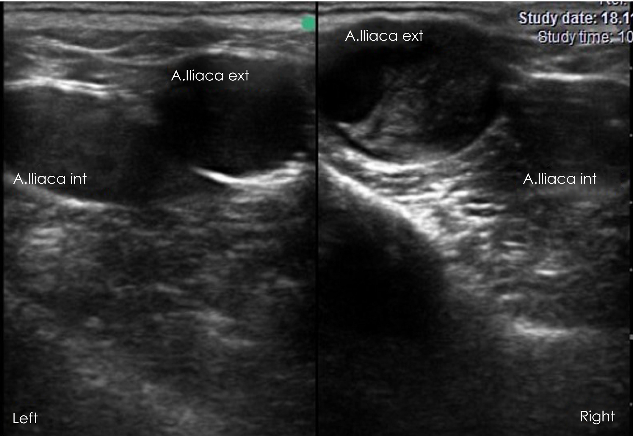
Tx
⭐ Early treatment is essential
NSAIDs
Long-term antithrombotic treatment:
Heparin
Warfarin
Larvicidal deworming if parasite-associated
Prognosis
⚠ Guarded
🧠 AORTOILIAC THROMBOSIS = HINDLIMB SIGNS DURING EXERCISE + ABSENT/BETTER AT REST
🪱 BABESIOSIS ⭐⭐⭐
Etiology
Babesia caballi, Babesia equi
Host/vector
Horse = vertebrate host
🕷 Ticks = vector
Life cycle:
In horse: → asexual reproduction in RBCs
In tick: → sexual development/sporogony
Pathogenesis
Parasites invade RBCs → RBC destruction → ⭐ hemolytic anemia
Blood stasis → clogged vessels → degeneration/damage of capillary endothelial cells → anoxia → accumulation of toxic metabolites
Macroscopic hemorrhages
Clinical signs
⭐ High fever, Hemolytic anemia, Icterus, ⭐ Hemoglobinuria
Macroscopic hemorrhages
Diagnosis
Blood smear → piroplasms in RBCs
Treatment
⭐ Imidocarb dipropionate
Symptomatic/supportive treatment
🧠 BABESIA = RBC DESTRUCTION → HEMOLYTIC ANEMIA + ICTERUS + HEMOGLOBINURIA → IMIDOCARB
THEILERIOSIS ⭐⭐⭐
Etiology
Theileria equi
Pathogenesis
Tick-borne protozoan
T. equi has stages involving leukocytes before erythrocytic infection.
⭐ Formation of Koch's blue bodies
Also invades RBCs → piroplasms → RBC destruction → hemolytic anemia
Clinical signs
Fever, Hemolytic anemia, Icterus may occur
Abortion
Diagnosis
⭐ Blood smear
Koch's blue bodies / piroplasms
Treatment
⭐ Imidocarb
🧠 THEILERIA EQUI = WBC + RBC → KOCH'S BLUE BODIES + HEMOLYTIC ANEMIA → IMIDOCARB
🪱 STRONGYLUS VULGARIS — ARTERITIS PARASITARIA ⭐⭐⭐
Etiology
Strongylus vulgaris (= large strongyle)
Adults:
→ cecum + colon
Larvae:
→ migrate through arteries, especially the ⭐ cranial mesenteric artery
Life cycle ⭐⭐⭐
Eggs shed in feces → Develop in environment → infective L3 → Horse ingests L3 while grazing → L3 penetrate intestinal mucosa → Larvae migrate in arteries, especially Cranial mesenteric artery (May also involve: Jejunal artery, Ileocolic artery, Caudal mesenteric artery) → Develop/molt toward L5 → Return to large intestine → Form nodules → Nodules rupture → adults emerge into intestinal lumen
Pathogenesis ⭐⭐⭐
Adults:
Large buccal capsule → attach to intestinal mucosa → blood feeding → bleeding ulcers
Migrating larvae:
Migration in arterial walls → inflammation → ⭐ THROMBOARTERITIS → thrombosis → arterial wall damage → aneurysm → reduced/interrupted intestinal blood supply → ischemia/infarction → colic
🧠 S. VULGARIS → CRANIAL MESENTERIC ARTERY → THROMBOARTERITIS → ↓ BLOOD TO INTESTINE → COLIC
CS
Colic
Anemia, Poor body condition, Diarrhea, Fever, Anorexia, Weight loss
Dx
Coprology - flotation method
Larval culture → species determination
Necropsy
Tx
Fenbendazole
⭐ Moxidectin → adults + migrating larvae
⚠ Problematic as resistance for pyrantel, ivermectin and benzimidazoles!
🦟🪱 FILARIOSIS — SETARIA EQUINA ⭐⭐
Etiology
Setaria equina
Hosts
Final host = horse
Intermediate host/vector = mosquito
Location
Adult worms:
→ abdominal/peritoneal cavity
Microfilariae:
→ circulate in blood
Life cycle
Adults in abdominal cavity
→ release microfilariae into blood → mosquito takes blood meal → ingests microfilariae → develop to infective L3 → mosquito transmits L3 to another horse
CS
Anemia, Hemorrhages, Skin edema
CNS signs → your lecture associates these with toxic metabolites/aberrant migration
Dx
⭐ Blood smear → microfilariae
Tx
⭐ Ivermectin
🧠 SETARIA = MOSQUITO → ADULTS ABDOMEN → MICROFILARIAE IN BLOOD → IVERMECTIN
🩸🪱 PARAFILARIOSIS ⭐⭐
Etiology
Parafilaria multipapillosa
Location
Adults:
→ subcutaneous tissue
Especially:
Neck, Chest
Transmission
🪰 Blood-feeding flies act as vectors.
CS
⭐ “SUMMER BLEEDING” / parasitic dermatorrhagia
Subcutaneous nodules
Spontaneously bleeding skin lesions
Most obvious during warm/fly season
Dx
Microfilariae/parasite stages in blood or lesion material
Tx
⭐ Ivermectin
Fly control
🧠 PARAFILARIA = SUBCUTANEOUS → SUMMER BLEEDING
🦠 VIRAL DISEASES OF BLOOD/VESSELS
AFRICAN HORSE SICKNESS ⭐⭐
Etiology
Reoviridae → Orbivirus
Severe disease with high mortality.
Transmission
⭐ Culicoides midges
❌ No direct horse-to-horse transmission
CS
🫁 Pulmonary form
Pulmonary edema
Cough
Lung congestion
❤ Cardiac form
Pyrexia
⭐ Edema of head + neck
Dx
PCR
ELISA
VNT
Prevention
💉 Vaccination in endemic areas
🧠 AHS = CULICOIDES → PULMONARY EDEMA / HEAD-NECK EDEMA
9. EQUINE VIRAL ARTERITIS — EVA ⭐⭐⭐
Etiology
Arteriviridae → Equine arteritis virus
Transmission
Aerosol
In utero
⭐ Venereal
⭐ Long-term carrier stallions
Pathogenesis
⭐ Vasculitis of small blood vessels
CS
Fever
Cough
Respiratory distress
Edema may occur
Abortion
Dx
Nasopharyngeal swab
PCR
ELISA
Tx
Supportive
Prevention
💉 Vaccination
🧠 EVA = VASCULITIS + VENEREAL + CARRIER STALLION + ABORTION
EQUINE INFECTIOUS ANEMIA — EIA ⭐⭐⭐
Etiology
Retroviridae → Lentivirus, equine infectious anemia virus
Transmission:
Mainly through infected blood, particularly:
Blood-feeding insects
Contaminated blood/equipment
Transplacental
Pathogenesis:
Persistent infection
→ immune-mediated destruction of RBCs/platelets → anemia + thrombocytopenia
CS
Disease can be acute, chronic or inapparent:
Recurrent fever, Anemia, Arrhythmia
Weakness, Weight loss
Edema of lower chest, abdomen and legs
Petechial hemorrhages of mm
Dx
⭐ Serology:
AGID / Coggins test
ELISA, PCR
Tx
❌ No curative treatment
Infected horses remain lifelong carriers, so management depends on official disease-control requirements. Euthanise!
🧠 EIA = LENTIVIRUS → BLOOD TRANSMISSION → FEVER + ANEMIA → COGGINS → LIFELONG INFECTION
🧠 EXAM MEMORY — QUESTION 13
For recall, think of the question as three blocks:
Vascular
→ Thrombophlebitis + Aortoiliac thrombosis
Parasites
→ Strongylus vulgaris + Babesia/Theileria + Setaria + Parafilaria
Viruses
→ AHS + EVA + EIA
⭐ Key associations
Thrombophlebitis → IV CATHETER + JUGULAR THROMBUS
Aortoiliac thrombosis → HINDLIMB LAMENESS DURING EXERCISE
Strongylus vulgaris → CRANIAL MESENTERIC ARTERY → THROMBOARTERITIS → COLIC
Babesia → RBC → HEMOLYTIC ANEMIA
Theileria → WBC/RBC + KOCH’S BLUE BODIES
Setaria → MOSQUITO + ABDOMINAL CAVITY + MICROFILARIAE
Parafilaria → SUMMER BLEEDING
AHS → CULICOIDES
EVA → VASCULITIS + STALLION + ABORTION
EIA → LENTIVIRUS + ANEMIA + COGGINS
For the actual oral answer, I’d put the most emphasis on thrombophlebitis, aortoiliac thrombosis, and especially Strongylus vulgaris, because S. vulgaris is the direct link to the “arteritis parasitaria” part of the question.
Equine dermatitis - infectious and parasitic origin
Main groups
Infectious:
🦠 Bacterial → Glanders, Staphylococcal dermatitis, Dermatophilosis
🧬 Viral → Papillomatosis, Vesicular stomatitis, Horsepox
🍄 Fungal → Dermatophytosis / ringworm
🦠 Protozoal → Besnoitiosis, Trypanosomosis/Dourine
Parasitic:
🕷 Mites → Sarcoptes, Psoroptes, Chorioptes
🪲 Lice → biting + sucking
🪰 Biting flies + ticks
🪱 Nematodes → Onchocerciasis, Habronemiasis, Parafilariosis
🦠 BACTERIAL
1. GLANDERS ⭐⭐⭐
Etiology
Burkholderia mallei
⚠ ZOONOTIC
Notifiable/regulated disease
Pathogenesis
→ Ulcerating nodules in URT, lungs + skin
Acute: rapid severe disease/death
Chronic: recurrent abscessation
3 forms
👃 Nasal: catarrhal → purulent discharge → nodules ulcerate → characteristic stellate scars
🫁 Pulmonary: tubercle-like lung nodules → break down into bronchioles → spread → lung consolidation + pneumonia
🦵 Cutaneous = FARCY ⭐
→ nodules along lymphatic vessels, especially extremities
→ ulceration
→ sticky, highly infectious dark-honey pus
Dx
CFT — complement fixation test
Intradermal mallein test
Tx
Doxycycline, Trimethoprim, Streptomycin
⭐ In Europe/control programs → mandatory culling/euthanasia rather than treatment
Prevention
❌ No vaccine
🧠 GLANDERS = B. mallei → ZOONOTIC → NASAL + PULMONARY + FARCY
🦠 2. FOLLICULITIS & FURUNCULOSIS / STAPHYLOCOCCAL DERMATITIS
Etiology
Staphylococcus aureus
CS
Inflammation of:
Hair follicles → folliculitis
Surrounding dermis/subcutis → deeper furunculosis
→ warm + painful skin
Common sites:
Saddle area, Pastern, Tail

Dx
Bacterial culture
Tx
Antiseptic shampoo:
Iodophor, Chlorhexidine
Antibiotics when indicated for bacterial infection
autovaccine
🧠 STAPH → HAIR FOLLICLES → warm painful saddle-area lesions
🌧 3. DERMATOPHILOSIS — “RAIN SCALD/RAIN ROT” ⭐⭐⭐
Etiology
Dermatophilus congolensis
Pathogenesis
Prolonged rain/moisture/sweat → weakens skin protective barrier → release/spread of dormant bacterium living in the skin → skin infection
CS
Papules + pustules → Exudative lesions → ⭐ Crusty lesions with hairs in
Sites:
Dorsal trunk, Muzzle, Distal limbs (areas exposed to rain, and sweaty regions)
Dx
Culture, Impression smear, Skin biopsy
Tx
Soak lesions in chlorhexidine soap and remove crusts
Topical antibacterial/antiseptic treatment
ATB spray/solution once daily × 1 week
🧠 RAIN + D. CONGOLENSIS → PAPULE → EXUDATE → CRUST

🧬 VIRAL SKIN DISEASES
4. EQUINE PAPILLOMATOSIS ⭐
Etiology
Papillomaviridae → Equine papillomavirus
Highly contagious amoung young horses by direct contact or shared items
Common:
⭐ Young horses below 3 years (immune system still developing)
Older immunosuppressed horses
CS
Warts on Muzzle, Lips, Inner surface of ear

Dx
Clinical signs, Biopsy, Histopathology
Tx
Usually self-limiting
→ spontaneously disappears after several monthsAutogenous vaccine may be used in selected cases
🧠 YOUNG HORSE + WARTS → PAPILLOMAVIRUS → usually SELF-LIMITING
🦠 5. VESICULAR STOMATITIS
Etiology
Rhabdoviridae → vesiculovirus
Transmission
Insect transmitted: Sand flies, Black flies
CS
⭐ Excessive salivation
Vesicles on Mouth, Tongue, Lips
Lesions may occur around hooves

Dx
Samples:
Vesicle fluid, Epithelium
Tests:
PCR, ELISA, CFT
Tx/control
No specific antiviral treatment
Isolation/quarantine + official control measures
🧠 VESICULAR = VESICLES IN MOUTH + SALIVATION + HOOF LESIONS
🦠 6. HORSEPOX - Extinct or nearly absent??
Etiology
Poxviridae → Horsepox virus
CS
Vesicles → rapidly ulcerate → progressively crust
Mainly:
Muzzle, Face
Dx
Virus isolation/cultivation
Tx
Usually spontaneous resolution in approximately 4 weeks
🍄 7. DERMATOPHYTOSIS / RINGWORM
⭐⭐⭐Etiology
Trichophyton equinum
Transmission
Direct contact
Fomites ⭐
CS
Red, Itchy, Scaly, ⭐ Circular lesions
Alopecia
Common sites:
Saddle area, Neck, Face

Dx
Fungal culture → Sabouraud agar
Skin scraping + hair → microscopy
Wood's lamp may be used, although it is not reliable for all equine dermatophytes
Tx
Often spontaneously resolves, BUT treatment:
→ speeds recovery
→ decreases spread/transmission
Topical antifungal treatment:
Thiabendazole
Lime sulfur
Other appropriate antifungals
⭐ Disinfect environment + equipment
Vaccination for prevention/treatment.
🧠 RINGWORM = CIRCULAR ALOPECIA + FOMITES → FUNGAL CULTURE
PARASITE ORIGIN
NB! Mention this list first, then she will ask to explain 2/3 of them:
PROTOZOA:
Besnoitiosis — Besnoitia bennetti
Trypanosomosis / Dourine — Trypanosoma equiperdum
NEMATODES:
Habronemiasis — Habronema spp.
Parafilariosis — Parafilaria multipapillosa
Onchocerciasis
Onchocerca cervicalis, O. gutturosa, O. reticulata
ECTOPARASITES:
Acarinosis = mites/mange
Sarcoptes scabiei var. equi, Psoroptes equi, Chorioptes equi
Entomosis:
Pediculosis / lice
Hippoboscidosis (Hippobosca equina),
Dermatitis caused by biting Diptera
Simuliotoxicosis
🦠 PROTOZOA
8. BESNOITIOSIS ⭐⭐
Etiology
Besnoitia bennetti, a cyst-forming coccidian parasite
FH: cats, IH: horse
Vector: flies
Life cycle
Cat sheds oocysts → contaminate grazing/water → horse ingests → tissue cyst formation of skin over nostrils etc.
Especially:
Skin, Nostrils, Eyes/sclera
Pathogenesis
Cysts cause:
Skin edema, Hyperkeratosis, Alopecia
Eye involvement:
→ small visible cysts on sclera
→ ⭐ “scleral pearls”
CS
Fever, Nasal discharge, Ocular discharge, Salivation
Stiff gait, Orchitis, Subcutaneous edema
⭐ Multifocal pinpoint cysts:
Nostrils, Ears, Face, Body
crusty and hard skin lesions
⭐ Scleral pearls
Dx
Skin biopsy - gold standard
Endoscopy of nasal cavity
ELISA
Tx
⚠ Difficult/problematic
No reliable treatment to eliminate established tissue cysts → life long carriers
Early infection → lecture suggests trimethoprim
Trimetoprim + sulfamethoxazole (Bactrim) stops cyst formation but not kill the parasite or already formed cysts, it is mainly symptomatic/supportive
🧠 BESNOITIA = SKIN CYSTS + SCLERAL PEARLS
2⃣ TRYPANOSOMOSIS / DOURINE ⭐⭐⭐
Etiology
Trypanosoma equiperdum
Transmission ⭐
Coitus / venereal transmission
CS
Initially:
Vaginal/genital discharge, Genital edema, Perineal edema
Characteristic skin lesion:
⭐ “SILVER DOLLAR PLAQUES”
→ transient cutaneous plaques, last maximum ~7 days, then it migrates to blood and release neurotoxins
Disease progresses to neurological involvement:
→ weakness, ataxia, neurological dysfunction, may progress to death
🧠 DOURINE = SEX → GENITAL EDEMA → SILVER DOLLAR PLAQUES → NEURO SIGNS
ECTOPARASITES — MITES
9. SARCOPTIC MANGE ⭐
Etiology
Sarcoptes scabiei var. equi
Burrowing mite → forms tunnels in skin
Very uncommon in horses
Transmission → direct contact
CS
⭐ Severe pruritus
Hard/thickened skin, Papules, Crusts, Alopecia
Dx
⭐ DEEP skin scraping (border between healthy and affected skin)
Tx
Pyrethroid dip/spray
Repeat every 7–10 days, ≥3 treatments
Macrocyclic lactones orally (ivermectin)
± ATB for secondary infection
Minerals + vitamin E to support skin healing
10. PSOROPTIC MANGE
Etiology
Psoroptes equi
NON-BURROWING
CS
Crusting, Serous exudation, Erythema, strong Pruritus, Alopecia
Dx
⭐ SUPERFICIAL skin scraping
Tx
Topical Pyrethroids, Ivermectin
11. CHORIOPTIC MANGE ⭐⭐
Etiology
Chorioptes equi
Non-burrowing
⭐ “LEG MANGE”
CS
Sticky scales, Strong pruritus, Restlessness, self mutilation, Lesions especially on distal limbs
Severe infestation → lameness
Skin lesions can progress through:
1. Eczema crustosum
dry scales, small nodules/pustules → dry → crusts/scales
⬇
2. Eczema madidans
→ loss of superficial skin layers → exposed corium / moist-weeping dermatitis
⬇
3. Eczema verrucosum
→ chronic proliferative skin → multiple wart-like lesions → ⭐ “cauliflower” appearance
🧠 CRUSTOSUM → MADIDANS → VERRUCOSUM
DRY → WET/RAW → CAULIFLOWER
Dx
Superficial skin scraping + microscopy
Tx
Clip hair, Remove scabs, Scrub/shampoo
Treat horse AND in-contact animals
Topical treatment is the main method → Selenium sulfide shampoo → appropriate topical acaricide
Oral ivermectin paste, Fipronil spray
Ivermectin, Topical synthetic pyrethroids
🧠 CHORIOPTES = LEG MANGE
9⃣ HORSE LICE — PEDICULOSIS
Biting louse: Werneckiella equi equi
Sucking louse: Haematopinus asini
CS
Pruritus, Rubbing, Hair loss, Skin sores
Loss of condition
Heavy sucking-lice infestation → anemia
Dx
→ lice/nits visible on hair + microscopy

Tx
→ ectoparasitic treatment, including synthetic pyrethroids
🪰 13. BITING FLIES/DIPTERA
Important examples:
Horse flies
Deer flies
Culicoides → biting midges
Simulium → black flies
Pathogenesis
⭐ Only females blood-feed/bite in many important biting-fly groups.
Blood feeding → saliva/anticoagulants injected → local skin injury → secondary bacterial infection possible→ saliva can cause allergic/hypersensitivity reactions → pruritus + dermatitis
Can also act as vectors, depending on species/pathogen.
Examples
African horse sickness, Equine infectious anemia, Equine onchocerciasis
Tx/prevention
Mainly Supportive
Fly control/repellents: ⭐ Synthetic pyrethroids
Treat secondary bacterial dermatitis if significant
1⃣1⃣ HIPPOBOSCIDOSIS
Etiology
H- ippobosca equina = forest fly / horse louse fly
Important
Blood-feeding ectoparasite, Mainly affects horses, Can also feed on cattle
CS
Bites can cause:
Irritation, Restlessness, Pruritus, Local dermatitis
🧠 HIPPOBOSCA EQUINA = FOREST FLY → BLOOD FEEDING
🪱 NEMATODES AFFECTING SKIN
14. CUTANEOUS ONCHOCERCIASIS ⭐⭐
Etiology
Onchocerca cervicalis, O. gutturosa, O. reticulata
Adult O. cervicalis live especially in ligamentous/connective tissue adjacent to the nuchal ligament
Migrating microfilariae affect the skin + eyes
Pathogenesis
Microfilariae migrate into the skin → hypersensitivity → dermatitis
Migration to the eyes → keratoconjunctivitis
Clinical signs
Alopecia, scaling and crusting, especially on the face + neck
Pruritus/dermatitis
Subcutaneous masses
Edema
Lameness
Eye problems
Diagnosis
Clinical signs
Skin biopsy → demonstration of microfilariae
Treatment
⭐ Ivermectin → very effective against microfilariae
Corticosteroids → inflammatory/hypersensitivity reaction when appropriate
🧠 ONCHOCERCA = NUCHAL LIGAMENT → MICROFILARIAE → SKIN + EYES → IVERMECTIN
15. HABRONEMIASIS ⭐⭐⭐
Etiology
Habronema muscae, Habronema microstoma → adults in stomach mucosa
Draschia megastoma → nodules in stomach wall
Final host = horse/equids
Intermediate host/vector = muscid flies
Life cycle ⭐
Eggs/L1 shed in feces → fly ingests eggs/L1 → develop to L3 inside fly → fly feeds around horse's eyes, genitalia, nostrils, lips or wounds → deposits L3 onto horse
Normal route: horse swallows L3 → adults develop in stomach
Aberrant route: larvae remain in skin/eyes → cannot complete life cycle → inflammation + hypersensitivity
🧠 FECES → FLY → L3 → HORSE → SWALLOW → STOMACH
3 Forms ⭐⭐⭐
1⃣ Gastric form — most common
Adults in stomach → gastritis
Large granulomas in gastric mucosa
2⃣ Cutaneous form — aberrant
Larvae deposited in skin/wounds → cannot complete life cycle
Local hypersensitivity
Non-healing ulcerative/granulomatous skin lesions
⭐ “SUMMER SORES”
3⃣ Conjunctival form — aberrant
Larvae around eyes → conjunctivitis
Thickened eyelids + granulomatous lesions
Aberrant lesions may also involve nostrils and genitalia
Diagnosis
Characteristic non-healing reddish cutaneous granulomas
Larvae may be demonstrated
Endoscopy → gastric form
ELISA
Eggs are difficult to detect by routine fecal flotation because they do not float well in standard flotation solutions
Treatment
⭐ Ivermectin
Moxidectin mentioned in lecture
Wound management + fly control
🧠 HABRONEMA = MUSCID FLY → GASTRIC / CONJUNCTIVAL / CUTANEOUS → SUMMER SORES ⭐⭐⭐
16. PARAFILARIOSIS ⭐⭐⭐
Etiology
Parafilaria multipapillosa
Location
Adults live in subcutaneous tissue
Form small nodules, especially on the neck and chest
Transmission
🪰 Blood-feeding flies act as vectors
Life cycle
Fly feeds on horse → transmits infective larvae → parasites develop in subcutaneous tissue → skin nodules/lesions develop → lesions bleed → flies acquire parasite stages while feeding → transmission continues
Clinical signs
Subcutaneous nodules
Spontaneously bleeding skin lesions
More common/prominent during the warm/fly season
Characteristically called:
⭐ “SUMMER BLEEDING”
⭐ “PARASITIC DERMATORRHAGIA”
Diagnosis
Demonstration of parasite stages/microfilariae in lesion or blood material
Lecture: microfilariae in blood smear
Treatment
Ivermectin
⭐ Fly control
🧠 PARAFILARIA = SUBCUTANEOUS TISSUE → BLOOD-FEEDING FLIES → SUMMER BLEEDING
🧠 EXAM MEMORY — QUESTION 14
You have a lot of diseases in this question, so learn them by groups:
🦠 BACTERIAL
Glanders → B. mallei → FARCY → dark sticky pus → CFT/mallein → euthanasia
Staph → S. aureus → folliculitis/furunculosis → warm painful skin
Dermatophilosis → D. congolensis → RAIN SCALD → crusts
🧬 VIRAL
Papilloma → young horse + warts + self-limiting
Vesicular stomatitis → mouth vesicles + salivation
Horsepox → vesicles → ulcers → crusts
🍄 FUNGAL
Ringworm → T. equinum → circular alopecia + fomites
🦠 PROTOZOA
Besnoitia → skin cysts + SCLERAL PEARLS
🕷 MITES
Sarcoptes → BURROWING → DEEP scraping
Psoroptes → NON-BURROWING → SUPERFICIAL scraping
Chorioptes → LEG MANGE
🪲 LICE
Biting + sucking → pruritus/hair loss; sucking → anemia
🪱 NEMATODES ⭐⭐⭐
Onchocerca → NUCHAL LIGAMENT → SKIN + EYES
Habronema → FLY → SUMMER SORES
Parafilaria → SUMMER BLEEDING
🔥 Three “summer” associations to separate
Habronema → SUMMER SORES
Parafilaria → SUMMER BLEEDING
Culicoides → insect-bite hypersensitivity / summer-associated pruritus
That distinction is particularly useful for an oral exam.
Equine skin tumors
Main skin tumours to know:
⭐ Sarcoid — most common equine skin neoplasm
⭐ Melanoma — especially older grey/white horses
⭐ Squamous cell carcinoma (SCC) — non-pigmented/mucocutaneous areas
Mastocytoma
Lymphoma
Lymphosarcoma
1. SARCOIDS ⭐⭐⭐
Tumour of fibroblastic cell origin
Unique to equids
⭐ Most common equine skin neoplasm
Most commonly affects young to middle-aged horses
Etiology
Associated with bovine papillomavirus (BPV)
Insects may contribute to transmission
Possible genetic predisposition
Clinical signs
Can have several appearances:
Verrucous → scaly, wart-like
Fibroblastic → fleshy, often ulcerated
Occult → flat lesion
Common locations:
Head, Legs, Ventrum
Dx
Appearance
Biopsy + histopathology
⚠ Sarcoids can be difficult because trauma/biopsy may sometimes stimulate aggressive growth, so biopsy should be planned carefully.
Tx
⭐ Difficult to treat + recurrence is common
Options:
Wide surgical excision
Location may make complete excision difficult, Recurrence can occur
Cryotherapy
BCG immunotherapy
Autogenous vaccines
Intralesional radiation/brachytherapy → e.g. iridium
Chemotherapy:
5-fluorouracil, Cisplatin, Topical/intralesional depending on protocol
🧠 SARCOID = MOST COMMON → BPV → YOUNGER HORSE → MANY FORMS → HIGH RECURRENCE

2. MELANOMA ⭐⭐⭐
Tumour arising from: Melanocytes, Melanoblasts
Can be benign or malignant.
⭐ Very common in older grey horses
Especially horses that become progressively: dappled grey → white with age
Pathogenesis
Abnormal proliferation of melanocytic cells and melanin production
Often slow-growing
May remain localized for years, But some become locally invasive or metastatic
CS
⭐ Pigmented nodules
Single or multiple, Dermal or subcutaneous, May ulcerate
Typical locations:
⭐ Under/around tail and perineum
Around anus/rectum, Genitalia
Around mouth, Around eyes
Distal limbs

Dx
Characteristic appearance
FNA → cytology
⭐ Biopsy + histopathology = definitive
Tx
If:
Few lesions, Small, Not growing, Not causing problems → monitor/observe
Treatment options:
Surgical excision
Laser, Cryotherapy
Intralesional chemotherapy → cisplatin
Therapeutic melanoma vaccine → canine melanoma vaccine has been used off-label
High-frequency irreversible electroporation
→ electrical pulses create pores in tumour cell membranes
→ irreversible cell damage/death
🧠 MELANOMA = OLD GREY HORSE → BLACK NODULES → TAIL/PERINEUM → OFTEN SLOW-GROWING
3. SQUAMOUS CELL CARCINOMA — SCC ⭐⭐⭐
Common epithelial neoplasm affecting:
Head, Mucocutaneous junctions, Genitalia
Predisposition
⭐ Older horses, Light-coloured horses, Particularly non-pigmented skin
Etiology
⭐ UV light-associated neoplasia
Pathogenesis
Tumour cells invade through the dermis as cords/islands of neoplastic cells.
Important:
⭐ Often locally aggressive/invasive
Metastasis is less common, particularly early in the disease
CS
Wart-like papules
Ulcerated nodules/masses
Usually solitary
May have pigment/colour changes depending on location
Typical sites:
Eyelids/ocular region, Mucocutaneous junctions, Genitalia

Dx
Location + appearance
⭐ Biopsy + histopathology
Tx
Surgical excision
Cryotherapy
Laser
⚠ Complete removal is important because:
→ incomplete excision → recurrence
Prognosis depends strongly on location, size, invasiveness and ability to completely remove the tumour.
🧠 SCC = OLD + LIGHT/NON-PIGMENTED + UV → ULCERATIVE/WART-LIKE → LOCALLY AGGRESSIVE
4. MASTOCYTOMA
= Mast cell tumour
Tx
⭐ Surgical resection
Generally:
Good outcome after complete excision, Low recurrence rate
🧠 MASTOCYTOMA = MAST CELLS → SURGERY → LOW RECURRENCE

5. LYMPHOMA
= Tumour arising from lymphoid cells/lymphocytes.
Cutaneous involvement can occur as part of lymphoid neoplasia.
Tx / Prognosis
Treatment generally not recommended
Poor prognosis
🧠 LYMPHOMA = LYMPHOCYTES → POOR PROGNOSIS

6. LYMPHOSARCOMA
Uncommon in horses
Usually affects middle-aged to older horses
CS
⭐ Typically:
Multiple subcutaneous nodules
Especially on the trunk
🧠 LYMPHOSARCOMA = OLDER HORSE → MULTIPLE SUBCUTANEOUS NODULES ON TRUNK
🧠 EXAM DIFFERENTIATION ⭐⭐⭐
Tumour | Typical horse | Typical appearance/location | Key fact |
|---|---|---|---|
Sarcoid | Young–middle-aged | Variable: flat, wart-like, fibroblastic | ⭐ Most common; BPV; recurrence |
Melanoma | Older grey horse | Pigmented nodules, especially tail/perineum | Often slow-growing |
SCC | Older, light-coloured | Non-pigmented/mucocutaneous areas | ⭐ UV; locally aggressive |
Mastocytoma | — | Cutaneous mass | Surgery → low recurrence |
Lymphoma | — | Lymphoid tumour | Poor prognosis |
Lymphosarcoma | Middle-aged/older | Multiple subcutaneous trunk nodules | Uncommon |
🔥 Three major tumours
SARCOID
→ Most common + BPV + younger horse + high recurrence
MELANOMA
→ Older GREY horse + pigmented nodules + tail/perineum
SCC
→ Older LIGHT horse + non-pigmented skin + UV + locally aggressive
If you can immediately distinguish those three, you have the core of this question.
Equine cystitis and hematuria
1. CYSTITIS ⭐⭐⭐
Cystitis = inflammation of the urinary bladder.
→ More common in mares because of the shorter urethra, which facilitates ascending infection.
Etiology
1. Cystic calculi
⭐ Mainly calcium carbonate stones

2. Ascending bacterial infection
E. coli, Proteus, Klebsiella, Enterococcus, Streptococcus, Staphylococcus, Pseudomonas
3. Neurological/viral disorders causing bladder dysfunction
Herpesvirus
Equine herpesvirus myeloencephalopathy
Polyneuritis equi
→ impaired bladder emptying → urine retention → predisposition to cystitis.
🧠 CYSTITIS = CALCULI + ASCENDING BACTERIA + NEUROLOGICAL BLADDER DYSFUNCTION

Clinical signs ⭐⭐⭐
Dysuria = difficult/painful urination
⭐ Pollakiuria = abnormally frequent urination
In mares, may resemble signs of oestrus
Urine dribbling
Haematuria, Pyuria
Perineal scalding in mares
Soiling of hindlegs in males
🧠 DYSURIA + POLLAKIURIA + DRIBBLING + BLOOD/PUS IN URINE
Diagnosis
Rectal palpation
→ assess bladderUltrasonography
→ ⭐ thickened bladder wall
→ may detect calculiEndoscopy/cystoscopy
Mucosa may be:
Thickened, Hyperaemic, Ulcerated
Urinalysis + sediment examination
May find:
→ ⭐ RBC → haematuria
→ WBC/leukocytes → inflammation/pyuria
→ Bacteria, Crystals
Bacterial culture
→ identify bacteria and guide antibiotic treatment.
🧪 Normal equine urine:
Important because horse urine normally looks unusual:
Often cloudy and thick
Contains microscopic calcium carbonate crystals
Alkaline
⭐ Normal pH approximately 7.5–8.5
So → Cloudy urine ≠ automatically cystitis in a horse!!
Blood analysis
May show increased:
Creatinine
BUN — blood urea nitrogen
Particularly if there is concurrent renal dysfunction/urinary obstruction.
Treatment
⭐ Treat/correct the underlying cause
Bacterial cystitis
→ antibiotics based preferably on culture + sensitivity
Lecture lists drugs excreted through kidneys/urinary tract:
Aminoglycosides
Trimethoprim/sulfadiazine
Fluoroquinolones
Penicillins
Cephalosporins
Cystic calculi
→ ⭐ surgical removal
🧠 CYSTITIS Tx = CORRECT CAUSE → CULTURE-BASED ATB → REMOVE CALCULI IF PRESENT
🩸 2. HAEMATURIA ⭐⭐⭐
Haematuria = presence of intact RBCs/blood in urine.
Blood can originate from:
Kidneys, Ureters, Bladder, Urethra
Reproductive tract contamination
Etiology
Important causes include:
⭐ Urinary tract infection / cystitis
Urolithiasis
Trauma, Neoplasia, Drug toxicity
Systemic disease, Exercise-induced haematuria
Urethral defects, Urethral rents
Idiopathic renal haemorrhage
⭐ Verminous nephritis → Halicephalobus gingivalis
Urethral rent
= tear in the urethra that communicates with the corpus spongiosum of the penis
→ blood enters the urethra → haematuria/urethral bleeding.
🧠 URETHRAL RENT = TEAR → CORPUS SPONGIOSUM ↔ URETHRA → BLOOD IN URINE
Clinical signs
Main sign:
⭐ Red/discoloured urine
Other signs depend on underlying cause:
Dysuria, Pollakiuria
Colic/discomfort
Urinary obstruction
Diagnosis
⭐ Urinalysis
Urine sediment:
→ presence of RBCs confirms haematuria
Then investigate source with:
USG
Cystoscopy/endoscopy
Rectal examination
Culture if infection suspected
Blood examination when indicated
Treatment
Depends entirely on the cause:
Bacterial infection → antibiotics
Dehydration/renal compromise → IV fluids when indicated
Urolithiasis → surgical removal
Trauma → appropriate management
Treat underlying renal/systemic disease
🧠 HAEMATURIA IS A SIGN → FIND WHERE THE BLOOD COMES FROM → TREAT THE CAUSE
🧪 HAEMATURIA vs HAEMOGLOBINURIA ⭐⭐⭐
This is probably one of the most important distinctions for the oral exam.
🩸 Haematuria = intact RBCs in urine
Centrifuge urine: → RBCs sediment at the bottom → supernatant becomes yellow/clear
⭐ RBCs visible in urine sediment
🔴 Haemoglobinuria
= free haemoglobin in urine due to intravascular haemolysis
Centrifuge urine:
→ no RBC pellet explaining the colour, → ⭐ supernatant remains red

Haematuria | Haemoglobinuria | |
|---|---|---|
What is present? | Intact RBCs | Free haemoglobin |
Sediment microscopy | ⭐ RBCs | No/very few RBCs |
After centrifugation | RBCs settle → supernatant clears | ⭐ Supernatant stays red |
Think | Urinary tract bleeding | Intravascular haemolysis |
🧠 CENTRIFUGE IT:
HAEMATURIA → RED CELLS GO DOWN ⬇ → SUPERNATANT CLEARS
HAEMOGLOBINURIA → RED COLOUR STAYS 🔴
Equine acute and chronic renal failure
⭐ Acute renal failure (ARF)
⭐ Chronic renal failure (CRF)
Renal parasites
Klossiella equi
Halicephalobus gingivalis
Leptospirosis
1. ACUTE RENAL FAILURE — ARF ⭐⭐⭐
= Rapid deterioration of renal function
→ ↓ filtration/excretion → accumulation of nitrogenous waste products → azotemia ± uremia
Often associated with:
→ ↓ urine production = oliguria
→ disturbances in:
Fluid balance, Electrolytes, Acid-base balance
🧠 ARF = RAPID LOSS OF RENAL FUNCTION → AZOTEMIA + OLIGURIA
Etiology ⭐⭐⭐
1. Endotoxemia
Especially associated with:
Colic
Acute diarrhea/colitis
→ renal hypoperfusion/ischemia
2. Acute septic pyelonephritis
Leptospira spp.
3. Nephrotoxins
🌱 Plants
Red maple, Oak, Onion, White snakeroot
⚙ Heavy metals
Mercury, Lead, Arsenic
💊 Antibiotics
⭐ Gentamicin
Neomycin, Tetracyclines, Sulfonamides, Cephalosporins
💊 NSAIDs
⭐ Flunixin meglumine
🩸 Pigments
Myoglobin, Hemoglobin
🧠 ARF = ENDOTOXEMIA + INFECTION + NEPHROTOXINS
Pathogenesis
Different etiological agents can cause:
→ renal tubular injury/necrosis
→ renal ischemia
→ inflammatory response
→ direct nephron damage
Examples:
NSAIDs
→ ↓ renal prostaglandins → ↓ renal perfusion → ischemic injuryNephrotoxic antibiotics
→ tubular damage/necrosisMyoglobin/hemoglobin
→ pigment-associated renal tubular injury
Result:
↓ GFR → azotemia → fluid/electrolyte/acid-base disturbances
Clinical signs
⭐ Anorexia
Oliguria
Depression, Colic
May occur with: Colitis, Myositis
⚠ Nephrotoxins are often not kidney-specific, so clinical signs from damage to other organs may predominate.
Diagnosis ⭐⭐⭐
Rectal palpation
Kidney may be enlarged/irregular
⭐ In the horse, only the caudal pole of the LEFT kidney is normally accessible rectally
Ultrasonography
Acute injury may show:
Enlarged/swollen kidney
Edema
Altered echogenicity
Laboratory
⭐ Most important:
↑ Creatinine, Azotemia
Inappropriately low specific gravity (= poorly concentrated urine)
Other abnormalities from your lecture:
↑ Globulins, ↑ Calcium
↓ Phosphorus, ↓ Albumin
Urinary casts
Enzymes
Glucosuria
Urine dipstick → measure pH, Protein, Glucose, Ketones, Bilirubin, Urobilinogen, Blood
Treatment ⭐⭐⭐
1. Treat the primary cause
2. Remove nephrotoxins
⭐ Stop suspected nephrotoxic drugs/toxins.
3. Correct fluid/electrolyte abnormalities
IV fluid therapy
Correct:
Dehydration, Electrolyte abnormalities, Acid-base abnormalities
Monitor carefully for:
⚠ Edema/fluid overload, especially if oliguria persists.
Your lecture lists methods to stimulate urine production:
Dextrose, Mannitol, Furosemide, Dopamine
4. Pyelonephritis
Use appropriate antimicrobials, ideally based on culture/sensitivity.
Lecture lists:
Aminoglycosides, Trimethoprim/sulfadiazine (equibactim oral powder)
Fluoroquinolones, Penicillins, Cephalosporins
⚠ Remember that aminoglycosides themselves can be nephrotoxic, so renal function matters when selecting treatment.
Leptospiral infection
Tetracycline, Streptomycin, Penicillin
Prognosis
Better prognosis when:
⭐ Underlying cause can be corrected
Creatinine stabilizes/decreases after treatment
Horse becomes polyuric
🧠 ARF Tx = REMOVE CAUSE → FLUIDS/ELECTROLYTES → MONITOR URINE + CREATININE
2. CHRONIC RENAL FAILURE — CRF ⭐⭐⭐
= Slow, progressive and irreversible loss of functional nephrons
→ progressively reduced renal function.
Important cause to consider in horses presented with: ⭐ WEIGHT LOSS + ANOREXIA
Etiology
Two main groups:
A. Tubulointerstitial causes
Toxins, Obstruction, Pyelonephritis, Neoplasia, Infiltrative disease
Chronic septic pyelonephritis → usually associated with ascending urinary tract infection
Renal neoplasia → SCC, Adenocarcinoma, Primary renal cell carcinoma
Chronic interstitial nephritis → Associated with: Toxins, Hemodynamic injury
B. Glomerular causes
Glomerulonephritis
Renal hypoplasia
Amyloidosis
Glomerulosclerosis
Chronic glomerulonephritis → Can be associated with: Immune-mediated injury, Ischemia, Infarction
Clinical signs ⭐⭐⭐
Think of a thin horse that drinks and urinates a lot:
⭐ Weight loss, Anorexia, Cachexia, Depression, Dehydration
⭐ Polyuria
⭐ Polydipsia
Edema, Fever
Oral ulceration
🧠 CRF = WEIGHT LOSS + PU/PD + DEHYDRATION
Diagnosis
Ultrasound: Chronic kidneys may become: ⭐ Small/shrunken + irregular with chronic structural changes.
Endoscopy: May demonstrate ureteral discharge: Blood, Pus (Especially with pyelonephritis)
Renal biopsy: ⭐ Provides definitive histopathological diagnosis of the underlying renal lesion.
Laboratory:
Anemia, Leukocytosis, Proteinuria, Hypochloremia, Hyponatremia, Hematuria
Renal function testing commonly demonstrates: ↑ Creatinine, ↑ BUN/azotemia, Poor urine concentrating ability (low specific gravity)
Treatment
⚠ CRF is progressive, so treatment is primarily supportive and aimed at maintaining quality of life/prolonging survival.
Nutritional/supportive management:
Supplement carbohydrates
Fat
Electrolytes:
Sodium, Potassium, Calcium, Bicarbonate
Diet:
Low-protein diet (<10%)
Also:
→ maintain hydration
→ treat underlying infection/other correctable causes where possible.
🧠 CRF Tx = SUPPORTIVE → HYDRATION + NUTRITION/ELECTROLYTES + MANAGE CAUSE
🪱 3. RENAL PARASITES
A. KLOSSIELLOSIS ⭐⭐
Etiology
Protozoan apicomplexa/coccidian: Klossiella equi → renal coccidiosis
Pathogenesis
Develops within renal tissue → lesions/granules in renal cortex → may impair renal function.
Meronts in endothelial cells of glomerular capsule. LC not understood.
Dx
⭐ Oocysts in urine
Tx
❌ No known effective treatment that reliably eliminates Klossiella.
🧠 KLOSSIELLA EQUI = RENAL COCCIDIA → KIDNEY CORTEX → OOCYSTS IN URINE → NO SPECIFIC Tx
B. HALICEPHALOBUS GINGIVALIS
Etiology
Free-living opportunistic nematode.
During abnormal migration: → may disseminate to kidneys (BUT it is very rare!)
It may also affect other tissues, particularly CNS and other organs.
🧠 HALICEPHALOBUS = ABERRANT MIGRATION → KIDNEY POSSIBLE BUT RARE
🦠 4. LEPTOSPIROSIS ⭐⭐⭐
Etiology
Bacterium: Leptospira interrogans
In horses, leptospirosis is especially associated with:
⭐ Uveitis
⭐ Abortion
Renal disease can also occur, particularly in affected foals.
Pathogenesis
Organism enters bloodstream
→ spreads hematogenously
→ affinity for:
Kidneys, Liver, Pregnant uterus
Kidney
→ colonization of renal tubules/nephrons → renal damage → possible renal failure
Liver
→ hepatitis/necrosis
Pregnant uterus
→ crosses placenta → fetal infection → ⭐ abortion
🧠 LEPTO = BLOOD → KIDNEY + LIVER + PREGNANT UTERUS
Clinical signs
⭐ Polydipsia
⭐ Polyuria
Fever, Dehydration
Icterus/jaundice
Depending on manifestation:
Uveitis, Abortion, Renal disease
Diagnosis
ELISA
⭐ MAT (microscopic agglutination test) (gold standard!!!)
Treatment
Antibiotics:
Tetracycline, Streptomycin, Penicillin
🧠 LEPTO = UVEITIS + ABORTION ± KIDNEY → MAT → ANTIBIOTICS
🔥 ARF vs CRF — KNOW THIS
ACUTE RENAL FAILURE | CHRONIC RENAL FAILURE | |
|---|---|---|
Onset | ⭐ Rapid | ⭐ Slow/progressive |
Urination | Often oliguria initially | ⭐ Polyuria/polydipsia |
Kidney | Often swollen/enlarged | Often small/shrunken/irregular |
Common causes | Endotoxemia, nephrotoxins, infection | Chronic nephritis, pyelonephritis, glomerular disease |
Creatinine | ↑ | ↑ |
Main approach | ⭐ Remove cause + restore fluid/electrolyte balance | ⭐ Long-term supportive management |
Reversibility | Can be reversible | Usually irreversible/progressive |
🧠 Final memory
ACUTE = BIG/WET KIDNEY + OLIGURIA → REMOVE CAUSE + FLUID THERAPY
CHRONIC = SMALL/IRREGULAR KIDNEY + WEIGHT LOSS + PU/PD → SUPPORTIVE CARE
KLOSSIELLA = OOCYSTS IN URINE
HALICEPHALOBUS = RARE ABERRANT KIDNEY MIGRATION
LEPTOSPIRA = UVEITIS + ABORTION + POSSIBLE RENAL FAILURE
Disorders of horse consciousness
The nervous system is divided into:
Brain, Spinal cord, Peripheral nerves
Clinical signs depend on which part of the nervous system is affected.
The neurological examination aims to:
Determine whether neurological dysfunction is present
Determine its severity
⭐ Localize the lesion
🧠 1. GENERAL NEUROLOGICAL EXAMINATION
A. History
Ask about:
Age, Breed
Duration of signs, Progression
History of trauma
Previous/current diseases
Medication, Vaccination status
Recent travel
Environment
B. Physical + neurological examination ⭐⭐⭐
1. Behaviour
Look for:
Seizures, Head pressing, Circling, Aggressiveness, Abnormal behaviour
2. Mental status/consciousness ⭐⭐⭐
Evaluate level of consciousness and awareness:
Normal/alert
Depression
Somnolence → abnormally sleepy
Semicoma/stupor → responds only to strong stimuli
Coma → unconscious, does not respond appropriately to stimuli
🧠 DEPRESSION → SOMNOLENCE → STUPOR/SEMICOMA → COMA
3. Head posture + coordination
Look for:
Head tilt, Head swaying
Abnormal position/movement
→ Head tilt especially suggests vestibular dysfunction!
4. Cranial nerves
Examine CN I–XII.
5. Gait + posture ⭐⭐⭐
Observe:
Ataxia → incoordination
Paresis → weakness
Spasticity
Hypermetria → exaggerated limb movement
Stumbling
Observe the horse:
Walking, Turning/circling, Backing
If appropriate, with additional gait challenges
6. Neck + forelimbs
Evaluate:
Symmetry, Malformations, Muscle atrophy, Patchy sweating
Strength of voluntary movement, Skin sensation, Spinal reflexes
7. Trunk + hindlimbs
Evaluate:
Symmetry, Muscle mass, Sensation, Strength, Coordination
8. Tail + anus
Evaluate:
⭐ Perineal reflex
Tail tone
Anal tone
Asymmetry
9. PROPRIOCEPTION ⭐⭐⭐
Proprioception = horse's awareness of the position of its limbs/body.
Test:
Make the horse walk in tight circles.
Normal horse → mildly crosses limbs appropriately.
Neurological horse → may Cross excessively, Step on itself, Swing/reach the outside limb too far, Misplace limbs
🧠 PROPRIOCEPTION = “DOES THE HORSE KNOW WHERE ITS LEGS ARE?”
10. LIMB STRENGTH
Forelimbs
Push the horse at the shoulder toward the opposite limb.
Normal → resists displacement → does not collapse.
Hindlimbs
⭐ Tail-pull test
Pull the tail laterally while horse:
Stands
Walks forward
Normal → resists → maintains balance and supports body weight.
Neurological weakness → easily displaced → difficulty recovering balance.
Also look for:
Stumbling
🧠 PUSH SHOULDER = FORELIMBS; PULL TAIL = HINDLIMBS
🐴 2. NEUROLOGICAL EXAMINATION OF FOALS ⭐⭐
Be careful because some findings that would be abnormal in adults can be normal in young foals.
Normal/possible in foals:
Menace response reduced or absent
Jerky head movements
Mild dysmetria
Mild incoordination
Slap-test response can be variable up to approximately 1 month
🧠 FOAL ≠ SMALL ADULT — immature neurological responses can be normal
👋 3. SLAP TEST — TLAR ⭐⭐⭐
= Thoracolaryngeal adductor reflex (TLAR)
Procedure:
→ Slap the saddle/thoracic region on one side
→ observe movement of the contralateral arytenoid cartilage using transnasal endoscopy.
⭐ the expected reflex is arytenoid adduction (moves inward)
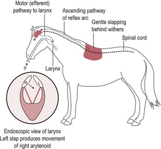
Used to assess neurological pathways involving:
Cervical spinal cord and Brainstem/vagal-recurrent laryngeal pathways
Can help detect neurological dysfunction/lesions affecting these pathways.
🧠 SLAP BACK → WATCH ARYTENOID ADDUCT
🔬 4. DIAGNOSTIC TESTS
Cerebrospinal fluid (CSF)
Can be collected from: Atlanto-occipital space, and Lumbosacral region
Imaging
Radiography, CT
MRI → particularly useful for intracranial disease
Other
Needle electromyography (EMG)
Necropsy
🧠 5. DISORDERS OF CONSCIOUSNESS
EPILEPSY ⭐⭐⭐
= recurrent seizures
A seizure results from abnormal electrical activity in the brain.
Etiology
Idiopathic
OR secondary to cerebral cortical damage caused by:
Ischemia, Trauma, Infectious disease, Neoplasia
Clinical signs
⭐ Loss/alteration of consciousness
Tonic-clonic seizures
Abnormal movements, Paddling, Urination, Defecation, Salivation
Treatment
Acute seizure:
⭐ DiazepamIdiopathic/recurrent cases: phenytoin
🧠 EPILEPSY = RECURRENT SEIZURES → LOSS OF CONSCIOUSNESS + TONIC/CLONIC MOVEMENTS → DIAZEPAM ACUTELY
🦠 6. CIZEK'S INFECTIOUS DISEASES OF THE NERVOUS SYSTEM
Know the list:
Equine encephalomyelitis ⭐⭐⭐
Eastern equine encephalomyelitis — EEE
Western equine encephalomyelitis — WEE
Venezuelan equine encephalomyelitis — VEE
Transmission:
Mosquitoes
Rodent/bird reservoir cycles depending on virus
Pathogenesis:
→ inflammation of brain/CNS
Dx:
Lecture: ELISA
Tx:
Supportive
Other infectious neurological diseases:
Getah virus — Alphavirus
Borna disease — Bornavirus
Pseudorabies/Aujeszky disease — Suid alphaherpesvirus 1
Louping ill — Flavivirus
⭐ Rabies — Lyssavirus
🧠 EEE/WEE/VEE + GETAH + BORNA + AUJESZKY + LOUPING ILL + RABIES
👁 7. CRANIAL NERVES I–XII ⭐⭐⭐
This part is much easier if you learn function + how to test it.
CN I — OLFACTORY
Function: smell
Test: → response to familiar/non-irritating odours
🧠 I = SMELL
CN II — OPTIC ⭐⭐⭐
Function:
Vision
Afferent component of menace response and pupillary light reflex
Test: Menace response → threatening hand gesture toward eye → horse should blink.
Pathway involves:
⭐ CN II → sees threat
⭐ CN VII → closes eyelid
Obstacle course → evaluates vision.
🧠 II SEES, VII BLINKS
CN III — OCULOMOTOR ⭐⭐⭐
Function:
Eye movement, Pupil constriction
Test: Observe Pupil size, Symmetry
Pupillary light reflex — PLR → shine bright light into eye → immediate pupil constriction
🧠 III = CONSTRICT PUPIL
CN IV — TROCHLEAR
Function:
Eye position/movement
Innervates dorsal oblique muscle
Assess normal eye position and movement.
🧠 IV = EYE POSITION
CN V — TRIGEMINAL ⭐⭐⭐
Three branches:
Ophthalmic
Maxillary
Mandibular
Function
⭐ Major sensory nerve of the face
Also motor function to muscles of mastication.
Test
Facial sensation/reflexes:
Touch/prick face, Corneal/palpebral region, Nostrils, Lips
Look for:
Ear movement, Eyelid closure, Nostril movement, Withdrawal of lip/labial commissure
🧠 V = FEEL THE FACE + CHEW

CN VI — ABDUCENS
Function:
Eye position/movement
Important for lateral movement of eye.
🧠 VI = ABDUCT EYE
CN VII — FACIAL ⭐⭐⭐
Branches include:
Auricular, Palpebral, Buccal
Function
Controls facial muscles.
Test
Observe ability to:
Move ears, ⭐ Blink eyelids, Move lips, Move nostrils, Eat normally
Remember:
CN V = sensory input from face
CN VII = facial motor response
🧠 VII = FACIAL MOVEMENT + BLINK
CN VIII — VESTIBULOCOCHLEAR ⭐⭐⭐
Two functions:
Cochlear → hearing
Vestibular → balance
Abnormality may produce:
Head tilt, Ataxia, Nystagmus
🧠 VIII = HEARING + BALANCE
CN IX — GLOSSOPHARYNGEAL
Function:
Pharynx, Swallowing, Component of gag/pharyngeal reflex
🧠 IX = PHARYNX + SWALLOW
CN X — VAGUS ⭐⭐⭐
Functions include:
Pharynx/larynx, Swallowing, Laryngeal function
Parasympathetic visceral functions
Your lecture particularly associates it with:
⭐ SLAP TEST / TLAR
🧠 X = VAGUS → LARYNX → SLAP TEST
CN XI — ACCESSORY
Function:
Motor function to muscles of:
Neck, Shoulder region
🧠 XI = NECK/SHOULDER MUSCLES
CN XII — HYPOGLOSSAL ⭐⭐
Function:
⭐ Tongue movement
Test
Inspect tongue size/symmetry, Pull tongue from mouth → Horse should provide resistance
Observe tongue movement
🧠 XII = TONGUE
🔥 CRANIAL NERVES — RAPID EXAM TABLE
CN | Nerve | Main thing to remember |
|---|---|---|
I | Olfactory | Smell |
II | Optic | Vision |
III | Oculomotor | Pupil constriction |
IV | Trochlear | Eye position |
V | Trigeminal | Facial sensation + chewing |
VI | Abducens | Eye position/abduction |
VII | Facial | Facial movement + blink |
VIII | Vestibulocochlear | Hearing + balance |
IX | Glossopharyngeal | Swallow/pharynx |
X | Vagus | Larynx + slap test |
XI | Accessory | Neck muscles |
XII | Hypoglossal | Tongue |
🧠 The highest-yield associations
II + VII → MENACE:
II sees → VII blinks
II + III → PLR:
II detects light → III constricts pupil
V + VII → FACIAL REFLEX:
V feels → VII moves
VIII → HEAD TILT/BALANCE
IX + X → SWALLOWING/PHARYNX
X → SLAP TEST
XII → TONGUE
Vestibular syndrome and brainstem disorders
🧠 Vestibular system
Responsible for:
⭐ Balance
Posture
⭐ Eye movements
It has two major parts:
Peripheral vestibular system
→ inner/middle ear + vestibular nerveCentral vestibular system
→ vestibular structures/pathways in the brainstem/CNS
Dysfunction causes:
→ loss of normal posture and balance
→ ataxia
→ abnormal eye movements/nystagmus
1. VESTIBULAR SYNDROME ⭐⭐⭐
Vestibular syndrome = group of diseases affecting the vestibular system.
Main question:
⭐ Is it PERIPHERAL or CENTRAL?
🦻 2. PERIPHERAL VESTIBULAR DISEASE ⭐⭐⭐
The lesion is associated with the inner/middle ear or peripheral vestibular nerve.
Etiology
Otitis media/interna ⭐
Other aural diseases → Polyps, Granulomas, Neoplasia
Trauma → Blunt trauma
Polyneuritis equi: Neurological inflammatory disease, Also associated with cauda equina syndrome
⭐ Temporohyoid osteoarthropathy (THO), Very important equine cause.
🦴 TEMPOROHYOID OSTEOARTHROPATHY — THO ⭐⭐⭐
Pathogenesis
The stylohyoid bone becomes pathologically fused/ankylosed to the petrous temporal bone.
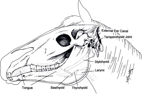
Normally the hyoid apparatus moves during:
Chewing, Swallowing
If fused:
→ normal movement creates excessive stress → pathological fracture can occur in:
Stylohyoid bone and Petrous temporal bone
→ nearby cranial nerves/vestibular structures can be damaged → vestibular signs ± facial paralysis.
Tx
⭐ Ceratohyoidectomy
→ removes part of the ceratohyoid bone
→ reduces mechanical forces transmitted through the hyoid apparatus.
🧠 THO = STYLOHYOID FUSES TO TEMPORAL BONE → FRACTURE/NERVE DAMAGE → VESTIBULAR SIGNS → CERATOHYOIDECTOMY
Clinical signs — PERIPHERAL ⭐⭐⭐
⭐ Head tilt
⭐ Circling toward the side of the lesion
Ataxia, Imbalance, Recumbency
⭐ Horizontal or rotatory nystagmus
Facial nerve paralysis (CN VII) may occur
Mental status is generally normal with a purely peripheral lesion.
🧠 PERIPHERAL = HEAD TILT + HORIZONTAL/ROTATORY NYSTAGMUS + NORMAL MENTATION
🧠 3. CENTRAL VESTIBULAR DISEASE ⭐⭐⭐
Lesion is within the CNS/brainstem.
Occurs less frequently than peripheral vestibular disease.
Etiology
🦠 Infectious diseases
Viral/protozoal
Equine protozoal myeloencephalitis — EPM
Eastern equine encephalomyelitis — EEE
Western equine encephalomyelitis — WEE
West Nile virus — WNV
Rabies
Equine herpesvirus-1 — EHV-1
⚠ EPM is protozoal, not viral.
Bacterial
Listeria, Salmonella
Streptococcus equi → strangles
Burkholderia mallei → glanders
Clostridium botulinum
Other
Trauma, Neoplasia, Brain abscess
Clinical signs — CENTRAL ⭐⭐⭐
⭐ Vertical nystagmus
Circling, Imbalance, Ataxia
⭐ Altered mentation
Other cranial nerve deficits
Because the lesion is in the brainstem/CNS, other neurological abnormalities are more likely.
🧠 CENTRAL = VERTICAL NYSTAGMUS + ALTERED MENTATION + MULTIPLE CN DEFICITS
🔥 PERIPHERAL vs CENTRAL
Peripheral | Central | |
|---|---|---|
Lesion | Inner/middle ear, CN VIII | Brainstem/CNS |
Head tilt | ⭐ Common | Can occur |
Circling | Toward lesion | Can occur |
Nystagmus | ⭐ Horizontal/rotatory | ⭐ Vertical possible |
Mentation | ⭐ Normal | ⭐ Altered |
CN deficits | VII/VIII commonly | Multiple CNs possible |
Examples | Otitis, THO | Encephalitis, EPM, trauma, neoplasia |
🧠 Easiest distinction
PERIPHERAL → EARS + HORIZONTAL/ROTATORY + NORMAL BRAIN
CENTRAL → BRAINSTEM + VERTICAL + ABNORMAL MENTATION
🔬 4. DIAGNOSIS ⭐⭐⭐
1.Full neurological examination
Use the examination from Q18:
Behaviour
Mental status
Head posture
Cranial nerves
Gait
Ataxia
Proprioception
Limb strength
Tail/perineal reflexes
Goal:
⭐ Localize lesion as peripheral or central.
2.Endoscopy
Guttural pouch endoscopy
Can help evaluate structures around:
Stylohyoid bone, Temporohyoid joint
Especially useful with suspected THO.
3.Imaging
X-ray:
Look for:
⭐ THO, Otitis, Masses, Fractures
CT
Very useful for:
Skull, Temporohyoid region, Middle/inner ear
MRI
Useful for:
Brain, Brainstem, Soft tissues
4.💉 CSF ANALYSIS
Collect and examine cerebrospinal fluid.
May demonstrate:
Inflammatory/cytological abnormalities
Changes associated with neurological disease
Your lecture specifically mentions:
Polyneuritis equi
Immunological testing for EPM
EPM can sometimes mimic peripheral vestibular disease.
5.🦠 INFECTIOUS DISEASE TESTING
Serology/PCR/specific testing depending on suspicion:
EPM
EEE
WEE
WNV
EHV-1
💊 TREATMENT
Treatment depends on the underlying cause.
Otitis media/interna →⭐ Prolonged antibiotic therapy
THO → ⭐ Ceratohyoidectomy
Infectious central vestibular disease → specific antimicrobial/antiprotozoal treatment when available, → supportive care
Trauma/inflammation → supportive treatment according to cause and severity.
🧠 EXAM MEMORY
First say:
“The vestibular system controls balance, posture and eye movements and can be divided into peripheral and central components.”
Then differentiate:
🦻 PERIPHERAL
OTITIS + THO
→ head tilt
→ circles toward lesion
→ horizontal/rotatory nystagmus
→ facial paralysis
→ normal mentation
🧠 CENTRAL
ENCEPHALITIS + EPM + TRAUMA + NEOPLASIA
→ vertical nystagmus
→ altered mentation
→ multiple cranial nerve deficits
→ imbalance/ataxia
⭐ THO
STYLOHYOID ↔ PETROUS TEMPORAL BONE FUSION → FRACTURE → VESTIBULAR SIGNS → CERATOHYOIDECTOMY
Spinal ataxia and equine protozoal myeloencephalitis
1. ATAXIA ⭐⭐⭐
Ataxia = incoordination caused by impaired proprioception (sense of body/limb position), often accompanied by some degree of weakness/paresis.
Types of ataxia
Cerebellar ataxia
Cerebral ataxia
Vestibular ataxia
Sensory/proprioceptive ataxia
⭐ Spinal ataxia → motor dysfunction with incoordination of the limbs and/or trunk
🧠 ATAXIA = INCOORDINATION; PARESIS = WEAKNESS
🧠 2. SPINAL ATAXIA
Etiology
Spinal cord dysfunction can result from:
Trauma, Myelopathy, Inflammation
Infectious disease:
EHV-1
Protozoal disease → EPM
Degenerative disease, Neoplasia, Toxins
Compression, e.g., Wobbler syndrome
Clinical signs
Motor dysfunction
⭐ Incoordination of extremities
Trunk instability
Paresis
Proprioceptive deficits
📍 3. SPINAL CORD LESION LOCALIZATION ⭐⭐⭐
This is the important pattern to understand rather than memorize randomly.
First remember:
UMN lesion
→ normal/increased tone and reflexes
→ spastic/strong-looking but poorly coordinated movement
LMN lesion
→ ⭐ weakness
→ decreased tone/reflexes
→ possible muscle atrophy
C1–C5
Lesion is above the limb LMNs.
Thoracic limbs → UMN
Pelvic limbs → UMN
→ all four limbs can be affected.
🧠 C1–C5 = UMN ALL 4
C6–T2
This region contains LMNs supplying the thoracic limbs.
Therefore:
Thoracic limbs → LMN
Pelvic limbs → UMN
🧠 C6–T2 = LMN FRONT + UMN BACK
T3–L3
Below the thoracic limb innervation:
Thoracic limbs → NORMAL
Pelvic limbs → UMN
🧠 T3–L3 = FRONT NORMAL + BACK UMN
L4–S1
Contains LMNs supplying pelvic limbs:
Thoracic limbs → NORMAL
Pelvic limbs → LMN
🧠 L4–S1 = FRONT NORMAL + BACK LMN
Sacral–caudal
Thoracic limbs → normal
Pelvic limbs → normal or LMN, depending on exact lesion.
May produce ⭐ cauda equina signs:
Urinary incontinence/dysfunction
Rectal/fecal incontinence
Reduced tail/anal tone
🔥 Localization table
Lesion | Thoracic limbs | Pelvic limbs |
|---|---|---|
C1–C5 | UMN | UMN |
C6–T2 | ⭐ LMN | UMN |
T3–L3 | Normal | UMN |
L4–S1 | Normal | ⭐ LMN |
Sacral/caudal | Normal | Normal/LMN + cauda equina signs |
🧠 C6–T2 controls FRONT LMN; L4–S1 controls BACK LMN
📊 4. GRADING OF ATAXIA — 0–5 ⭐⭐⭐
Grade 0
→ No neurological deficit detected
Grade 1
→ Very mild deficit
→ not readily detected during normal gait/posture
→ may become apparent with challenging manoeuvres
Grade 2
→ ⭐ Deficit easily detected
→ Exaggerated when challenging the horse
Grade 3
→ ⭐ Deficit prominent at normal gait
Grade 4
→ Severe
Stumbling
Tripping
Spontaneously falling
Grade 5
→ ⭐ Recumbent
→ unable to stand/get up without assistance
🧠 0 NONE → 1 SUBTLE → 2 OBVIOUS WHEN CHALLENGED → 3 OBVIOUS NORMALLY → 4 FALLS → 5 RECUMBENT
🔬 5. DIAGNOSIS OF SPINAL ATAXIA
Neurological examination
Assess: Gait, Proprioception, Paresis, UMN/LMN signs, Tail/anal tone, Cranial nerves where relevant
Radiography
Especially cervical spine: C1–T1
Lateral, Oblique, Neutral, Flexed, Extended views
Myelography
→ evaluates spinal cord compression
Slap test
→ evaluates thoracolaryngeal reflex
As discussed in Q18: ⭐ normal response = brief contralateral laryngeal/arytenoid adduction
Laboratory
CSF, Hematology, Biochemistry, Serology
🦴 6. WOBBLER SYNDROME ⭐⭐⭐
Cervical vertebral stenotic myelopathy — CVSM
= Compression of the cervical spinal cord
Etiology
Can result from:
Vertebral malformation, Malarticulation, Abnormal vertebral development, Displacement, Vertebral canal stenosis
Synovial cysts/other compressive lesions → spinal cord compression → proprioceptive/UMN dysfunction.
Clinical signs
⭐ Usually pelvic limbs affected more severely initially:
Hindlimb ataxia
Paresis, Wide-based stance, Incoordination
As disease progresses → forelimbs may also become affected
🧠 WOBBLER = CERVICAL CORD COMPRESSION → HINDLIMBS WORSE THAN FORELIMBS
Diagnosis
Neurological examination
Cervical radiographs
⭐ Myelography
Advanced imaging where available
Abnormal slap test
Treatment
Glucocorticoids
Neck braces
⭐ Surgical treatment/decompression/stabilization in selected cases
🐴 7. OCCIPITO-ATLANTO-AXIAL MALFORMATION — OAAM ⭐⭐⭐
= Congenital developmental abnormality particularly associated with Arabian horses.
Affects: Occipital bone, Atlas C1 and Axis C2
Typical abnormality:
⭐ Atlas is fused to the occipital bone → abnormal upper cervical anatomy → compression/damage of the cranial cervical spinal cord.
Clinical signs
Foals may:
Be stillborn
Be ⭐ ataxic from birth
Develop progressive ataxia during first months of life
Characteristic posture:
Extended neck
Reduced ability to flex the atlanto-occipital joint
Dx
⭐ X-ray
Genetic testing
Tx
❌ No effective treatment
🧠 OAAM = ARABIAN FOAL → OCCIPUT + C1 + C2 MALFORMATION → ATAXIA FROM BIRTH
🦠 8. EQUINE PROTOZOAL MYELOENCEPHALITIS — EPM ⭐⭐⭐
Etiology
Protozoan:
⭐ Sarcocystis neurona
Mainly important in:
🇺🇸 North America
Hosts
Definitive host (FH):
⭐ Opossum
Intermediate hosts can include:
Raccoons
Other wildlife
Horse:
⭐ Aberrant/dead-end host
🧠 EPM = SARCOCYSTIS NEURONA → OPOSSUM → HORSE = ABERRANT HOST
🔄 Life cycle
Opossum sheds sporocysts in feces → Contaminates Pasture, Feed, Water → Horse ingests sporocysts → Sporozoites released → Parasite disseminates and can enter the CNS → Lesions develop in:
Brain
Brainstem
Spinal cord
→ neurological signs depend on location of CNS lesions
⚠ For your exam, I would avoid saying that S. neurona routinely “forms cysts in the horse brain.” The key point is multifocal inflammatory lesions/meronts in the CNS; horses are aberrant hosts.
🧠 Pathogenesis
Parasite reaches CNS → inflammation + neuronal/tissue damage → multifocal neurological dysfunction.
Because lesions can occur in different places: ⭐ Clinical signs are highly variable and often asymmetric.
🚨 Clinical signs ⭐⭐⭐
Very important characteristic:
⭐ ASYMMETRIC NEUROLOGICAL SIGNS
Can include:
Asymmetric ataxia, Weakness, Incoordination
⭐ Muscle atrophy
Facial sensory deficits, Lameness, Urinary incontinence, Cranial nerve deficits depending on lesion, Other signs depending on CNS localization
incoordination, gait abn, head tilt, facial nerve paralysis, diffic swallowing, loss of sensation, seizures, lameness, blindness, muscle atrophy of gluteus muscle etc.
🧠 EPM = ASYMMETRIC ATAXIA + FOCAL MUSCLE ATROPHY
🔬 Diagnosis
Diagnosis can be difficult because exposure does not necessarily mean neurological disease.
Your lecture lists:
Serology, PCR, Western blot
For the exam, remember:
⭐ Serum + CSF antibody testing can support diagnosis when interpreted together with neurological findings.
So:
neurological examination + compatible signs + laboratory testing
💊 Treatment
Your lecture lists:
Antiprotozoal
Trimethoprim-sulfadiazine
Toltrazuril
Supportive
NSAIDs
⭐ Vitamin E
Vitamin B complex
🧠 EPM Tx = ANTIPROTOZOAL + ANTI-INFLAMMATORY/SUPPORTIVE CARE
🔥 EXAM STRUCTURE FOR Q20
If you draw this question, I would answer it in this order:
1. Define ataxia
→ incoordination + loss of proprioception ± paresis
2. Name types
→ cerebral, cerebellar, vestibular, sensory, spinal
3. Localize spinal lesion
→ C1–5 = UMN/UMN
→ C6–T2 = LMN/UMN
→ T3–L3 = normal/UMN
→ L4–S1 = normal/LMN
4. Grade 0–5
→ 0 normal → 5 recumbent
5. Important spinal disease
→ Wobbler = cervical spinal cord compression
6. Congenital disease
→ OAAM = Arabian foal + C1/C2/occiput
7. EPM
→ S. neurona → opossum → horse aberrant host → asymmetric ataxia + muscle atrophy
🧠 Final memory line
SPINAL ATAXIA = PROPRIOCEPTIVE INCOORDINATION ± PARESIS
WOBBLER = CERVICAL COMPRESSION
OAAM = ARABIAN FOAL
EPM = OPOSSUM → S. NEURONA → ASYMMETRIC ATAXIA + MUSCLE ATROPHY
Peripheral nerve disorders
Peripheral nerve disorders in horses can result from:
⭐ Trauma
Compression/ischemia during anaesthesia
Fractures, Deep injections, Infection/inflammation
Neuromuscular junction disorders, Neurotoxins
⭐ Nerves commonly affected during anaesthesia
Facial nerve, Radial nerve, Suprascapular nerve

🧠 1. PERIPHERAL NERVE INJURIES
Peripheral nerve injuries are common after trauma or prolonged compression.
Main nerves to know:
Ischiatic/sciatic, Femoral, Suprascapular, Radial, Tibial, Facial
🦵 ISCHIATIC / SCIATIC NERVE
Anatomy/function:
Origin → approximately L5–S1 region
Runs from lower back/pelvis into the hindlimb.
Provides major motor and sensory innervation to the pelvic limb and divides into:
→ Tibial nerve and Common fibular/peroneal nerve (n.peroneus communis)
Causes of damage
⭐ Deep IM injections, Pelvic fractures, Femoral fractures, Trauma
Clinical signs
weakness/paresis or partial paralysis of affected hindlimb
Severity depends on the location and extent of nerve damage.
🧠 SCIATIC = DEEP INJECTION + PELVIC/FEMORAL TRAUMA → HINDLIMB PARESIS
🦵 FEMORAL NERVE ⭐⭐
Function:
Innervates the:⭐ Quadriceps
Quadriceps: → extends the stifle
Causes
Dystocia/birth trauma, Prolonged tissue pressure, Anaesthesia in dorsal recumbency, Pelvic trauma
Clinical signs
Damage → quadriceps dysfunction
→ inability to properly extend/fix the stifle
→ limb may collapse/flex during weight bearing
→ difficulty supporting weight.
🧠 FEMORAL → QUADRICEPS → EXTENDS STIFLE
🦴 SUPRASCAPULAR NERVE ⭐⭐⭐
Very important because it produces:
⭐ SWEENEY SHOULDER

Causes:
Kicks, Collision/trauma, Compression/injury, Connective tissue/scar formation after injury
Muscles affected:
Suprascapular nerve supplies: M. supraspinatus, M. infraspinatus → These muscles stabilize the shoulder joint.
Clinical signs:
Early:
→ loss of shoulder stabilization → ⭐ lateral shoulder subluxation / “popping” during weight-bearing
After approximately 2–4 weeks:
→ obvious atrophy of Supraspinatus and Infraspinatus muscles
→ scapular spine (spina scapulae) becomes very prominent = ⭐ Sweeney shoulder
Treatment:
Stall rest, Physiotherapy, Long recovery period
Severe/chronic compression → surgery may be performed to Remove scar tissue, Decompress/reduce tension on nerve
🧠 SUPRASCAPULAR → SUPRA + INFRA ATROPHY → PROMINENT SCAPULAR SPINE = SWEENEY SHOULDER
🦵 RADIAL NERVE ⭐⭐⭐
Function
Innervates muscles involved in:
Elbow extension, Carpal extension, Digital extension
It also contributes to movement around the shoulder.
Causes
⭐ Ischemic/compressive injury during anaesthesia in lateral recumbency
Humeral fractures, Trauma
Clinical signs
⭐ Dropped elbow
Failure/inability to extend the limb, Carpus/digit remain flexed
Difficulty advancing/positioning limb normally. ⭐ Unable to properly bear weight
🧠 RADIAL = EXTENSOR NERVE → DAMAGE = DROPPED ELBOW + CANNOT EXTEND/BEAR WEIGHT
🦵 TIBIAL NERVE
Major terminal branch/continuation of the sciatic nerve.
Innervates → Gastrocnemius, Digital flexors, Other caudal crus muscles
Gastrocnemius contributes strongly to: → ⭐ hock extension
Damage → gastrocnemius weakness/atrophy → inability to maintain normal hock extension → hock flexion/dropped hock
🧠 TIBIAL → GASTROCNEMIUS → EXTENDS HOCK → DAMAGE = FLEXED/DROPPED HOCK

🙂 2. FACIAL NERVE PARALYSIS ⭐⭐⭐
CN VII — Facial nerve
A relatively common peripheral nerve injury.
Etiology
Most classically:
⭐ Horse lies for prolonged period with pressure on side of face
Especially:
Anaesthesia
Halter left on during recumbency
Other causes:
Facial surgery, Rough handling, Otitis media, Trauma, Guttural pouch infection
Clinical signs
A unilateral proximal facial nerve lesion causes paresis/paralysis of facial muscles.
Affected side may show:
⭐ Drooping eyelid
Drooping ear, drooping lip, drooping nostril
Facial asymmetry
⭐ Inability to blink

Auriculopalpebral branch
If only the auriculopalpebral branch is damaged:
→ primarily affects: Ear movement, Eyelid movement/blinking
🧠 FACIAL NERVE = MOVE FACE → DAMAGE = DROOPING EAR/EYELID/LIP/NOSTRIL
Diagnosis
Electromyography — EMG
Can help determine:
Location, Severity, Extent of nerve damage
Treatment
Supportive methods from your lecture:
Acupuncture, Massage, Heat to affected muscles, Laser therapy, Electrotherapy
Peripheral nerves regenerate slowly.
⚠ Protect the eye if the horse cannot blink → risk of corneal damage.
Prognosis → Poor prognosis if there is no improvement within 6 months.
💪 3. MYASTHENIA GRAVIS ⭐⭐⭐
= Disorder of neuromuscular transmission → nerve signal cannot efficiently activate skeletal muscle.
Pathogenesis
Autoantibodies are directed against Acetylcholine receptors — AChR at the neuromuscular junction.
→ ↓ number/function of ACh receptors → impaired neuromuscular transmission → muscle weakness.
🧠 MG = ANTIBODIES AGAINST ACh RECEPTORS
Clinical signs
Characteristic: ⭐ Muscle weakness worsens with exercise and improves with rest
Can also cause:
Megaesophagus, Dysphagia
Megaesophagus:
→ food/material may be aspirated → secondary aspiration pneumonia
🧠 MG = EXERCISE → WORSE; REST → BETTER
Diagnosis
AChR antibodies
Lecture:
IFAT for circulating antibodies against AChR
Tensilon test: Administration of a short-acting anticholinesterase → transient improvement in muscle strength supports neuromuscular transmission dysfunction.
Treatment
⭐ Prednisolone → immunosuppressive treatment.
🦠 4. TETANUS ⭐⭐⭐
Clostridium tetani
→ produces a powerful neurotoxin → ⭐ SPASTIC PARALYSIS
🧠 TETANUS = TIGHT/STIFF
Transmission
C. tetani spores are found in soil.
Enter through wounds, especially where anaerobic conditions develop.
Examples:
Castration
Skin wounds/lesions
Surgical wounds
Other deep contaminated wounds
→ bacteria proliferate under anaerobic conditions → produce neurotoxin → toxin affects nervous system.
Clinical signs ⭐⭐⭐
Initial stiffness/lameness, Progressive generalized muscle stiffness
⭐ Lockjaw — trismus
Elevated/stiff ears, Elevated/stiff tail, Hyperresponsiveness, Arched back
Generalized spastic paralysis
Later → lateral recumbency → legs held rigidly extended
Death may occur due to: ⭐ respiratory muscle spasm/paralysis
Diagnosis
Often sufficient:
Clinical signs
History of recent wound/trauma

Gram stain
C. tetani has characteristic: ⭐ “Tennis-racket” appearance due to terminal spores. G+ → purple color in gram stain
Toxin may also be demonstrated, although diagnosis is usually clinical.
Treatment ⭐⭐⭐
Wound treatment
Very important:
→ open/aerate wound → remove necrotic tissue → clean/debride → your lecture mentions hydrogen peroxide → local antimicrobial management
Antibiotics
Penicillin (kills G+ bacteria)
Tetanus antitoxin
→ neutralizes unbound circulating toxin
⚠ Cannot reverse toxin already bound to nervous tissue.
Supportive
Sedatives, Muscle relaxants, Quiet/dark environment, Supportive nursing
Prevention ⭐⭐⭐
⭐ VACCINATION
🧠 TETANUS = WOUND → TOXIN → SPASTIC PARALYSIS + LOCKJAW → ANTITOXIN + WOUND CARE + ATB → VACCINATE
☠ 5. BOTULISM ⭐⭐⭐
Etiology: Clostridium botulinum
Exposure can be associated with:
⭐ Decaying/contaminated hay, Silage, Contaminated feed
Neurotoxin interferes with neuromuscular transmission → ⭐ FLACCID PARALYSIS
Clinical signs
Incoordination, Muscle tremors, Progressive weakness
⭐ Flaccid paralysis, Drooling, Dysphagia, Recumbency, Colic
🧠 TETANUS = SPASTIC 🔒 vs BOTULISM = FLACCID 🫠
🌱 6. EQUINE GRASS SICKNESS ⭐⭐⭐
= Highly fatal neuropathy of horses.
→ Causes degeneration of neurons of the:
⭐ Autonomic nervous system, especially the⭐ Enteric nervous system
Therefore, the disease is predominantly manifested by GIT signs.
Etiology
The exact cause is not fully established.
Your lecture mentions a proposed association with: Clostridium botulinum type C/toxicoinfection → intestinal toxin production → neuronal damage.
For the exam, phrase it as:
“The exact cause is unknown, but clostridial toxicoinfection has been proposed.”
Clinical consequences:
Autonomic/enteric neuronal degeneration → severe gastrointestinal dysfunction.
Can result in:
Dysphagia, Reduced/absent intestinal motility, Colic, Gastric distension, Constipation, Weight loss depending on form
Diagnosis
Presumptive
Clinical signs, History, Physical examination
Definitive
⭐ Histopathology/biopsy
Post-mortem findings may include:
Gastric distension, Colon containing hard, dry feces
Treatment
Primarily: Supportive care
Severe/acute cases: → poor prognosis → euthanasia often recommended.
🧠 GRASS SICKNESS = AUTONOMIC/ENTERIC NEURON DEGENERATION → GIT FAILURE
☠ 7. ORGANOPHOSPHATE POISONING ⭐⭐⭐
Etiology
Exposure to:
Insecticides, Contaminated food, Contaminated water
Overdosing of antiparasitic/insecticidal preparations
Pathogenesis
Organophosphates → ⭐ INHIBIT ACETYLCHOLINESTERASE
Normally:
ACh (acetylcholine) released → signal → acetylcholinesterase breaks down ACh.
With organophosphate:
❌ AChE inhibited → acetylcholine accumulates → continuous cholinergic stimulation → excessive:
Autonomic activity
Neuromuscular activity
🧠 ORGANOPHOSPHATE = ↓ AChE → ↑ ACh → TOO MUCH CHOLINERGIC ACTIVITY
Clinical signs
Anxiety, ⭐ Salivation, Bradycardia, Stiff gait, Muscle tremors, Incoordination, Ataxia, Respiratory dysfunction/failure
Treatment
⭐ Atropine → blocks muscarinic effects of excess acetylcholine.
Plus:
⭐ Oximes, e.g. pralidoxime → can reactivate acetylcholinesterase if administered appropriately/early.
🧠 ORGANOPHOSPHATE → AChE INHIBITION → ATROPINE + OXIME
🔥 EXAM SUMMARY — KNOW THESE ASSOCIATIONS
Disorder/nerve | Key association |
|---|---|
Sciatic nerve | Deep injection / pelvic trauma → hindlimb paresis |
Femoral nerve | ⭐ Quadriceps → cannot extend/stabilize stifle |
Suprascapular nerve | ⭐ Sweeney shoulder |
Radial nerve | ⭐ Dropped elbow + cannot extend limb |
Tibial nerve | Gastrocnemius → flexed/dropped hock |
Facial nerve | ⭐ Drooping face + inability to blink |
Myasthenia gravis | ⭐ AChR antibodies → exercise-induced weakness |
Tetanus | ⭐ Spastic paralysis + lockjaw |
Botulism | ⭐ Flaccid paralysis |
Grass sickness | ⭐ Autonomic/enteric neuron degeneration |
Organophosphate | ⭐ AChE inhibition → cholinergic overstimulation |
🧠 Final memory line
SWEENEY = SUPRASCAPULAR
DROPPED ELBOW = RADIAL
CAN'T EXTEND STIFLE = FEMORAL
DROPPING FACE = FACIAL
MG = AChR ANTIBODIES
TETANUS = SPASTIC
BOTULISM = FLACCID
GRASS SICKNESS = AUTONOMIC GUT
ORGANOPHOSPHATE = AChE ↓ → ACh ↑
Equine endocrinopathies
The main endocrinopathies to know:
⭐ PPID / Equine Cushing's disease
⭐ Insulin resistance / insulin dysregulation
⭐ Equine metabolic syndrome — EMS
Diabetes mellitus — rare
Hypoadrenocorticism
Hypothyroidism
🧠 1. PPID — PITUITARY PARS INTERMEDIA DYSFUNCTION ⭐⭐⭐
Also called: Equine Cushing's disease — ECD
Epidemiology
⭐ Most common endocrinopathy of older horses, Especially horses >10 years (Prevalence increases with age)
Etiology + pathogenesis
→ tumor/enlargement of the pars intermedia of the pituitary gland → excessive production of ACTH → adrenal stimulation → ↑ cortisol → Cushing-like clinical signs.
Loss of dopaminergic inhibition of the pars intermedia → hyperplasia/adenoma → excessive production of POMC-derived hormones, including ACTH.
This explains why: ⭐ Pergolide = dopamine agonist works.
🧠 PPID = OLD HORSE + PARS INTERMEDIA + ↑ ACTH
Clinical signs ⭐⭐⭐
1. Abnormal hair coat
Your lecture calls this: ⭐ HIRSUTISM
→ More modern term: Hypertrichosis
Retention of winter coat, Abnormally long hair, Delayed/incomplete shedding
This is a highly characteristic sign of advanced PPID!!
🧠 OLD HORSE THAT DOESN'T SHED → THINK PPID

2. Laminitis ⭐⭐⭐
PPID horses have increased risk of → endocrinopathic laminitis
This is one of the most clinically important complications.
3. Abnormal fat distribution
Fat can accumulate:
⭐ Neck → cresty neck
Around tail head
May also have muscle wasting despite regional fat deposition.
4. PU/PD
Polyuria, Polydipsia
5. Recurrent/chronic infections
Increased susceptibility to:
Tooth root infections, Sinusitis, Sole abscesses, Other infections
Also:
⭐ Delayed wound healing
Other signs
Lethargy, Muscle weakness/wasting, ⭐ Excessive sweating, Abnormal body condition
🔬 Diagnosis
Clinical signs
An older horse with: ⭐ hypertrichosis + laminitis + muscle wasting/abnormal fat distribution → strongly suggests PPID.
Resting plasma ACTH ⭐⭐⭐ Measure: plasma ACTH concentration (>35pg/ml → positive)
⚠ For understanding: ACTH varies substantially with season, so modern interpretation uses seasonally adjusted reference intervals rather than one fixed cutoff throughout the year.
CBC
Lecture abnormalities:
Neutrophilia, Lymphopenia, Monocytosis
Glucose: May show Hyperglycemia, Glucosuria, Especially when insulin dysregulation is also present.
Endocrine tests
⭐ Resting ACTH
TRH stimulation test
Dexamethasone suppression test — in your lecture
ACTH stimulation test — in your lecture
💊 Treatment
⭐ Pergolide mesylate
Pergolide = dopamine agonist
→ restores dopaminergic inhibition of pars intermedia activity → reduces excessive hormone production.
🧠 PPID → PERGOLIDE
Trilostane
→ inhibits adrenal steroid synthesis → ↓ cortisol production.
But pergolide is the key PPID treatment to know!
Management
Very important: ⭐ Prevent/manage laminitis
Also:
Appropriate diet, Hoof care, Treat infections, Dental care, Clip excessive hair if necessary, Monitor body condition
🍬 2. INSULIN RESISTANCE / INSULIN DYSREGULATION ⭐⭐⭐
Insulin resistance = decreased tissue response to circulating insulin.
Therefore insulin is less effective at promoting normal glucose metabolism in:
Skeletal muscle, Adipose tissue, Liver
Important distinction:
⭐ Horses with EMS (equine metabolic syndrome) commonly have insulin dysregulation, but insulin resistance/dysregulation can occur without the full EMS phenotype.
Etiology/mechanisms
↓ density of insulin receptors
Malfunction of insulin receptors
Problems with glucose transporter translocation/function
Result: → tissues respond poorly to insulin.
Pathogenesis
Compensated insulin resistance ⭐⭐⭐
Most common situation:
Tissues respond poorly to insulin → Pancreas produces MORE insulin → ⭐ Hyperinsulinemia → Blood glucose may initially remain relatively normal.
🧠 COMPENSATED IR = ↑ INSULIN TO KEEP GLUCOSE CONTROLLED
Uncompensated IR / β-cell exhaustion
Eventually: → pancreatic β-cells cannot compensate adequately → insulin secretion becomes insufficient relative to need → persistent hyperglycemia → possible type 2 diabetes mellitus
🍬 Diabetes mellitus
Very rare in horses.
More likely in horses with severe/advanced endocrine disease such as PPID with insulin dysregulation.
When hyperglycemia becomes severe → glucose exceeds renal threshold → glucosuria → osmotic diuresis → ⭐ polyuria → polydipsia.
🧠 DIABETES = HYPERGLYCEMIA → GLUCOSURIA → OSMOTIC DIURESIS → PU/PD
🐴 3. EQUINE METABOLIC SYNDROME — EMS ⭐⭐⭐
EMS is an endocrine/metabolic disorder characterized principally by:
⭐ Insulin dysregulation
⭐ Regional/generalized adiposity in many affected horses
⭐ High risk of endocrinopathic laminitis
Predisposed horses:
Often affects “thrifty” equids:
⭐ Ponies, Donkeys, Arabians, Mustangs
These animals are genetically/metabolically efficient at storing energy.
Pathogenesis
Adipose tissue → production of adipokines → hormonal/metabolic disturbance → impaired normal insulin response → hyperinsulinemia ± altered glucose metabolism.
The key exam concept is:
EMS triad
ADIPOSITY + INSULIN DYSREGULATION/HYPERINSULINEMIA + LAMINITIS RISK
🚨 Laminitis ⭐⭐⭐
This is the major clinical consequence.
Insulin dysregulation → prolonged/excessive hyperinsulinemia → lamellar dysfunction → ⭐ endocrinopathic laminitis
⚠ Your notes say hyperglycemia causes the lamellar damage. For the exam, if you want the more accurate key association, remember: HYPERINSULINEMIA → LAMINITIS
rather than hyperglycemia itself being the main mechanism.
🧠 EMS → HIGH INSULIN → HIGH LAMINITIS RISK
Clinical signs
Adiposity: Can be Generalized obesity or ⭐ Regional adiposity. Typical locations:
→ Cresty neck: fat along neck crest
→ Tail head: abnormal fat deposits
Other: Prepuce, Under skin of trunk, Other regional deposits
⭐ LAMINITIS
Some horses may have insulin dysregulation without being generally obese.
🔬 Diagnosis
Resting serum insulin: ⭐ Important screening test.
Dynamic testing: Combined glucose-insulin test → assesses insulin/glucose regulation.
💊 Treatment of EMS ⭐⭐⭐
1. MANAGEMENT — MOST IMPORTANT
Weight loss
For obese horses: → controlled reduction of body condition.
Diet
⭐ Restrict pasture access
Low sugar, Low starch, Reduce non-structural carbohydrates
Appropriate forage management
Exercise
⭐ Regular exercise when safe.
⚠ Do not exercise a horse with active painful laminitis until appropriately managed.
🧠 EMS Tx = DIET + WEIGHT LOSS + EXERCISE
2. Medical treatment
For selected horses not responding adequately to management:
Metformin
→ lowers blood glucose
→ improves insulin sensitivity/metabolic control.
Levothyroxine sodium
→ promotes weight loss
→ can improve insulin sensitivity in obese horses.
But remember:
⭐ Medication does not replace dietary/management changes.
🔥 PPID vs EMS — VERY IMPORTANT
PPID | EMS | |
|---|---|---|
Typical horse | ⭐ Older horse | Often younger/middle-aged “thrifty” horse |
Main problem | Pituitary pars intermedia dysfunction | ⭐ Insulin dysregulation |
Hair coat | ⭐ Hypertrichosis | Usually normal |
Body condition | Muscle wasting ± abnormal fat | ⭐ Obesity/regional adiposity common |
Cresty neck | Can occur | ⭐ Common |
Laminitis | ⭐ Common | ⭐ Common |
PU/PD | Common | Not classic unless marked metabolic disturbance |
Main diagnostic test | ⭐ ACTH / TRH stimulation | ⭐ Insulin testing |
Main treatment | ⭐ Pergolide | ⭐ Diet + weight loss + exercise |
🧠 Easy distinction
OLD + HAIRY → PPID
FAT/CRESTY + HIGH INSULIN → EMS
BOTH → LAMINITIS
🟠 4. HYPOADRENOCORTICISM
Also called:
Adrenal insufficiency → “Steroid let-down syndrome” → insufficient adrenal corticosteroid activity.
Treatment
⭐ Prednisolone
🧠 HYPOADRENOCORTICISM → CORTICOSTEROID REPLACEMENT
🦋 5. HYPOTHYROIDISM
True primary hypothyroidism is uncommon in adult horses.
Clinical sign from your lecture
⭐ Goitre
Treatment
⭐ Levothyroxine sodium → thyroid hormone supplementation.
🧠 HYPOTHYROIDISM → GOITRE → LEVOTHYROXINE
🔥 EXAM STRUCTURE FOR Q22
Start with the two important diseases:
1⃣ PPID
Old horse → pars intermedia → ↑ ACTH → hypertrichosis + laminitis + PU/PD → ACTH/TRH → PERGOLIDE
2⃣ INSULIN RESISTANCE
↓ tissue response to insulin → compensatory hyperinsulinemia
3⃣ EMS
Thrifty horse → regional adiposity + insulin dysregulation → LAMINITIS → insulin testing → DIET + WEIGHT LOSS + EXERCISE
Then briefly mention:
Diabetes → rare → hyperglycemia + glucosuria + PU/PD
Hypoadrenocorticism → prednisolone
Hypothyroidism → goitre → levothyroxine
🧠 Final memory line
PPID = OLD + HAIRY → ACTH → PERGOLIDE
EMS = CRESTY/FAT + HIGH INSULIN → LAMINITIS → DIET/EXERCISE
DIABETES = RARE
HYPOADRENOCORTICISM = PREDNISOLONE
HYPOTHYROIDISM = GOITRE → LEVOTHYROXINE
Equine myopathies
Main conditions:
⭐ Exertional rhabdomyolysis — “Monday morning disease”
⭐ Polysaccharide storage myopathy — PSSM
⭐ Hyperkalemic periodic paralysis — HYPP
Post-anesthetic myopathy/myoneuropathy
Nutritional myopathy — Vit E/Se deficiency
Sweeney shoulder — suprascapular nerve damage
🔬 1. DIAGNOSTIC APPROACH TO THE MUSCULAR SYSTEM
Physical examination
Inspection
Symmetry, Size, Shape of muscle groups, Muscle wasting/atrophy
Palpation
Muscle tone, Sensitivity/pain, Asymmetry, Atrophy, Swelling, heat
Gait
Walk the horse and look for: Lameness, Weakness, Stiffness, Pain, Reluctance to move
🩸 Clinical pathology ⭐⭐⭐
Three important muscle enzymes:
⭐ CK — Creatine kinase/creatine phosphokinase/CPK.
Found mainly in:
Skeletal muscle, Cardiac muscle
Muscle damage → ↑ CK
⚠ Training, transport and exercise can cause mild increases.
🧠 CK = sensitive indicator of RECENT muscle damage
LDH — Lactate dehydrogenase
Not tissue-specific, Found in: Muscle, Liver, Erythrocytes
→ can increase with muscle damage, but not specific.
AST — Aspartate aminotransferase
High activity in Skeletal muscle, Liver, Erythrocytes
→ ↑ with muscle damage but also other tissue injury.
🧠 MYOPATHY BLOODWORK = CK + AST + LDH
🔬 Muscle biopsy
Percutaneous biopsy commonly from:
⭐ Gluteus medius
Semitendinosus
→ sample normal and abnormal muscle where appropriate.Useful for diagnosing structural/metabolic myopathies.
⚡ Electromyography — EMG
Used to investigate: ⭐ Neuromuscular disease
🏇 2. EXERTIONAL RHABDOMYOLYSIS ⭐⭐⭐
Also called:
Paralytic myoglobinuria
Monday morning disease
“Tying-up”
Classical presentation:
Horse in training → rested for >1 day, BUT continues receiving a full/high-carbohydrate diet → returned to exercise → develops painful muscle disease.
🧠 REST + FULL GRAIN → EXERCISE → TYING-UP
Etiology
⭐ Dietary imbalance
Overexertion
Defects in intracellular Ca²⁺ regulation
Breed predisposition
Different underlying myopathies can produce exertional rhabdomyolysis.
Pathogenesis
During rest + high carbohydrate feeding → ↑ glycogen storage in muscles
During exercise → increased energy demand → muscle metabolic dysfunction/injury → muscle fiber damage → intracellular contents released.
One very important product: ⭐ MYOGLOBIN
Muscle damage → myoglobin enters blood → filtered by kidneys → myoglobinuria → urine can become dark/red-brown.
Severe muscle damage/dehydration: → risk of acute kidney injury
Your lecture also describes accumulation of lactic acid causing muscle irritation, pain and damage.
For the exam, the safest core pathway is:
Exercise → muscle fiber injury/rhabdomyolysis → CK/AST ↑ + myoglobin release → myoglobinuria ± renal injury
Clinical signs ⭐⭐⭐
Severity is variable:
⭐ Stiff gait, Reluctance to move, Painful muscles, Sweating
Tachycardia, Tachypnea
Pyrexia
Muscle cramping,
Dark urine/myoglobinuria in severe cases
🧠 TYING-UP = PAINFUL + STIFF AFTER EXERCISE
Diagnosis
Clinical signs/history
⭐ ↑ CK, ↑ AST, ↑ LDH
⭐ Myoglobinuria
Fluid abnormalities
Electrolyte abnormalities
💊 Treatment
Goals:
⭐ Limit further muscle damage
Maintain fluid balance
Correct electrolytes
⭐ Prevent renal damage/failure
Control pain
Mild cases:
Phenylbutazone OR flunixin meglumine
Acepromazine
Rest
Vitamin E + Selenium
Acepromazine:
→ sedation + vasodilation → may help reduce anxiety/stiffness.
Severe cases:
⭐ IV fluids
Corticosteroids, Phenylbutazone
Xylazine
Rest — 6–8 weeks
⚠ Return to exercise should depend on the cause and clinical/biochemical recovery rather than automatically applying the same 6–8 week period to every rhabdomyolysis case.
Prevention ⭐⭐⭐
Appropriate exercise management
Avoid sudden exercise after prolonged rest
⭐ High-fat, low-starch diet
Adapt carbohydrate intake to workload
🧠 RHABDO PREVENTION = REGULAR EXERCISE + ↓ STARCH + ↑ FAT
🧬 3. POLYSACCHARIDE STORAGE MYOPATHY — PSSM ⭐⭐⭐
PSSM is an important cause of:
⭐ Exertional rhabdomyolysis
Predisposed breeds:
⭐ Quarter Horses, Draft horses, Warmbloods
Pathogenesis
Abnormal muscle carbohydrate/glycogen metabolism/storage → abnormal accumulation of glycogen/polysaccharide in skeletal muscle → predisposition to muscle dysfunction → exertional rhabdomyolysis.
Clinical signs
Similar to exertional rhabdomyolysis:
Muscle stiffness, Pain, Reluctance to move
Exercise intolerance, Episodes of tying-up
Treatment/management ⭐⭐⭐
Key nutritional principle:
⭐ SUPPLEMENT FAT — NOT SUGAR
→ reduce starch/sugar intake
→ use fat as alternative energy source
→ regular controlled exercise.
🧠 PSSM = POLYSACCHARIDE STORAGE → TYING-UP → LOW STARCH + HIGH FAT
⚡ 4. HYPERKALEMIC PERIODIC PARALYSIS — HYPP ⭐⭐⭐
Inherited muscle disorder particularly associated with: ⭐ Quarter Horses
Characterized by intermittent episodes of:
Muscle tremors, Weakness, Possible collapse
Pathogenesis ⭐⭐⭐
HYPP is a disorder of skeletal muscle sodium channels (Na+)
Abnormal ion movement → excessive/persistent sodium influx → abnormal membrane depolarization → muscle fiber excitability → fasciculations/tremors, followed by:
→ weakness ± paralysis.
🧠 HYPP = Na⁺ CHANNEL DEFECT → ABNORMAL DEPOLARIZATION → TREMOR + WEAKNESS
Clinical signs
Often begins in relatively young horses.
⭐ Recurrent episodes
Muscle fasciculations/tremors, Weakness, Collapse, Possible recumbency
Between attacks: → horse may appear normal.
Diagnosis ⭐⭐⭐
DNA test: ⭐ Genetic test for HYPP mutation. Very important.
Clinical signs, Signalment/history
Potassium chloride challenge test: → administer KCl → provoke clinical signs.
⚠ This is historical and potentially dangerous; genetic testing is the key diagnostic method to remember!
Treatment
Mild attack
Horse trembling but not recumbent:
Light exercise, Feed grain, ⭐ Acetazolamide
Severe attack
Recumbency:
IV catheter/access
⭐ Calcium gluconate
If insufficient response, lecture lists:
Sodium bicarbonate, Dextrose
These help stabilize membrane excitability and/or shift potassium intracellularly.
🧠 HYPP Dx = DNA; Tx = ACETAZOLAMIDE / severe → IV therapy
🛏 5. POST-ANESTHETIC MYOPATHY / MYONEUROPATHY ⭐⭐⭐
Pathogenesis
During prolonged anesthesia: Body weight + immobility → prolonged muscle compression → ↓ blood flow → ⭐ ischemia + hypoperfusion → muscle injury/necrosis.
Large, heavy, well-muscled horses are especially susceptible.
🧠 ANAESTHESIA → PRESSURE → ISCHEMIA → MUSCLE DAMAGE
Muscles commonly affected
Triceps, Quadriceps, Hindlimb extensors, Longissimus, Masseter, Gluteal muscles
Position matters ⭐⭐⭐
Lateral recumbency → Triceps, Deltoid, Dependent hindlimb muscles
Dorsal recumbency → Longissimus, Gluteal muscles
Clinical signs
Localized
Affected muscle may be ⭐ Hard OR flaccid, Hot, Painful, Swollen
Horse may:
Be weak, Have difficulty standing, Be unable to stand
Generalized
Weakness, Paresis, Restlessness, Anxiety, Sweating, Colic-like signs
Diagnosis
Important history: ⭐ Recent/prolonged general anesthesia
Plus:
Clinical signs
↑ CK, ↑ AST, ↑ LDH
Treatment
Mild
Phenylbutazone OR flunixin
Dimethyl sulfoxide — DMSO
Severe
NSAIDs, Xylazine, Butorphanol, Acepromazine
⭐ Fluid therapy
Mannitol
Also supportive nursing and assistance with standing where appropriate.
🍃 6. NUTRITIONAL MYOPATHY
⭐ Vitamin E + selenium deficiency
These are important antioxidant nutrients.
Deficiency:
→ oxidative muscle damage → myopathy.
🧠 NUTRITIONAL MYOPATHY = ↓ VIT E + ↓ Se
🦴 7. SWEENEY SHOULDER
This was also covered in Q21 — peripheral nerve disorders.
Cause
Damage to:
⭐ SUPRASCAPULAR NERVE
→ denervation/atrophy of:
M. supraspinatus
M. infraspinatus
→ prominent scapular spine
= ⭐ Sweeney shoulder
🧠 SWEENEY = SUPRASCAPULAR NERVE → SUPRA + INFRA ATROPHY
🔥 EXAM DIFFERENTIATION
Disease | Key association |
|---|---|
Exertional rhabdomyolysis | ⭐ Exercise → painful stiff muscles + ↑ CK ± myoglobinuria |
PSSM | ⭐ Abnormal glycogen/polysaccharide storage → tying-up |
HYPP | ⭐ Quarter Horse + Na⁺ channel defect → episodic tremor/weakness |
Post-anesthetic myopathy | ⭐ Compression/ischemia after GA |
Nutritional myopathy | ⭐ Vitamin E + Se deficiency |
Sweeney shoulder | ⭐ Suprascapular nerve → supra/infraspinatus atrophy |
🧠 Final memory
RHABDO = EXERCISE → MUSCLE BREAKDOWN → CK ↑ + MYOGLOBINURIA
PSSM = GLYCOGEN/POLYSACCHARIDE → LOW STARCH + HIGH FAT
HYPP = QUARTER HORSE + Na⁺ CHANNEL → TREMORS/WEAKNESS → DNA TEST
POST-ANAESTHETIC = PRESSURE → ISCHEMIA
NUTRITIONAL = VIT E + Se DEFICIENCY
SWEENEY = SUPRASCAPULAR NERVE
Hepatopathies and intoxications
Main parts:
⭐ Diagnostic approach to liver disease
General treatment of liver failure
Theiler's disease
Chronic active hepatitis
Cirrhosis/fibrosis
Cholangiohepatitis + biliary calculi
Tyzzer's disease
Liver flukes
⭐ Important intoxications
🔬 1. DIAGNOSTIC APPROACH TO HEPATOPATHIES
History
Age
Foals → congenital/neonatal diseases
Older horses → chronic/fibrotic disease
Single vs multiple animals affected
Multiple → think toxicosis/feed problem or infectious/parasitic disease
Duration, Progression, Feed/pasture, Drugs/treatments, Exposure to toxic plants
🩸 Laboratory diagnosis ⭐⭐⭐
A. Hepatocellular damage
Enzymes associated with hepatocellular injury:
⭐ GLDH — glutamate dehydrogenase
SDH — sorbitol dehydrogenase
LDH — lactate dehydrogenase
AST — aspartate aminotransferase
🧠 HEPATOCYTES → GLDH + SDH
B. Biliary tract damage / cholestasis
Important enzymes:
⭐ GGT — gamma-glutamyl transferase
ALP/AP — alkaline phosphatase
🧠 BILE DUCT → GGT + ALP
C. Liver function
With impaired liver function:
⭐ ↑ Direct/conjugated bilirubin
⭐ ↑ Serum bile acids
↑ Blood ammonia
↓ Albumin
↓ Urea production
Hypoglycemia can occur
↑ Triglycerides/cholesterol may occur
⚠ Your notes have both “decreased blood urea” and “increased BUN.” With true severe hepatic insufficiency, urea synthesis can decrease, while BUN can vary with hydration, renal function, protein metabolism, etc. For the exam, remember ↓ urea synthesis as the direct liver-function concept.
🩸 Clotting function
The liver synthesizes many clotting factors.
Liver failure:
→ impaired coagulation
→ ⭐ prolonged prothrombin time — PT
🔬 Liver biopsy ⭐⭐⭐
Used to determine:
Fibrosis, Inflammation, Type of lesion, Distribution/location of disease, Severity/prognosis
🧠 BIOPSY = WHAT TYPE + HOW MUCH FIBROSIS?
💻 Ultrasound
Liver can be examined mainly on the right side, caudal/ventral to the lung field, approximately: 6th–15th intercostal spaces
Can identify:
Abscesses, Masses, ⭐ Choleliths, Biliary sludge, Dilated bile ducts, Chronic fibrosis, Hepatomegaly
2. GENERAL TREATMENT OF LIVER FAILURE
Treatment is primarily:
⭐ SUPPORTIVE + TREAT UNDERLYING CAUSE
Fluid therapy
IV balanced electrolyte solutions as appropriate.
Correct:
Dehydration, Electrolyte abnormalities, Glucose abnormalities, Acid-base disturbances
🧠 Hepatic encephalopathy
Liver failure → inadequate detoxification of ammonia → ↑ blood ammonia → CNS dysfunction → hepatic encephalopathy
Methods to reduce intestinal ammonia production/absorption:
Lactulose, Neomycin, Mineral oil, Vinegar/acidification in older lecture protocols
🧠 LIVER FAILURE → ↑ NH₃ → BRAIN → HEPATIC ENCEPHALOPATHY
🟠 3. ACUTE HEPATIC NECROSIS — THEILER'S DISEASE ⭐⭐⭐
Theiler´s disease = Acute/peracute severe hepatic necrosis resulting in ⭐ Acute liver failure
Historically also called serum hepatitis.
Etiology
Unknown
Classically, there may be a history of ⭐ Tetanus antitoxin 4–10 weeks previously
So the exam association is:
HORSE + TETANUS ANTITOXIN WEEKS AGO + ACUTE LIVER FAILURE → THEILER'S DISEASE
Clinical signs
Peracute liver failure:
⭐ Hepatic encephalopathy
Icterus, Depression/neurological signs, Red/discolored urine, Rapid deterioration
Diagnosis
History, Acute onset
⭐ ↑ liver enzymes, Liver function abnormalities
Histopathology
🧠 THEILER = ANTITOXIN HISTORY → PERACUTE NECROSIS → ICTERUS + ENCEPHALOPATHY
🟠 4. CHRONIC ACTIVE HEPATITIS ⭐⭐⭐
= Chronic, active, progressive inflammatory liver disease
Can eventually progress to → fibrosis, liver failure, major CNS involvement/hepatic encephalopathy, death.
Lesion location: ⭐ Often periportal
Etiology
Not fully determined. Possible: ⭐ Immune-mediated/autoimmune mechanism
Clinical signs
Often intermittent/nonspecific:
Depression, ⭐ Weight loss, variable icterus, Recurrent/intermittent signs
Diagnosis
Laboratory
⭐ ↑ GGT, ↑ ALP, ↑ Bilirubin
Bilirubin in urine — from your lecture
Leukocytosis, Monocytosis, ↑ Total plasma protein
Liver biopsy
Important to assess:
Inflammation, Fibrosis, Distribution
Treatment
Supportive care, Plus lecture corticosteroid protocol:
Dexamethasone
→ approximately 5 days → gradually reduce over ~10 days → change to oral prednisolone for ~5 weeks.
🧠 CHRONIC ACTIVE HEPATITIS = PROGRESSIVE INFLAMMATION → PERIPORTAL → SUPPORT + CORTICOSTEROIDS
🟤 5. CIRRHOSIS / HEPATIC FIBROSIS
End-stage consequence of:
→ hepatocyte death, chronic inflammation, ⭐ extensive fibrosis.
Liver architecture becomes distorted:
Depressed fibrous bands, Alternating regenerative nodules of parenchyma
Clinical signs
Most characteristic:
⭐ Chronic progressive weight loss
Later:
Icterus, Hepatic encephalopathy, Other signs of liver failure
Diagnosis
⭐ Liver biopsy → determines degree of fibrosis.
Treatment
❌ Fibrosis cannot generally be reversed once advanced, → supportive treatment only.
Prognosis
🔴 Poor
🧠 CIRRHOSIS = END-STAGE FIBROSIS → BIOPSY → POOR PROGNOSIS
🦠 6. CHOLANGIOHEPATITIS + BILIARY CALCULI ⭐⭐⭐
Importance
⭐ One of the most important/common biliary tract diseases in horses.
Pathogenesis:
Probably starts as Cholangitis ( inflammation/infection of bile ducts) → extends into liver → Cholangiohepatitis
Chronic disease may be associated with:
⭐ Intrahepatic biliary calculi
⭐ Extrahepatic biliary calculi
Severe cases:
→ suppurative inflammation, → bridging fibrosis.
Clinical signs
Nonspecific:
Fever, ⭐ Icterus, Weight loss, Hepatic encephalopathy
Diagnosis
Ultrasound
Look for:
Choleliths, Dilated bile ducts, Biliary abnormalities
Laboratory
⭐ ↑ GGT, ↑ ALP, ↑ Bilirubin
Hematological inflammatory changes
Treatment
⭐ Long-term antibiotics
Preferably: → based on bacterial culture + sensitivity
🧠 CHOLANGIOHEPATITIS = BILE DUCT INFECTION → GGT ↑ + ICTERUS → LONG-TERM ATB
🐴 7. TYZZER'S DISEASE ⭐⭐⭐
= ⭐ Acute, highly fatal hepatitis of foals
Etiology
Correct key organism: ⭐ Clostridium piliforme
(now commonly called Clostridium piliforme; historically Bacillus piliformis)
⚠ Your notes also list Clostridium perfringens, but Tyzzer's disease specifically = C. piliforme.
Transmission
Spores in environment → fecal-oral → infection → severe hepatic disease/necrosis.
Clinical signs
Acute onset:
Depression, Anorexia, Recumbency, Ataxia, ⭐ Icterus, Fever, Often rapid death
Diagnosis
⭐ Liver biopsy/histopathology
PCR
Treatment
Lecture: IV penicillin
But: ⚠ Disease progresses rapidly, so treatment is often unsuccessful.
🧠 TYZZER = FOAL + C. PILIFORME + ACUTE FATAL HEPATITIS
🪱 8. LIVER FLUKE
Rare in horses.
Etiology
Trematodes:
⭐ Fasciola hepatica, Fasciola gigantica
IH: ⭐ Snail
🔄 Life cycle
Adult flukes in bile ducts → eggs passed in feces → miracidium develops/hatches → penetrates snail → develops through larval stages → cercariae leave snail → attach to vegetation and become metacercariae → horse eats contaminated vegetation → larvae released in intestine → penetrate/migrate to liver → young flukes tunnel through liver tissue → after approximately 8 weeks, enter bile ducts → mature adults.
🧠 FASCIOLA = SNAIL → GRASS/METACERCARIA → INTESTINE → LIVER → BILE DUCT
Clinical signs
Fever, ⭐ Icterus, Weight loss
Diagnosis
Coprology:⭐ Sedimentation, because trematode eggs are heavy.
Blood: May have increased: SDH, AST, ALP, LDH
Serology → ELISA
Treatment
Your lecture lists:
⭐ Triclabendazole
Praziquantel
Snail control
For your exam, triclabendazole is the drug I would strongly associate with Fasciola.
🧠 LIVER FLUKE → SEDIMENTATION → TRICLABENDAZOLE
☠ 9. INTOXICATIONS
Important groups:
⭐ Pyrrolizidine alkaloids
Castor bean
Maple
Cyanogenic plants
Organophosphates/carbamates
⭐ Mycotoxins
🌿 A. PYRROLIZIDINE ALKALOID TOXICITY ⭐⭐⭐
Plants
Lecture lists:
⭐ Ragwort, Fiddleneck, Rattleweed, Hound's tongue, “Salivation Jane” in your notes
Pathogenesis
Repeated ingestion over: ⭐ weeks → months
→ cumulative hepatotoxicity → progressive hepatocyte damage → fibrosis/liver failure → hepatic encephalopathy.
Clinical signs
⭐ Weight loss, Inappetence, Liver failure, ⭐ Hepatic encephalopathy
🧠 PYRROLIZIDINE = CHRONIC PLANT INGESTION → LIVER DAMAGE → ENCEPHALOPATHY
🌱 B. CASTOR BEAN
Seeds contain ⭐ RICIN, Highly toxic.
Clinical signs
Incoordination, Sweating, Muscle spasms, Tachycardia, Tachypnea
Diarrhea, Colic, Convulsions, Shock
Severe poisoning → rapid death, potentially within approximately 35 hours according to your lecture.
🧠 CASTOR BEAN = RICIN → SEVERE GI + SYSTEMIC TOXICITY/SHOCK
🍁 C. MAPLE TOXICITY ⭐⭐⭐
Certain maple exposure can cause severe oxidative damage to RBCs.
→ ⭐ Methemoglobinemia
→ Heinz body formation
→ hemolysis
Clinical signs
Icterus, ⭐ Cyanosis, Respiratory distress, Weakness
Treatment
Your lecture:
⭐ Methylene blue
🧠 MAPLE → RBC OXIDATION → METHEMOGLOBIN + HEINZ BODIES
☠ D. CYANOGENIC GLYCOSIDES ⭐⭐⭐
Plants
Lecture:
Hydrangea, Flax, Cherry, Sudan grass
Contain cyanogenic glycosides.
Pathogenesis
Glycosides → release hydrogen cyanide (HCN) → ⭐ inhibits cytochrome c oxidase → cells cannot effectively utilize oxygen → histotoxic hypoxia → rapid respiratory/cellular failure.
Clinical signs
⭐ Dyspnea, Ataxia, Trembling, Recumbency, Rapid death, Respiratory arrest
Treatment
Lecture:
⭐ IV sodium nitrite + sodium thiosulfate
🧠 CYANIDE = BLOCKS CYTOCHROME OXIDASE → TISSUES CAN'T USE O₂
🧪 E. ORGANOPHOSPHATES + CARBAMATES ⭐⭐⭐
AChE — acetylcholinesterase inhibitors
Pathogenesis
Inhibit:
⭐ ACETYLCHOLINESTERASE → acetylcholine accumulates → excessive cholinergic stimulation → muscarinic + nicotinic signs.
Clinical signs
Colic, Diarrhea, Dyspnea, Convulsions
⭐ Hypersalivation
Sweating, Muscle tremors
⭐ Miosis, Weakness
Your lecture also associates carbamates with methemoglobinemia/cyanosis.
Diagnosis
EDTA blood
⭐ Measure cholinesterase activity
Examine:
GI contents, Liver, Feed
Treatment
⭐ Atropine sulfate
Lecture says “alloxine”; this almost certainly refers to an oxime, such as:
⭐ Pralidoxime (2-PAM)
particularly relevant for organophosphate poisoning.
🧠 ORGANOPHOSPHATE = AChE ↓ → ACh ↑ → CHOLINERGIC SIGNS → ATROPINE + OXIME
🍄 F. MYCOTOXICOSIS ⭐⭐⭐
= Toxicity caused by toxins produced during fungal/mould growth
Etiology
Ingestion of: Contaminated grains, Contaminated forage containing fungal toxic metabolites.
Effects
Damage liver
Impair metabolism
Impair nutrition
Affect endocrine/exocrine systems
↓ Growth, ↓ Feed efficiency
⭐ Suppress immunity
Alter microorganisms
Irritate/ulcerate mucous membranes and skin
Stomatitis, Hyperkeratosis
🍄 FUMONISIN ⭐⭐⭐
Very important in horses. Neurotoxic
Clinical signs:
Anorexia, ⭐ Ataxia, Blindness, ⭐ Head pressing, Depression, Seizures
🧠 FUMONISIN = BRAIN → ATAXIA + BLINDNESS + HEAD PRESSING
🍄 AFLATOXIN
Mainly:
⭐ Hepatotoxic
Can cause:
Liver dysfunction, Icterus, Neurological signs secondary to severe hepatic dysfunction.
🧠 AFLATOXIN = LIVER
Diagnosis of mycotoxicosis
Often nonspecific.
Use:
Clinical signs, History
⭐ Analysis of feed
Removal/change of suspect feed, Improvement after feed change supports suspicion
Treatment
⭐ Remove ALL contaminated feed
Supportive therapy, Replace with safe feed
Lecture: add adsorbent/binding agents to feed
🔥 EXAM SUMMARY
Disease/toxin | ⭐ Key association |
|---|---|
Theiler's disease | Tetanus antitoxin weeks earlier → acute hepatic necrosis |
Chronic active hepatitis | Progressive inflammation → periportal disease |
Cirrhosis | End-stage fibrosis → poor prognosis |
Cholangiohepatitis | GGT ↑ + biliary inflammation/calculi |
Tyzzer's | Foal + C. piliforme + fatal hepatitis |
Fasciola | Snail → liver → sedimentation → triclabendazole |
Pyrrolizidine | Chronic plant ingestion → liver failure |
Castor bean | Ricin |
Maple | Methemoglobin + Heinz bodies |
Cyanide | Cytochrome oxidase inhibition |
Organophosphate | AChE inhibition → atropine + oxime |
Fumonisin | ⭐ Neurological disease |
Aflatoxin | ⭐ Hepatotoxicity |
🧠 Final memory line
GLDH/SDH = HEPATOCYTES; GGT/ALP = BILE DUCTS
THEILER = ACUTE NECROSIS
CIRRHOSIS = FIBROSIS
TYZZER = FOAL
FASCIOLA = SNAIL
PYRROLIZIDINE + AFLATOXIN = LIVER
FUMONISIN = BRAIN
CYANIDE = CELLS CAN'T USE O₂
ORGANOPHOSPHATE = TOO MUCH ACh
Foal diseases: infectious and parasitic
For the exam, divide them into:
1. Bacterial
⭐ Rhodococcus equi
Neonatal salmonellosis
Septic arthritis + osteomyelitis
Bacterial pneumonia
⭐ Tyzzer's disease
Bacterial causes of diarrhea
2. Viral
Adenovirus
EHV-1
EVA
⭐ Rotavirus
Coronavirus / parvovirus
3. Parasitic
⭐ Parascaris equorum
⭐ Strongyloides westeri
Eimeria leuckarti
🦠 1. BACTERIAL DISEASES
A. RHODOCOCCUS EQUI ⭐⭐⭐
One of the most important causes of severe pneumonia in foals!
Typical age: ⭐ 1–4 months
Pathogenesis
Primarily causes:→ pyogranulomatous pneumonia → ⭐ lung abscesses
Can disseminate and cause extrapulmonary disease, including:
→ ⭐ Polysynovitis
→ Diarrhea/intestinal lesions
→ Other abscesses
🧠 R. EQUI = 1–4 MONTH FOAL + PNEUMONIA + LUNG ABSCESSES
Clinical signs
Lethargy, Fever, ⭐ Cough, Nasal discharge, Tachypnea, Dyspnea, Diarrhea, Colic, Weight loss
Severe disease: → high mortality if untreated.
Diagnosis ⭐⭐⭐
Tracheal aspirate / transtracheal wash → bacterial culture/PCR
Ultrasound → detects peripheral pulmonary abscesses/consolidation.
X-ray → pulmonary lesions/abscesses.
🧠 R. EQUI Dx = TRACHEAL SAMPLE + USG/X-RAY
Treatment
Requires prolonged antimicrobial therapy.
Your lecture lists:
⭐ Azithromycin, Erythromycin
Treatment may last ~2 months
Penicillin + streptomycin — lecture alternative
Supportive:
Expectorants, Bronchodilators, e.g. salbutamol, Mucolytics, NSAIDs
🧠 R. EQUI = LONG-TERM ANTIBIOTIC THERAPY
B. NEONATAL SALMONELLOSIS ⭐⭐⭐
Etiology/transmission
Mare may be an asymptomatic carrier
→ sheds Salmonella in feces → contaminates environment → foal exposed around birth.
Clinical signs
Your lecture emphasizes very early onset:
⭐ 12–72 hours after birth
Severe watery diarrhea, Fever, Dehydration, Limb edema, Septicemia, Possible death
🧠 SALMONELLA = NEONATAL FOAL + EARLY WATERY DIARRHEA + SEPSIS
Diagnosis
History, Clinical signs, CBC
⭐ Fecal culture/PCR as appropriate
Blood culture if septicemia suspected
Treatment
⭐ IV fluids
Antibiotics for septicemic foals, based on culture where possible
Lecture: trimethoprim/sulfadiazine
NSAIDs/supportive treatment
Prevention
⭐ Adequate colostrum / passive transfer
Plus:
Clean mare, Clean foaling environment, Identify/manage carrier animals, Good hygiene
🦴 C. SEPTIC ARTHRITIS + OSTEOMYELITIS ⭐⭐⭐
Joint and/or bone infection.
Especially in: ⭐ Neonatal foals <30 days
Often secondary to: Bacteremia/septicemia
Etiology
E. coli, Actinobacillus, Klebsiella, Salmonella
Entry can occur through:
⭐ Intestine, Umbilicus, Lungs, Penetrating wounds → bacteremia → organisms localize in joints/bones.
Clinical signs
⭐ Sudden severe lameness
Swollen joint, Pain, Fever, Depression, Anorexia
Diarrhea/systemic septic signs
Diagnosis
Ultrasound
→ synovial effusion, → synovial membrane proliferation.
⭐ Joint aspiration
Synovial fluid: Cloudy/turbid, ↓ viscosity, ↑ WBC, Fibrin/clots, Culture
Blood culture
→ especially septic foals.
X-ray
May demonstrate:
Osteolysis, Sclerosis, Soft-tissue swelling
Treatment ⭐⭐⭐
⚠ EMERGENCY — rapid aggressive treatment is required to prevent irreversible cartilage/bone damage.
Systemic antibiotics
Lecture: ampicillin
Intra-articular antibiotics
Lecture: gentamicin
⭐ Regional limb perfusion with gentamicin
Joint drainage/lavage
Analgesia
🧠 FOAL + HOT SWOLLEN JOINT + LAMENESS = SEPTIC ARTHRITIS UNTIL PROVEN OTHERWISE
🫁 D. BACTERIAL PNEUMONIA
Etiology
Often mixed infection.
E. coli, Salmonella, Klebsiella, Citrobacter, Actinobacillus, Pasteurella, Streptococcus, Rhodococcus
Routes of infection
Placenta/in utero, Aspiration during birth
Umbilical infection → hematogenous spread
⭐ Inhalation
Clinical signs
⭐ Increased respiratory rate
Fever, Cough, Nasal discharge, Depression, Dyspnea in severe cases
Diagnosis
Auscultation → abnormal lung sounds
⭐ Ultrasound
X-ray
⭐ Transtracheal aspirate/wash → cytology + culture
Treatment
⭐ Antibiotics based on culture/sensitivity
supportive treatment.
🟡 E. TYZZER'S DISEASE ⭐⭐⭐
Definition
Acute, highly fatal hepatitis of foals
Etiology
⭐ Clostridium piliforme
Spores in environment.
Transmission: → fecal-oral
⚠ As in Q24: C. perfringens can cause foal enterocolitis, but Tyzzer's disease specifically = C. piliforme.
Clinical signs
Acute depression, Anorexia, Recumbency, Ataxia, ⭐ Icterus, Fever, Rapid deterioration/death
Diagnosis
Liver biopsy/histopathology
⭐ PCR
Treatment
Lecture:
IV penicillin
But treatment is often unsuccessful because disease progresses rapidly.
🧠 TYZZER = FOAL + C. PILIFORME + ACUTE FATAL HEPATITIS
💩 BACTERIAL CAUSES OF FOAL DIARRHEA
Know the list from your lecture:
⭐ E. coli
Lawsonia intracellularis
Salmonella
Clostridium perfringens
Rhodococcus equi
Actinobacillus equi
🧠 FOAL DIARRHEA → E. COLI + SALMONELLA + CLOSTRIDIUM + LAWSONIA + RHODOCOCCUS + ACTINOBACILLUS
🦠 2. VIRAL DISEASES
A. ADENOVIRUS
Generally opportunistic.
Especially associated with:
Foals, ⭐ Immunocompromised animals
Uncommon in normal healthy horses.
B. EHV-1
⭐ Important herpesvirus in foals.
Can cause:
Respiratory disease, Abortion in mares, Neonatal disease, Neurological disease
→ See your Q10B for complete EHV notes.
C. EQUINE VIRAL ARTERITIS — EVA
Foals can become infected:
⭐ In utero
→ severe neonatal disease → high mortality.
See Q10B for complete EVA.
💩 D. ROTAVIRUS ⭐⭐⭐
Important cause of foal diarrhea.
Your lecture: ⭐ 2–5 months old
Clinical signs
Fever, ⭐ Watery diarrhea, Dehydration
Diagnosis
Rota test / fecal antigen testing
🧠 ROTAVIRUS = YOUNG FOAL + WATERY DIARRHEA + DEHYDRATION
Other viruses causing diarrhea
Coronavirus
Adenovirus
Parvovirus
🪱 3. PARASITIC DISEASES
A. PARASCARIOSIS ⭐⭐⭐
Etiology
Nematode: Parascaris equorum
Large roundworm: → up to approximately 40 cm
Location of adults:
⭐ Small intestine
Very common particularly in foals and young horses.
🔄 Life cycle
⭐ DIRECT
Important:
ENTERIC → HEPATIC → PULMONARY MIGRATION
Horse ingests infective egg → larvae hatch in intestine → penetrate intestinal wall → liver → lungs → migrate up respiratory tract → swallowed → return to small intestine → mature adults.
🧠 PARASCARIS = GUT → LIVER → LUNG → GUT
Clinical signs
During migration/adult infection:
Inappetence, Poor growth, Fever
⭐ Cough
Respiratory signs
Pot-bellied appearance possible
Severe burdens → intestinal obstruction/colic
Diagnosis
⭐ Coprology/fecal egg examination

Treatment
Your lecture emphasizes:
⚠ Anthelmintic resistance is important in Parascaris.
Resistance can occur particularly to:
Macrocyclic lactones such as ivermectin
Benzimidazoles in some populations
Pyrantel in some populations
Treatment should therefore be based on appropriate parasite-control strategy and known efficacy.
⚠ I would not learn “require larger doses” as the solution to resistance. Increasing the dose is not the general answer to anthelmintic resistance.
🧠 PARASCARIS = YOUNG HORSE + LIVER/LUNG MIGRATION + RESISTANCE
🪱 B. STRONGYLOIDES WESTERI ⭐⭐⭐
Etiology
Nematode: Strongyloides westeri
Location:
⭐ Duodenum/small intestine
Important association:
⭐ One of the earliest parasites to infect foals
Adult biology
Interesting exam point:
Only parasitic females occur in the horse.
In the environment:
→ free-living males + females can occur.
🔄 Life cycle
Can involve several routes.
1. Percutaneous / tracheal route
Infective L3 → penetrate skin → subcutaneous tissues → lymphatics/blood → ⭐ lungs → pharynx → coughed up + swallowed → intestine → adult female → eggs passed.
🧠 SKIN → LUNG → SWALLOW → GUT
2. Peroral infection
L3 ingested → penetrate GI mucosa → migrate → eventually reach intestine and mature.
3. Somatic/lactogenic route ⭐⭐⭐
Larvae can become arrested/hypobiotic in tissues of mare.
During pregnancy/lactation:
→ larvae reactivate → ⭐ transmammary/lactogenic infection → foal infected through milk.
Your lecture also mentions transplacental infection.
Most important association:
⭐ Lactogenic transmission from mare → foal
Clinical signs
Young foals:
⭐ Enteritis, Watery diarrhea
Particularly early in life
Weakness/dehydration if severe
🧠 STRONGYLOIDES = VERY YOUNG FOAL + MILK + WATERY DIARRHEA
Diagnosis
⭐ Coprology

Treatment
Benzimidazoles, Ivermectin
🦠 C. EIMERIA LEUCKARTI
Etiology
Protozoan/coccidian: ⭐ Eimeria leuckarti
Location: Small intestine
Primarily affects:
→ young horses/foals, generally up to around 1–1.5 years
Pathogenesis
Parasite develops/reproduces in intestinal epithelial cells
→ multiplication → cell destruction → intestinal inflammation.
Can cause:
Catarrhal enteritis
More severe/hemorrhagic inflammation of small intestine
Clinical signs
Often mild/subclinical, but your lecture lists:
⭐ Mild, watery diarrhea
Anorexia
Fever
Weight loss
Emaciation
Jaundice
Diagnosis
⭐ Coprology
→ characteristic large coccidial oocysts.
Treatment
Your lecture:
❌ No established specific treatment
→ symptomatic/supportive therapy.
Lecture states:
→ recovery/healing may occur after 2–3 weeks.
🧠 EIMERIA LEUCKARTI = COCCIDIA + YOUNG HORSE + SMALL INTESTINE + OOCYSTS IN FECES
🔥 VERY IMPORTANT DIFFERENTIATION
Disease | Age/association | ⭐ Key sign |
|---|---|---|
Rhodococcus equi | 1–4 months | Lung abscesses + pneumonia |
Neonatal Salmonella | 12–72 h in lecture | Severe watery diarrhea/sepsis |
Septic arthritis | <30 days | Sudden lameness + swollen joint |
Tyzzer's | Young foal | Acute fatal hepatitis + icterus |
Rotavirus | Young foal | Watery diarrhea + dehydration |
Parascaris | Older foals/young horses | Liver-lung migration + cough |
Strongyloides | ⭐ Very young foal | Early watery diarrhea + lactogenic transmission |
Eimeria leuckarti | Up to ~1–1.5 yr | Usually mild intestinal coccidiosis |
🧠 FINAL EXAM MEMORY
RHODOCOCCUS = 1–4 MONTHS → LUNG ABSCESSES
SALMONELLA = NEONATE → DIARRHEA + SEPSIS
SEPTIC ARTHRITIS = <30 DAYS → SWOLLEN JOINT
TYZZER = C. PILIFORME → FATAL HEPATITIS
ROTAVIRUS = WATERY DIARRHEA
PARASCARIS = GUT → LIVER → LUNG → GUT
STRONGYLOIDES = MILK → VERY YOUNG FOAL → DIARRHEA
EIMERIA = COCCIDIA → SMALL INTESTINE