WSU Anatomy Bio 315 Lab Exam 2 2023
 Call Kai
 Learn
Learn Practice Test
 Spaced Repetition
 Match
Match Flashcards
 Knowt Play
1/291

There's no tags or description
Looks like no tags are added yet.
Name | Mastery | Learn | Test | Matching | Spaced | Call with Kai | Chat |
|---|
No analytics yet
Send a link to your students to track their progress
292 Terms
Medulla Oblongata (Medulla)

Olives

Pyramids

Inferior Cerebellar Peduncles (S1 Brain Model)

Glossopharyngeal nerve (CN IX) (S1 Brain Model)

Vagus nerve (CN X) (S1 Brain Model)
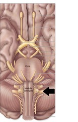
Hypoglossal nerve (CN XII) (S1 Brain Model)

Spinal Accessory nerve (CN XI) (S1 Brain Model)

Pons (S1 Brain Model)

Middle cerebellar peduncles (S1 Brain Model)

Superior cerebellar peduncles (S1 Brain Model)

Fourth ventricle (S1 Flat Half Brain)

Rhomboid fossa (S1 Flat Half Brain)

Cerebellum (S1 Flat Half Brain)

Abducens nerve (CN VI) (S1 Brain Model)

Facial nerve (CN VII) (S1 Brain Model)
7

Vestibulocochlear nerve (CN VII) (S1 Brain Model)

Cranial Nerves that attach to Pons (S1 Brain Model)
Trigeminal nerve (CN V) (S1 Brain Model)

Midbrain (S1 Brain Model)

Cerebral peduncles (S1 Brain Model)

Corpora quadrigemina (S1 Brain Model)

Superior colliculi (S1 Brain Model)

Inferior colliculi (S1 Brain Model)

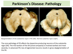
Substantia nigra
Cerebral aqueduct

Oculomotor nerve (CN III) (S1 Brain Model)

Trochlear nerve (CN IV) (S1 Brain Model)

Diencephalon (S1 Brain Model)

Thalamus (S1 Brain Model)

Interthalamic adhesion (S1 Brain Model)
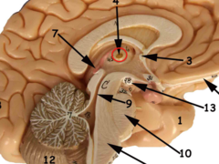
Epithalamus (S1 Brain Model)

Pineal gland (S1 Brain Model)

Hypothalamus (S1 Brain Model)

Infundibulum of the brain (S1 Brain Model)

Mammillary bodies (S1 Brain Model)

Optic tracts (S1 Brain)

Optic chiasm (S1 Brain)
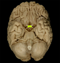
Optic nerves

Cerebellum (S1 Brain)
Under back part of brain, doesn't have as big ridges

Cerebellar hemispheres (S1 Brain)
Two halves of cerebellum

Folia and Fissures of the Cerebellum (S1 Brain)
Folia = ridges
Fissures = depressions
Vermis of the cerebellum (S1 Brain)
structure coming down that separates hemispheres

Cerebellar cortex (S1 Brain Model)
"leaves of tree", surrounding Arbor vitae

Arbor vitae (S1 Brain Model)
"trunks & branches of tree"

Cerebral hemispheres (S2 Split Brain)
*tested on whole structure, not just left or right hemispheres*

Longitudinal fissure (S2 Split Brain)
Space/line between cerebral hemispheres (space)

Cerebral cortex (S2 Brain Steak)
Gray matter
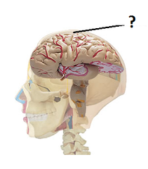
Cerebral gyri (S2 Full Brain)
ridges on surface of brain (ridge)

Cerebral sulci
Depressions between Cerebral gyri (ridges) on surface of brain (depression)

Frontal lobes (S2 Split Brain)
(region), most anterior portion of the brain

Parietal lobes (S2 Split Brain)
Region on top and in middle of brain

Temporal lobes (S2 Split Brain)
Region on side and underneath brain

Occipital lobes (S2 Split Brain)
Posterior/back portion of brain

Insula (S2 Hemi Brain)
hidden lobe

Central sulcus (S2 Hemi Brain)
depression
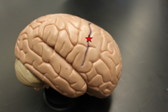
Precentral gyrus (S2 Hemi Brain)
ridge in front of Central sulcus

Postcentral gyrus (S2 Hemi Brain)
ridge in back of Central sulcus
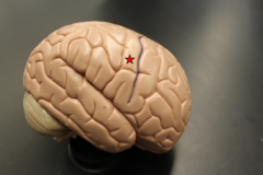
Precentral sulcus (S2 Hemi Brain)
depression

Postcentral sulcus (S2 Hemi Brain)
depression

Lateral sulcus (S2 Hemi Brain)
depression

Corpus callosum (S2 Hemi Brain)
"Hat on top of seahorse"

Cingulate gyrus (S2 Hemi Brain)
On top of "hat of seahorse" or corpus callosum, (ridge)

Cingulate sulcus (S2 Hemi Brain)
depression above Cingulate gyrus

Parieto-occipital sulcus (S2 Hemi Brain)
(depression), between parietal & occipital lobes

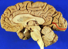
Calcarine sulcus (S2 Hemi Brain)
(depression), occipital lobe
Fornix of the Brain (S2 Big Brain Steak)
(structure), arch-way, inferior/on bottom

Septum pellucidum (S2 Big Brain Steak)
(structure), vertical ridge/line above fornix

Olfactory bulbs (S2 Full Brain)
on bottom of brain, (feature)

Olfactory tracts (S2 Full Brain)
on bottom of brain, (structure)

Lateral ventricles (S2 Big Brain Steak)
(space), space divided by Septum pellucidum

Septal nuclei (S2 Big Brain Steak)
(feature), top of Fornix, meets Septum pellucidum
*Board Question: One of the "pleasure centers" affected by addictive drugs*
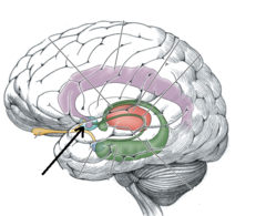
Hippocampus (S2 Brain Steak)
(structure), swirly structure, "cinnamon roll"
*Board Question: Functions in memory and understanding spatial-relationships*

Cranial dura mater (S2 Covered Brain)
(covering), flap on top of brain

Cranial arachnoid mater (S2 Split Brain)
(covering), webbing that covers the entire brain surface

Cranial pia mater (S2 Split Brain)
(covering), sits directly on brain, under Cranial arachnoid mater

Cranial Meningeal Spaces (S2 Space)
"potential space"
Epidural "space" (S2 Covered Brain)
On top of Cranial dura mater
*Board Question: "potential space"*

Subdural "space" (S2 IDK)
In between Cranial dura mater & Cranial arachnoid mater
*Board Question: "potential space"*

Subarachnoid "space" (S2 Split Brain)
In between Cranial arachnoid mater & Cranial pia mater
*Board Question: "real space"*

N/A *** Ventral median fissure
N/A ***

Conus medullaris (S3 Idk)
Narrowing in spinal cord, middle section

Central canal (S3 Drawing)

N/A *** Gray matter
N/A ***

Ventral gray columns (S3 Drawing)
On left side, dark green pointer

Ventral gray horns (S3 Drawing)

Dorsal gray columns (S3 Drawing)
purple pointer

Dorsal gray horns (S3 Drawing)

Intermediate gray columns (S3 Drawing)
orange pointer

Lateral gray horns (S3 Drawing)

N/A *** White matter
N/A ***

Ventral funiculus (S3 Drawing)

Lateral funiculi (paired) (S3 Drawing)

Dorsal funiculus (S3 Drawing)

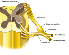
Dorsal roots (S3 18-57)
where rootlets meet

Spinal ganglion (S3 18-57)
bump on dorsal roots

Spinal nerve (S3 18-57)
strings in middle of spine

Ventral ramus (S3 Idk)

Dorsal ramus (S3 Idk)
stringy, coming out and around
Cervical spinal nerves (S3 Drawing)
C-1 to C-8