A&P Exam 3 - Nervous System
 Call Kai
 Learn
 Practice Test
 Spaced Repetition
 Match
 Flashcards
 Knowt Play
1/209

There's no tags or description
Looks like no tags are added yet.
210 Terms
Magnetic Resonance Imaging (MRI)
technology that can reconstruct a 3-D map of brain activity
Brain imaging & other methods = groups of neurons function in specialized circuits dedicated to different tasks
Neurons
transmit impulses
Neuroglia Cells
support, protect, & maintain the chemical & physical equilibrium of neurons
do not generate electrical impulses
Central Nervous System (CNS)
brain & spinal cord
Peripheral Nervous System (PNS)
cranial nerves, spinal nerves, & ganglia
Cell Bodies of Neurons
In CNS or in ganglia
Ventral Root = motor
Dorsal Root = sensory
Migraines/Headaches
painful & rob quality of life → migraines = 15% of population (1 billion people)
pounding headache, nausea, vomiting & photo/phono phobia → sometimes triggered by environment
genetics/hormones - 2-3x more in women
neurovascular - abnormal control of pain neurons in the trigeminal nucleus of brainstem
Sensory Function
(in)
sensory receptors gather information
information is carried to CNS
Integrative Function
sensory information used to create sensation, memory, thoughts, decisions
Motor Function
(out) decisions are acted upon, impulses carried to effectors
division of motor functions of PNS: somatic & autonomic
Somatic
transmit impulses to skeletal msucles
Autonomic
transmit impulses to smooth muscles, cardiac muscles, & glands
Transduce
(energy from 1 form to another)
sensory receptors transduce stimulus energy & transmit signals to CNS
sensory & preceptors begin with sensory reception: detection of stimuli by sensory receptors
Stretch receptor is an example of a sensory receptor
Sensations
action potentials that reach the brain via sensory neurons
the brain interprets sensations, giving the perception of stimuli
Exteroceptors
detect outside stimuli
Interoceptors
detect internal stimuli
Sensory Transduction
conversion of stimulus energy into a change in the membrane potential of a sensory receptor
many sensory receptors are very sensitive, able to detect the smallest physical unit of stimulus
Transmission
after energy has been transduced into a receptor potential, same senesory cells generate action potentials, which are transmitted to the CNS
Dendrites
receiving ends, increase surface area, tapered, & unmyelinated

Cell Body
contains the nucleus
large, ACTIVE, protein binding
large pale nucleus with nuceoli
rough ER called Nissl bodies - give gray color

Axon
transmits impulses & releases neurotransmitters to another neuron or effector

Do neurons divide?
Neurons do not generally divide
organelles turn over regularly in neurons
substances (like neurotransmitters) travel down axons a long distance (some local synthesis)
there are rare neural stem cells…need to harness those
Multipolar Neurons
99% of neurons
many processes
most neurons of CNS

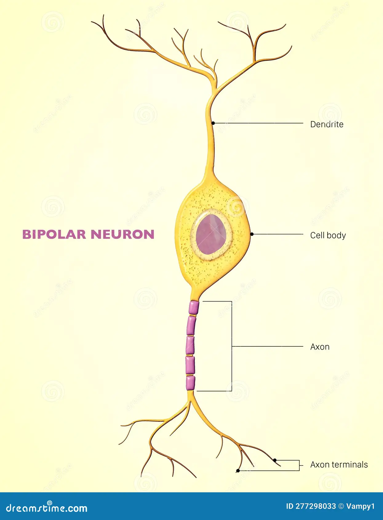
Bipolar Neurons
two processes
eyes, ears, & nose
only in retina of eye & ganglia of cranial nerves of hearing & smell
Unipolar Neurons
one process, ganglia of PNS, sensory
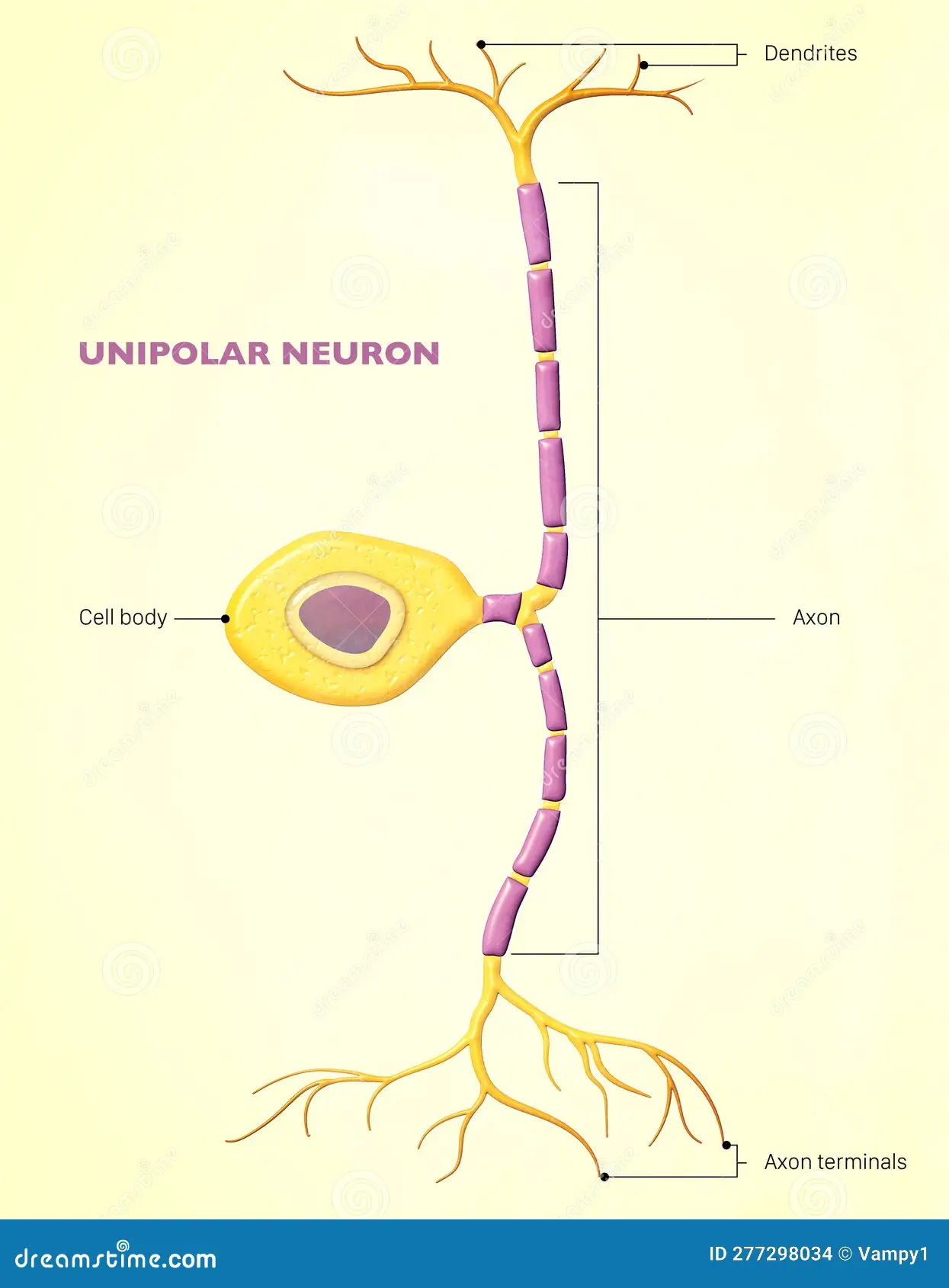
Sensory Neurons
afferent, carry impulses to CNS, most unipolar but some bipolar
Interneurons
link neurons, multipolar, only in CNS
Motor Neurons
multipolar, carry impulses away from CNS, carry impulses to effectors
Efferent = effect
motor neuron cell body = big in CNS
Myelinated Axons
axons which are tightly wrapped by neuroglial cells
myelinated nerve = faster
bigger nerve = faster
White Matter
contains myelinated axons, considered fiber tracts
Gray Matter
contains unmyelinated structures
cell bodies, dendrites
Schwann Cells
PNS: produce myelin sheath - lipid rich insulation
isolates axon from extracellular fluid
also clean up debris & guide regrowth of axons
Nodes of Ranvier
gives us saltatory (jumping) conclusion → electricity jumping = faster signals
gap between myelin sheaths → area of polarity reversal
Unmyelinated Axons (in PNS)
still enveloped by Schwann cells - fit in grooves
large Schwann cells serving - 20 axons
Demyelinating Diseases
immune symptom goes after myelin → cause unknown
Multiple Sclerosis (MS) - attacks myelin = breaks down myelin → CNS
born with genetic susceptibility to react to certain environmental factors
more prominent in northern regions
lack of vitamin D
Guillain-Barre Syndrome - attacks myelin in PNS
no cure = interferon, steroids, immunosuppressive drugs
Oligodendrocytes
form myelin in CNS (Schwann cells of CNS)
produce & maintain the myein sheath in CNS
several tongue-like processes to several axons
larger nodes than in PNS = more efficient conduction
Astrocytes
physical + metabolic support for neurons
largest glial cells, scaffolding at brain
blood vessels to neurons - maintain environment = blood-brain barrier
covering for “bare areas” of axons, such as at synapses - many remove excess neurotransmitters
form barrier around CNS
scar tissue
Microglia
macrophages of CNS
phagocytic cells, remove debris
5% of glial cells normally
from bone marrow, blood
Ependymal Cells
columnar cells lining ventricles of brain, canal of spinal cord
epithelial like lining of the ventricles of brain & spinal cord
cuboidal - columnar
lines spaces in the CNS
choroid plexus in ventricles secretes cerebral spinal fluid
Neuroglia
only nuclei seen w/ standard staining → neurons dependent on them (physical & chemical)
Blood Brain Barrier
prevents things in blood (ex. alcohol) getting into the brain
Origin of Nerve Cells
neurons from ectodermal cells of neural tube
microglia type of white blood cell from bone marrow
other glial cells from same cells as neurons
neurons reach destination, don’t divide
glial cells = slow turnover
small population
Schwann Cells
produce myelin found on peripheral myelinated neurons
speed up neurotransmission
in PNS
Satellite Cells
support clusters of neuron cell bodies (ganglia)
in PNS
Response of Neurons to Injury
portion of neuron distal to injury degenerates because no axonal transport
cell body changes - nucleus, Nissl bodies
neuron regenerates if Schwann cell makes cylinder so axon can grow back
Scar Formation
glial cells fill in = NO regeneration
Glial scar formed when astrocytes proliferate = blocking axonal growth
Synapse
nerve impulses pass from neuron to neuron at the synapses, moving from pre-synaptic to a post-synaptic neuron
when an action potential reaches a terminal, final result is release of neurotransmitters into synaptic cleft
Synaptic Transmission
neurotransmitters - released when impulse reaches synaptic know
effects fall into 2 categories:
excitatory postsynaptic potential
inhibitory postsynaptic potential
Summation of Postsynaptic Potentials
most neurons have many synapses on their dendrites & cell body
a single EPSP is usually too small to trigger an action potential
Cell Membrane Potential
a cell membrane is usually electrically charged, or polarized so that the inside of the membrane is negatively charged with respect to the outside of the membrane (which is then positively charged)
result of unequal distribution of ions on the inside + outside of the membrane
voltage difference can do WORK
Resting Potential
caused by an uneven distribution of ions (electrically charged molecules) of potassium (K+) and sodium (Na+) and chloride (Cl-)
Caused by Na+/k+ ion pumps that move 3 Na+ ions out of the cell for every 2 K+ it movies in
more + ions outside the cell than the inside is negatively charged with respect to the outside
Diffusion Pressure
molecules want to move from areas of high concentration to low concentration
membrane more permeable to K so K+ leaves the cell
Electrostatic Charge
ions with like charge are repelled & ions with different charges are attract → operation of ion pumps & ion channels
Ion Channel
large protein molecule that the crosses the membrane & allows specific ions to pass through at a specific rate
allow enough leakage of ions to mostly neutralize the effect of the ion pump but ion pumps use ATP
cell membrane = more permeable to K+, cytoplasm has negatively charged big ions
Local Potential Changes
neurons are excitable = caused by various stimuli (temp change, light, & pressure)
environmental changes affect the membrane potential by opening a gated ion channel
as ions flow through the membrane the membrane potential changes
graded or proportional to intensity of simulation reaching threshold potential
reaching threshold stimulation triggers voltage gated channels to open causing action potential
sub threshold depolarization will not result in action potential
channels are chemically gated, voltage gated, & mechanically gated
What is Action Potential?
blimp of electricity that runs through an ion channel
anything that alters the functioning of the ion channels can change the resting potential
depolarization = change that causes resting potential to be reduced
hyper-polarization = change that causes resting potential to increase
Action Potentials
at rest membrane is polarized -70 mV
threshold stimulus reached -55 mV
sodium channels open & membrane depolarizes (toward 0, up to 30 mV)
potassium leaves cytoplasm & membrane re polarizes
brief period of hyper-polarization -90 mV
Action Potentials - Voltage Gated
Trigger zone at first part of axon contains many voltage gated sodium channels
1) voltage gated Na+ channels open in response to threshold
2) as Na+ moves in the membrane depolarizes until it reaches +30 mV (action potential)
3) Na+ channels close & K+ channels open
4) K+ moves out & membrane repolarizes
5) as membrane potential drops below -70 mV, membrane is hyperpolarized
6) active transport re-establishes the resting potential of -70 mV as Na+ and K+ concentrations are maintained
All or None Law
Action potentials are always the same when they occur
once initiated it must run its course, nothing can stop or change it
How do action potentials move across a membrane?
when action potential occurs at one place on the membrane of an axon, surrounding membrane is depolarized past threshold causing an action potential → depolarizes neighboring membrane
action potential sweeps across a membrane as fast as 100 m/s
All or None Response
if a neuron axon responds completely - with an action potential (nerve impulse)
nerve impulse is conducted whenever a stimulus of threshold intensity or above is applied to an axon
all impulses carried on an axon are the same strength
Absolute Refractory Period
(1/2,500 sec) time when threshold stimulus does not start another action potential
Na+ channels unresponsive
Relative Refractory Period
time when stronger threshold stimulus can start another action potnetial - theority 700 impulses/sec
Impulse Conduction
the speed of impulse conduction varies on different types of neurons
myelinated axons transmit impulses through saltatory conduction, which is faster than impulses along unmyelinated axons (jump across nodes)
thick axon fibers transmit faster impulses than thin axon fibers
myelinated = bigger & faster nerves
Factors Affecting Impulse Conduction
Extra K+ ions outside the cell
Resting potential less negative, threshold reached easier
Excitable fibers = convulsions
Fewer K+ ions outside the cell
resting potential so negative that action potential not made
muscles become paralyzed
Anesthetic drugs such as procaine decrease membrane permeability to Na+ ions → keeps impulses from touch/pain from reaching brain
Lethal injection = potassium chloride
Postsynaptic Potentials
the membranes of dendrites & cell bodies do not have action potentials → instead only depolarizing stimulus causes a post synaptic potential (PSP) which spreads out across a membrane → the depolarization is weaker the further it gets from the stimulus → when stimulus turned off the PSP disappears
postsynaptic potentials can either be excitatory (depolarization) or inhibitory
Excitatory & inhibitory potentials can summate both in time (temporal summation) & across the membrane (spatial summation)
Net effect of summation is reflected at the axons hillock where action potentials are generated
The Synapse
cell bodies stimulated by either:
stimuli in the environment, such as sensory cells like the rods & cones in the eye
connections form other nerve cells to synapses
Synaptic Transmission
released neurotransmitters cross the synaptic cleft & react w/ specific molecules called receptors in the postsynaptic neuron membrane
effects of neurotransmitters vary
some neurotransmitters may open ion channels & others may close ion channels
chemically gated ion channels respond in neurotransmitters creating synaptic potentials
Excitatory Postsynaptic Potentials (EPSP)
graded, depolarizes membrane of postsynaptic neuron, action potential of postsynaptic neuron becomes more likely (like a greenlight)
Inhibitory Postsynaptic Potential (IPSP)
graded, hyper-polarizes membrane of postsynaptic neuron, action potentials of postsynaptic neuron becomes less likely (like a redlight)
Summation of EPSPs & IPSPs
added together in summation
more EPSPs lead to a greater probability of an action potential
summations usually occurs at the trigger zone - usually in hillock
Neurotransmitters
at least 100 different ones in brain
must be rapidly decomposed & recycled
Events leading to neurotransmitter release:
1) action potential passes along an axon & over the surface of its synaptic knob
2) synaptic knob membrane becomes more permeable to calcium ion & they diffuse inward
3) in the presence of calcium ions, synaptic vesicles fuse to synaptic knob membrane
4) synaptic vesicles release their neurotransmitter by exocytosis into the synaptic cleft
5) synaptic vesicles become part of membrane
6) the added membrane provides material for endocytotic vesicles
Neuropeptides
neurons in the brain or spinal cord synthesize neuropeptides
some neuropeptides act as neurotransmitters
other neuropeptides act as neuromodulators (substances which alter a neuron’s response to a neurotransmitter or block the release of a neurotransmitter)
Enkephalins
stress, bind to opiate receptors
Beta Endorphin
acts longer, more potent pain reliever
Substance P
neurotransmitter in pain impulse, 2 above may inhibit release
Opiates in the Human Body
morphine, heroin, codeine…potent painkillers from the poppy plant ‘
alter pain perception & elevate mood
similar structure to endorphins = released after exercise
addiction w/ painful withdraw → body makes less endorphins → body feels pain when stopped
Drug Addiction
Egyptians 3,500 years ago described opium addiction
nicotine today
drugs alter activity of neurotransmitters
antagonist = blocks, agonist = helps or activates receptor
with repeated use, # of receptors can decline = need higher dose
Nicotine
highly addictive, binds postsynaptic receptors that normally receive acetylcholine
neurons release dopamine = pleasurable feeling
number of receptors increased with increased cigarettes, nicotine binding impairs recycling of receptor proteins so more are produced
with time it takes more nicotine to produce same effect
Impulse Processing
the way the nervous system processes nerve impulses & acts upon them reflects the organization of neurons & axons in the CNS
Neuronal Pools
groups of interneurons that make synaptic connections with each-other
interneurons work together to perform a common function
each pool receives input from other neurons
each pool generates output from other neurons
facilitation may occur - sub-threshold but another impulse to threshold
Convergence
neuron receives input from several neurons
incoming impulses represent information from different types of sensory receptors
allows nervous system to collect, process, & respond to information
makes it possible for neuron to sum impulses from different sources
Divergence
one neuron sends impulses to several neurons
can amplify an impulse
impulse from a single neuron in CNS may be amplified to activate enough motor units needed for muscle contraction
Dura Mater
outer layer of meninges in the CNS
primarily composed of tough, white, dense connective tissue & contains many blood vessels & nerves
attaches to inside of of the cranial cavity & forms the internal periosteum of the surrounded skull bones
Arachnoid
“spider webs”
middle layer
subarachnoid space = cerebral spinal fluid (gap between arachnoid & pia mater)
does not have blood vessels
Pia Mater
inner layer
thin & contains many nerve & blood vessels that nourish the underlying cells of the brain & spinal cord
attached to the surfaces of these organs
Falx Cerebri
sheet of connective tissue that goes from crista galli & makes a “tent” over cerebellum → separates left & right cerebellar hemispheres
Falx Cerebri
extends downward into the longitudinal fissure, & separates the right & left cerebral hemispheres
Tentorium Cerebelli
separates the occipital lobes from the cerebrum form the cerebellum
Dural Sinus
collects all the blood from the brain
blood-filled channel formed by the division of the dura mater into two layers
Epidural Space
outside dura mater (between dura mater & bone) in spinal cord
space between the dural sheath of the spinal cord & the bone of the vertebral canal
contains blood vessels, loose connective tissue, & adipose tissue that pad the spinal cord
Epidural Hematoma
bleeding in the space between the dura mater & skull
Subdural Hematoma
bleeding into the space between the dura mater & the brain
Blood collecting outside or beneath the dura mater
increase pressure = death
Ventricles
there are 4 ventricles - lateral ventricles (1st & 2nd), 3rd ventricle, & 4th ventricle
interconnected cavities within cerebral hemisphere & brainstem
ventricles are continuous w/ the central canal of the spinal cord
filled with cerebral spinal fluid (CSF)

Cerebral Spinal Fluid (CSF)
secreted by the choroid plexus
circulates in the ventricles, central canal of spinal cord, & the subarachnoid space
Completely surrounds brain & spinal cord
Excess or wasted CSF is absorbed by the arachnoid granulations
140 mL in system at a time, 500 mL a day
Nutritive & protective
Helps maintain stable ion concentrations in CNS
Spinal Cord
Slender column of nerve tissue = continuous w/ brain & brainstem
extends downward through vertebral canal
begins at the foramen magnum & terminates at the 1st & 2nd vertebrae interspace (L1/L2)
vertebrae grows faster than spinal cord
Cauda equina
Safe for lumbar puncture - 3rd & 4th, 4th&5th
cervical & lumbar enlargements for nerves in arms & legs

Brain Herniation
Potentially deadly side effect of very high intracranial pressure when a part of the brain is squeezed across structures within the skull
Brain can shift across structures of falx cerebri, the tentorium cerebelli, & even through foramen magnum
Cuts off blood supply to part of the brain = often fatal
Gray Matter - Spinal Cord
w/ cell bodies (Nissl)
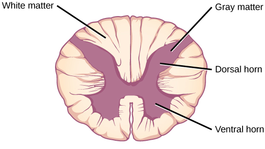
Whit Matter - Spinal Cord
outer of axons
myelinated = white

Spinal Cord Anatomy
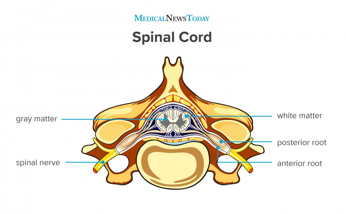
Spinal Cord Function
center for spinal reflexes
conduct (pathway) for impulses to & from the brain