Disorders of the Digestive System & Accessory Organs of Digestion
 Call Kai
 Learn
 Practice Test
 Spaced Repetition
 Match
 Flashcards
 Knowt Play
1/78

Earn XP
Description and Tags
Week 7
79 Terms
Structural and neural abnormalities can…
slow, obstruct or accelerate movement of intestinal contents at any level of GI tract.
Inflammatory and ulcerative conditions of GI wall…
disrupt secretion, motility, absorption
Inflammation or obstruction of liver, pancreas, or gallgladder….
can alter metabolism and result in local and systemic symptoms
What do the liver, gallbladder and pancreas do for digestion?
secrete substances
disorders of the liver, gallbladder and pancreas are called:
inflammatory disease,
obstruction of ducts
tumors
Intestinal obstructions and paralytic ileus
Any condition preventing flow of chyme (Food + Stomach acid + Digestive enzymes) through small or large intestinal lumen
5 ways of classification of intestinal obstructions
by onset : acute vs chronic
by extent: partial vs complete
by location: intrinsic vs extrinsic
by effects on intestinal wall: simple vs strangulated vs closed loop
by casual factors: mechanical vs functional (ileus)
Intrinsic: The blockage originates within the intestinal wall or lumen (inside the bowel). Examples include a tumour growing into the intestine or a stricture (narrowing).
Extrinsic: Something outside the intestine presses on or traps it. Examples include adhesions (scar tissue) or a hernia.
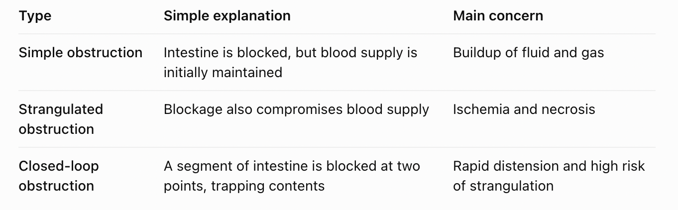

herniation (intestinal obstruction example)

adhesion (intestinal obstruction example)

volvulus (intestinal obstruction example, Twisted intestine.)
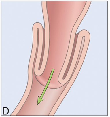
intussusception (intestinal obstruction example, Intestine slides inside itself.)
What is the overall pathophysiology of mechanical SBO in order?
mechanical causes: adhesion, hernia, tumor, intussusception
proximal distension from gas and fluid accumulate before obstruction (bowel wall stretches)
increases peristaisis (early): attempts to push contents forward → worsens distension and colicky pain
third spacing of fluid: secretion into lumen + impaired absorption → intravascular volume loss → dehydtaion + hypovolemia
electrolyte imbalance: loss of H+ or loss of HCO3; hypokalemia
vomiting worsens F&E loss
bowel wall edema and ischemia from compression of blood flow → necrosis + perforation
bacterial overgrowth and translocation → sepsis
systemic response: hypotension, tachycardia. shock if untreated
Why does SBO cause intestinal distension and colicky pain?
Gas and fluid accumulate proximal (before) the obstruction, stretching the bowel wall.
Increased peristalsis (early): Intestines contract harder to push contents past the blockage.
This causes colicky abdominal pain (cramping pain that comes and goes).
Continued accumulation worsens distension.
What is third spacing in SBO, and what does it cause?
Third spacing: Fluid becomes trapped in the bowel lumen, bowel wall, or abdominal cavity instead of being available in the bloodstream.
Increased intestinal secretions + impaired absorption cause fluid accumulation.
Less circulating fluid → Hypovolemia and dehydration.
Severe hypovolemia → Tachycardia, hypotension, and possible shock.
What electrolyte and acid-base imbalances occur in SBO?
Loss of H⁺ (acid): Metabolic alkalosis, commonly from vomiting stomach acid.
Loss of HCO₃⁻ (base): Metabolic acidosis from bicarbonate-rich intestinal fluid loss.
Loss of K⁺: Hypokalemia (low potassium).
Vomiting worsens fluid and electrolyte losses.
T/F large bowel obstruction is less common then SBO
True
Large bowel obstruction is commonly related to
colon cancer
Other causes for large bowel obstruction include…
diverticulitis (inflammation of small pouches in the colon), IBD (long-term inflammation of the intestines), and intrinsic/extrinsic lesions (abnormal growths or changes inside or outside the bowel that cause blockage).
T/F - the patho processes (distension → vascular compromise → systemic effects) are similar to SBO
True
What are the consequences of intestinal obstruction, and what causes each?
Fluid/electrolyte disturbances: Third spacing + vomiting → Dehydration and hypovolemia.
Metabolic alkalosis: Loss of H⁺ from vomiting.
Metabolic acidosis: Loss of HCO₃⁻ or lactic acid buildup from ischemia.
Hypokalemia: Potassium loss from vomiting and increased renal excretion.
Inflammation: Intestinal distension + reduced blood flow → Bowel wall injury and edema.
Metabolic alkalosis:
Loss of H⁺ from vomiting.
Metabolic acidosis::
Loss of HCO₃
Respiratory complications - bowel obstruction
Pressure on diaphragm → ↓ Respiratory volume → Atelectasis → Possible pneumonia
Gastrointestinal and metabolic complications - bowel obstruction
Colicky pain → Nausea/vomiting → Fluid/electrolyte loss → Alkalosis or acidosis
Circulatory complications - bowel obstruction
Third spacing + Vomiting → Dehydration → Hypovolemia → Tachycardia → Shock
Bowel injury and infection - bowel obstruction
Increased pressure → ↓ Venous drainage → Edema → ↓ Arterial flow → Ischemia → Necrosis/perforation → Peritonitis
Clinical manifestations
depend on the location of the blockage, extent of teh obstruction, degree of ischemia, and the stage of progression (refer to text for detail)
pylorus: profuse, early vomiting. proximal small intestine: mild distenstion, bile stained vomiting. Lower small intestine: pronounced distension, delayed vomiting. alrhe intestine: hypogastric pain, distension, vomiting occurs late in the process
partial obstruction: diarrhea or constipation; less severe symptoms compared to complete obstruction. complete obstruction: constipation, increased bowel sounds, cramping pain, signs of hypovolemia and metabolic acidosis
with ischemia: pain becomes constant and severe as blood supply is compromised, leading to complications liek necrosis
late stage (necrosis and perfoation): fever, peritonitis, severe abdo pain and signs of systemic inflammation.
As intestinal obstruction becomes more severe
the concern shifts from cramping, vomiting, and distension to constant severe pain, ischemia, necrosis, and perforation.
Manifestation based on location - Pylorus (opening between stomach and small intestine)
Profuse, early vomiting: Large amounts of vomiting occur quickly.
Why? Food and fluid cannot leave the stomach normally, so they build up and are vomited.
Remember: Blockage near stomach → Vomiting early.
Manifestation based on location - Proximal small intestine (upper small intestine)
Mild distension: The abdomen is not extremely swollen.
Bile-stained vomiting: Vomit may appear green or yellow-green because it contains bile.
Why? The blockage is relatively high, so contents back up and are vomited before large amounts accumulate throughout the intestine.
Remember: Upper SBO → Mild distension + Early, bile-stained vomiting.
Manifestation based on location - Lower small intestine (distal small bowel)
Pronounced distension: Significant abdominal swelling.
Delayed vomiting: Vomiting occurs later.
Why? There is more intestine above the blockage where gas and fluid can accumulate before vomiting begins.
Remember: Lower SBO → More distension + Later vomiting.
Manifestation based on location - Large intestine (colon)
Hypogastric pain: Pain in the lower abdomen.
Distension: Abdomen becomes swollen because gas and stool accumulate.
Vomiting occurs late: The blockage is far from the stomach, so vomiting usually takes longer to develop.
Remember: Large bowel obstruction → Lower abdominal pain + Distension + Late vomiting.
Partial = The intestine is not completely blocked.
Some stool and gas can still pass through.
Diarrhea: Liquid stool may still move past the narrowed area.
Constipation: Stool may move more slowly.
Symptoms are generally less severe than complete obstruction.
Remember: Partial obstruction → Some contents can still pass → Diarrhea OR constipation.
Complete = Nothing can pass through the blocked section.
Constipation: Stool cannot move past the blockage.
Increased bowel sounds: Early on, the intestines contract harder to push contents forward.
Cramping pain: Increased peristalsis causes colicky abdominal pain.
Hypovolemia: Vomiting and third spacing decrease circulating fluid volume.
Metabolic acidosis: May develop with severe dehydration and reduced tissue blood flow.
Remember: Complete obstruction → No passage of contents → Cramping, fluid loss, and potentially severe complications.
What happens when ischemia develops?
Ischemia = Reduced blood flow and oxygen to the intestine.
Normally, intestinal obstruction causes colicky pain (pain that comes and goes).
But if ischemia develops:
Increased pressure or twisting compresses intestinal blood vessels.
Less blood and oxygen reach the bowel.
Intestinal tissue becomes damaged.
Pain becomes constant and severe instead of coming and going.
Prolonged ischemia can cause necrosis (tissue death).
Important exam distinction:
Colicky pain: Intestine is contracting against the obstruction.
Constant, severe pain: May indicate ischemia and bowel injury.
Late-stage obstruction: Necrosis and perforation
Prolonged ischemia
Necrosis (intestinal tissue dies)
Perforation (hole develops in intestine)
Bacteria and intestinal contents leak into abdomen
Peritonitis (inflammation of abdominal lining)
Fever + Severe abdominal pain + Systemic inflammation
Treatment for intestinal obstructions (6)
replacement of fluid and electrolytes
gastric or intestinal suction
laparoscopic procedures for adhesions
immediate surgical intervention for strangulation and complete obstruction
colonic stent for malignant obstruction
iv antibiotics, fluid resuscitation, surgery for intestinal perforation
Fluid and electrolyte replacement: IV fluids correct dehydration, hypovolemia, and electrolyte imbalances.
Gastric or intestinal suction: Removes trapped gas and fluid (usually through an NG tube) to relieve distension and vomiting.
Laparoscopic surgery: Removes adhesions (scar tissue) causing the blockage.
Colonic stent: Opens the blocked colon in some cases of malignant obstruction (cancer).
When is emergency surgery required for intestinal obstruction, and what is the treatment?
Strangulation: Blood supply to the intestine is compromised → Immediate surgery to prevent necrosis.
Complete obstruction: May require urgent surgery to remove the blockage.
Intestinal perforation: Hole in the intestine allows contents to leak into the abdomen → IV antibiotics + fluid resuscitation + emergency surgery.
Appendicitis
inflamnmation of vermiform appendix
cause: most commonly due to luminal obstruction (stool,fecalith, tumor, foreign body)
bacterial infection: obstruction → stasis → bacterial overgrowth
secretion + pressure: continued mucosal secretion → incrased intraluminal pressure
vascular compromise: pressure decreases mucosal blood flow → hypoxia → mucosal ulceration
invasion: bacetria/microbes invade → inflammation, edema of appendix wall
progression:
inflammation can involve distal or entire appendix
Gangrene may develop if thrombosis of blood vessel occurs
may progress to perforation → peritonitis
exam takeaway for appendicitis patho
Appendicitis usually starts with a blockage, which traps bacteria and mucus. Pressure increases, blood flow decreases, and inflammation develops. If untreated, severe cases can progress to gangrene, perforation, and peritonitis.
review order for appendicitis

clinical manifestations of appendicitis
*appendicitis is a medical emergency!!!
epigastric and periumbical pain, rebound tenderness
n/v, fever, anorexia
complications can lead to → perforation, peritonitis, abscess formation
treatment for appendicitis (3)
Antibiotics
Appendectomy
Supportive care
Supportive care = Treatments that help stabilize the patient and relieve symptoms while the underlying condition is treated.
Examples:
IV fluids: Prevent or treat dehydration from vomiting and poor oral intake.
Pain medications: Relieve abdominal pain.
Antiemetics: Reduce nausea and vomiting.
NPO (nothing by mouth): Often ordered when preparing for surgery or while awaiting assessment.
Monitoring: Check vital signs, abdominal pain, and signs of worsening infection or perforation.
What is pancreatitis?
inflammation of pancreas
associated with several clinical disorders (alcohol intake and cholelithiasis)
caused by injury or damage to pancreatic cells and ducts
caused by injury or damage to pancreatic cells and ducts
pancreatic enzymes leak into pancreatic tissue
enzymes cause autodigestion of pancreatic tissue and leak into bloodstream and cause injury to blood vessels and other organs
What is the pathophysiology of acute pancreatitis?
Alcohol intake or gallstones → Injury to pancreatic cells and ducts.
Digestive enzymes become activated too early and damage pancreatic tissue.
Autodigestion: Pancreatic enzymes begin digesting the pancreas itself.
Causes inflammation, edema, and possible necrosis.
Enzymes and inflammatory substances enter bloodstream → Blood vessel injury, third spacing, and possible organ damage.
Most important concept: In acute pancreatitis, digestive enzymes become activated inside the pancreas instead of the small intestine, causing the pancreas to digest itself.
What is the overall pathophysiology of acute pancreatitis in order?
Initiating event: Gallstones block pancreatic duct OR alcohol, drugs, or hypertriglyceridemia cause pancreatic cell injury.
Premature enzyme activation: Trypsinogen activates into trypsin → Autodigestion of pancreas.
Inflammatory response: Calcium overload → NF-κB activation → Cytokine release → Neutrophil recruitment.
Tissue damage: Inflammation causes edema, fat necrosis, hemorrhage, and possible pancreatic necrosis.
Systemic effects: Increased vascular permeability → Third spacing → Hypovolemia → Possible shock and organ failure.
Which pancreatic enzyme is activated prematurely and causes autodigestion?
Trypsinogen (inactive enzyme) is prematurely converted into trypsin (active enzyme).
These enzymes begin digesting pancreatic tissue → Autodigestion, inflammation, and cell injury.
What is the inflammatory cascade in acute pancreatitis?
Pancreatic cell injury causes intracellular calcium overload.
This activates the NF-κB pathway, which promotes inflammation.
Inflammatory cytokines are released.
Neutrophils and other inflammatory mediators are recruited.
This causes tissue edema and necrosis.

What are the roles of pancreatic enzymes in acute pancreatitis?
Trypsin: Activates other digestive enzymes and contributes to pancreatic cell injury.
Chymotrypsin: Breaks down proteins → Tissue damage (proteolysis).
Phospholipase: Damages cell membranes → Cell injury.
Elastase: Damages blood vessel walls → Hemorrhage.
Lipase: Breaks down fat → Fat necrosis.
What are the local complications of acute pancreatitis?
Edema: Swelling of pancreatic tissue.
Fat necrosis: Destruction of fat tissue by lipase.
Hemorrhage: Bleeding from damaged blood vessels.
Pancreatic necrosis: Death of pancreatic tissue.
Peripancreatic fluid collections: Fluid accumulates around the pancreas.
Pseudocyst: A collection of pancreatic fluid surrounded by inflammatory tissue.
Pancreatic abscess: Localized collection of infection or pus.
Why does acute pancreatitis cause hypovolemia and shock?
Pancreatic inflammation causes massive release of inflammatory mediators.
These increase vascular permeability (blood vessels become leaky).
Plasma fluid leaks out of blood vessels into surrounding tissues (third spacing).
Circulating blood volume decreases → Hypovolemia.
Severe hypovolemia reduces organ perfusion → Shock.
What are the systemic complications of severe acute pancreatitis?
SIRS: Systemic inflammatory response syndrome (widespread inflammation throughout the body).
ARDS: Acute respiratory distress syndrome (severe lung inflammation causing impaired oxygen exchange).
ATN: Acute tubular necrosis (kidney tubule damage due to reduced blood flow or injury).
MODS: Multiple organ dysfunction syndrome (several organs begin failing).
Sepsis: Life-threatening organ dysfunction caused by infection.
Myocardial depression: Reduced ability of the heart to pump effectively.
Coagulation disorders: Abnormal blood clotting or bleeding.
Manifestations of acute pancreatitis
pain: sudden onset severe constant epigastric or mid abdominal pain radiating to the back
assoictaed findings:
n/v, abdo distension, paralyctic ileus,
fever, leukocytosis
tachycardia , hypotension (due to third spacing and shock)
jaundice (if bilary obstrcution)
severe cases: flank/periumbilical ecchymosis (cullens/grey turners sign) → poor prognosis
What are the clinical manifestations of acute pancreatitis and why do they occur?
Severe, constant epigastric pain radiating to back: Pancreatic inflammation and tissue damage.
Nausea, vomiting, distension, paralytic ileus: Inflammation disrupts intestinal motility.
Fever and leukocytosis: Inflammatory cytokine release.
Tachycardia and hypotension: Third spacing → Hypovolemia.
Jaundice: Biliary obstruction → Bilirubin buildup.
Cullen's sign: Bruising around umbilicus.
Grey Turner's sign: Bruising along flanks.
Both bruising signs: May indicate severe hemorrhagic pancreatitis and poor prognosis.
Why does acute pancreatitis cause tachycardia and hypotension?
Pancreatic inflammation → Release of inflammatory cytokines.
Increased vascular permeability → Fluid leaks out of blood vessels (third spacing).
Decreased circulating blood volume → Hypovolemia.
Heart compensates → Tachycardia.
Severe hypovolemia → Hypotension and possible shock.
What are Cullen's sign and Grey Turner's sign in acute pancreatitis?
Cullen's sign: Bruising around the umbilicus (belly button).
Grey Turner's sign: Bruising along the flanks (sides of abdomen).
Both can indicate severe hemorrhagic pancreatitis and are associated with a poor prognosis.
What are the major complications of appendicitis vs. acute pancreatitis?
Appendicitis:
Perforation: Appendix ruptures.
Peritonitis: Inflammation of abdominal lining from leakage.
Abscess: Localized collection of pus.
Acute pancreatitis:
Local: Edema, fat necrosis, hemorrhage, pancreatic necrosis, pseudocyst.
Systemic: SIRS, ARDS, acute tubular necrosis, shock, MODS, and possible sepsis.
What is the primary goal and supportive treatment for acute pancreatitis?
Stop autodigestion of the pancreas and prevent systemic complications.
Address Cause: Remove gallstone if obstructive; stop offending drug/alcohol.
supportive care:
IV fluids: Restore circulating blood volume and prevent hypovolemic shock from third spacing.
Pain management: Opioids (narcotics) or epidural analgesia for severe pain.
Oxygen support: Given as needed to maintain adequate oxygenation.
What is GI rest in acute pancreatitis, and how is it managed?
NPO initially: Nothing by mouth when necessary due to severe vomiting or inability to tolerate food.
Enteral feeding: Start feeding through the GI tract when tolerated, generally within 24–72 hours if oral intake is not possible and there is no contraindication such as significant ileus.
NG suction: Removes gastric contents if severe nausea, vomiting, or paralytic ileus occurs.
Address underlying cause: Remove an obstructing gallstone when indicated and stop alcohol or offending drugs.
What complications should be monitored for in acute pancreatitis, and what additional treatments may be used?
Complications:
Hypocalcemia: Low calcium → May cause tetany (involuntary muscle spasms).
ARDS: Severe lung inflammation causing impaired oxygen exchange.
Renal failure: Kidney dysfunction from reduced blood flow or inflammation.
Pseudocyst: Collection of pancreatic fluid surrounded by inflammatory tissue.
Sepsis: Life-threatening organ dysfunction caused by infection.
Additional treatments:
H₂ blockers: Reduce stomach acid secretion (listed on your slide as reducing pancreatic stimulation).
Antibiotics: Used when infection is confirmed or strongly suspected, not routinely for uncomplicated pancreatitis.
What is cholelithiasis?
Gallstone formation in bile that is supersaturated with cholesterol (too much cholesterol in bile causes gallstones to form).
Enzyme defect (problem with an enzyme that affects cholesterol production).
Increased cholesterol synthesis (body produces too much cholesterol).
Decreased secretion of bile acids to emulsify fats (less bile acid is released to help break down fats).
Decreased resorption of bile salts from ileum (less bile salt is reabsorbed from the last part of the small intestine).
Gallbladder smooth muscle hypomotility → stasis (gallbladder does not contract properly → bile sits too long).
Increased secretion of gallbladder mucin and biliary calcium (more mucus-like substance and calcium in bile help crystals form and stick together).
Genetic predisposition (inherited increased risk of developing gallstones).
What is the overall pathophysiology of cholelithiasis (gallstone formation)?
Imbalance of cholesterol, bile salts, and calcium → Supersaturation of bile with cholesterol (too much cholesterol in bile).
Cholesterol precipitates and forms crystals called microliths (tiny stones).
Crystals aggregate into macrostones (larger gallstones).
Gallbladder hypomotility → Stasis (bile remains still) → Promotes stone growth.
What factors contribute to cholelithiasis?
Increased cholesterol synthesis (more cholesterol produced).
Decreased secretion of bile acids (less bile acid available to keep cholesterol dissolved).
Decreased resorption of bile salts from ileum (less bile salt is recycled).
Gallbladder hypomotility (poor gallbladder contractions → bile stasis).
Increased gallbladder mucin and biliary calcium (helps crystals stick together).
Genetic predisposition (inherited increased risk).
What are the three types of gallstones?
Cholesterol stones: Most common, yellow-green.
Black pigment stones: Associated with chronic hemolysis (red blood cell breakdown) and cirrhosis (liver scarring).
Brown stones: Associated with bacterial or parasitic infections in the bile ducts.
What happens when a gallstone obstructs the cystic duct?
Obstruction of cystic duct → Cholecystitis (inflammation of the gallbladder).
Bile becomes trapped in the gallbladder → Increased pressure and inflammation.
Cystic duct obstruction → Cholecystitis. Common bile duct obstruction → Jaundice, cholangitis, and possible pancreatitis.
What happens when a gallstone obstructs the common bile duct?
Obstruction of common bile duct → May cause:
Jaundice: Bilirubin buildup → Yellowing of skin and eyes.
Cholangitis: Inflammation/infection of the bile ducts.
Pancreatitis: Inflammation of the pancreas if the stone also blocks pancreatic drainage.
Manifestations of cholelithiasis
asymptomatic: many patients have silent gallstones discoveted incidentally
symptomatic disease:
epigastric or right hypochondrium pain (bililary colicP)
triggered by fattu meal (due to gallbladder contraction from cholecystokinin)
pain is intermittent, steady and may radiate to right shoulder or scapula
may last 30 min to several hrs after eating
associated symptoms:
heartburn, flatulence, indigestion, food intolerance (esp. to fats/cabbage), nausea, vomiting during biliary colic attacks
jaundice: indicates stone in the common bile duct causing obstruction
What medical therapy is used for cholelithiasis?
Oral bile acids (ursodeoxycholic acid, chenodeoxycholic acid) can dissolve cholesterol stones/sludge (medications help dissolve certain cholesterol gallstones and thickened bile).
What is the limitation of medical therapy for cholelithiasis?
Stones often recur after drug discontinuation (gallstones can form again after stopping the medication).
What lifestyle changes help prevent gallstone formation?
Increase intake of polyunsaturated and monounsaturated fats, vitamin C, fiber (choose healthier fats and eat more fibre-rich foods).
Maintain healthy weight with gradual weight loss (lose weight slowly if needed).
Rapid weight loss ↑ risk (losing weight too quickly increases gallstone risk).
Regular exercise to prevent gallstone formation (physical activity helps lower risk).
What is the preferred definitive treatment for uncomplicated gallstones causing obstruction?
Laparoscopic cholecystectomy (surgical removal of the gallbladder using small incisions).
Preferred treatment for uncomplicated gallstones causing obstruction (removes the gallbladder to prevent further gallstone episodes).
What is extracorporeal shock wave lithotripsy used for in cholelithiasis?
Extracorporeal shock wave lithotripsy (uses shock waves from outside the body to break stones into smaller pieces).
Can fragment large stones before endoscopic removal if surgery is not immediately feasible (breaks stones into smaller pieces to make removal easier).
What are the main management strategies for cholelithiasis?Symptomatic relief:
Medical therapy: Pain control for biliary colic.
Oral bile acids to dissolve cholesterol stones/sludge.
Lifestyle/prevention: Healthy fats, vitamin C, fiber, gradual weight loss (rapid can cause ingresed risk), regular exercise.
Definitive treatment: Laparoscopic cholecystectomy.
Alternative procedure: Extracorporeal shock wave lithotripsy in selected cases.
Remember that laparoscopic cholecystectomy is the preferred definitive treatment for symptomatic gallstones requiring surgery, while oral bile acids may dissolve certain cholesterol stones but have a risk of recurrence.
•Laparoscopic cholecystectomy
•preferred treatment for uncomplicated gallstones causing obstruction
•Extracorporeal shock wave lithotripsy
•can fragment large stones before endoscopic removal if surgery is not immediately feasible
What is cholecystitis?
inflammation of gallbladder
When does cholecystitis develop?
after a gallstone becomes lodged in cystic duct
What are the clinical manifestations for cholecystitis (4)
fever
leukocytosis
rebound tenderness
abdo muscle guarding
What is the treatment for cholecystitis? (5)
pain control
replacement of fluids and electrolytes
fasting
antibiotic administration
laparoscopic cholecystectomy