lab 05 diagrams AP 2
0.0(0)
Studied by 0 people Call Kai
 Learn
 Practice Test
 Spaced Repetition
 Match
 Flashcards
 Knowt Play
Card Sorting
1/68

There's no tags or description
Looks like no tags are added yet.
Last updated 9:08 PM on 10/16/26
Name | Mastery | Learn | Test | Matching | Spaced | Call with Kai | Chat |
|---|
No analytics yet
Send a link to your students to track their progress
69 Terms
1
New cards
2
New cards


3
New cards


4
New cards


5
New cards

6
New cards


7
New cards

8
New cards

9
New cards


10
New cards
Trachea and esophagus: transverse section
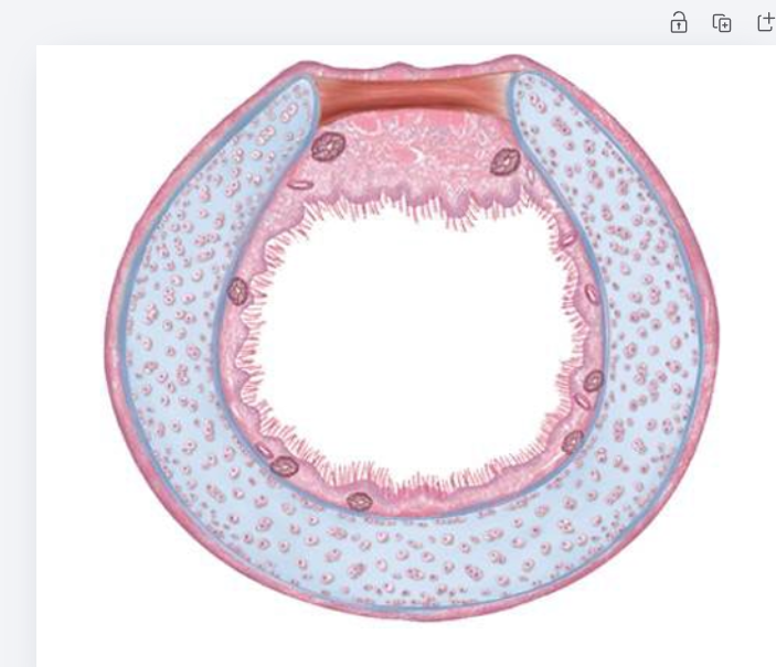
Trachea is a round tube with C-shaped hyaline cartilage, open side facing back. The esophagus is a flattened tube directly behind (posterior to) the trachea, touching the open side where the trachealis muscle is.
11
New cards
Trachea and esophagus: sagittal section
The trachea runs in front (anterior) and the esophagus lies behind (posterior) it, both in front of the vertebral column.
12
New cards
Essential difference: conducting vs respiratory division
Conducting division only carries air to the gas-exchange area (no gas exchange; warms, humidifies, filters). Respiratory division is where gas exchange actually occurs across thin walls.
13
New cards
Significance of pleural membrane and fluid in ventilation
Pleural fluid creates surface tension that holds the visceral pleura (lung) to the parietal pleura (chest wall), so the lungs expand and recoil with the thoracic cavity. It also lubricates to reduce friction.
14
New cards
Location of lungs relative to heart and diaphragm
Lungs fill the thoracic cavity on both sides of the heart (in the mediastinum). The bases rest on the relaxed, dome-shaped diaphragm; the apices extend slightly above the clavicles.
15
New cards
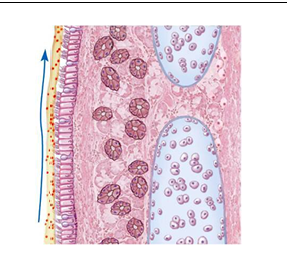
Trachea histology
Lined by pseudostratified ciliated columnar epithelium with goblet cells. Beneath it: lamina propria, mucus glands, and C-shaped hyaline cartilage rings (with chondrocytes). The trachealis (smooth muscle) closes the gap at the back.
16
New cards
Full pathway of O2: outside the body to the cells
Air -> nose/mouth -> pharynx -> larynx -> trachea -> bronchi -> bronchioles -> alveoli -> across respiratory membrane into pulmonary capillary -> pulmonary veins -> left atrium -> left ventricle -> aorta -> systemic arteries -> systemic capillaries -> across capillary wall into body cells
17
New cards
Full pathway of CO2: cells to outside the body
Body cells -> systemic capillaries -> veins -> vena cava -> right atrium -> right ventricle -> pulmonary trunk -> pulmonary arteries -> pulmonary capillaries -> across respiratory membrane into alveoli -> bronchioles -> bronchi -> trachea -> larynx -> pharynx -> nose/mouth -> exhaled
18
New cards
Five stages of respiration
1) Ventilation 2) External respiration 3) Transport 4) Internal respiration 5) Cellular respiration
19
New cards
Ventilation
Movement of air into and out of the lungs (atmosphere <-> alveoli). O2 moves in, CO2 moves out. Occurs in the airways and lungs.
20
New cards
External respiration
Gas exchange in the lungs between alveoli and pulmonary capillaries. O2 moves from alveoli into blood; CO2 moves from blood into alveoli.
21
New cards
Transport
Blood carries O2 and CO2 between the lungs and body tissues via the cardiovascular system. O2 is mostly bound to hemoglobin; CO2 is mostly carried as bicarbonate.
22
New cards
Internal respiration
Gas exchange between systemic capillaries and body cells. O2 moves from blood into cells; CO2 moves from cells into blood.
23
New cards
Cellular respiration
Cells use O2 to break down glucose and make ATP, producing CO2 and water. C6H12O6 + 6 O2 -> 6 CO2 + 6 H2O + ATP
24
New cards
Relationship between CO2 and cellular respiration
CO2 is a waste product of cellular respiration (made in the mitochondria when glucose is broken down) and must be carried away and exhaled.
25
New cards
Relaxed diaphragm shape and position
Dome-shaped, bulging up into the thoracic cavity. The lungs sit superior to it on either side of the heart (mediastinum).
26
New cards
Left lung vs right lung (lobes)
Left lung has 2 lobes (superior, inferior); right lung has 3 lobes (superior, middle, inferior).
27
New cards
Why is the left lung smaller?
It has a cardiac notch (indentation) to make room for the heart, which tilts toward the left.
28
New cards
Parietal pleura
Outer layer of the pleural membrane; lines the thoracic wall, diaphragm, and mediastinum.
29
New cards
Visceral pleura
Inner layer of the pleural membrane; covers the surface of the lungs.
30
New cards
Pleural cavity
Thin space between parietal and visceral pleura, filled with pleural (serous) fluid.
31
New cards
Three purposes of the pleural membranes
1) Pleural fluid reduces friction as lungs expand and recoil. 2) Fluid surface tension holds the lungs to the thoracic wall so they expand with it during ventilation. 3) Compartmentalizes each lung, limiting spread of infection and protecting the lungs.
32
New cards
What resists excessive expansion of the lungs?
The elastic fibers (elastic connective tissue) in the lungs, which recoil, along with stretch receptors that signal the brain to stop inhaling.
33
New cards
What actually prevents food from entering the larynx during swallowing?
The vocal folds and vestibular folds (false vocal cords) close the glottis, and the larynx elevates so the airway is sealed. The epiglottis only helps a little.
34
New cards
Purpose of cartilaginous rings in trachea and bronchi
Keep the airway open (patent) so it doesn't collapse when pressure changes during breathing.
35
New cards
Why are tracheal rings C-shaped (incomplete)?
The open part faces posteriorly toward the esophagus so the esophagus can expand into the trachea when food is swallowed. The trachealis muscle closes the gap.
36
New cards
Position of trachea vs esophagus
The trachea is anterior to the esophagus; the esophagus lies posterior to the trachea, in front of the vertebral column.
37
New cards
Left vs right primary bronchus
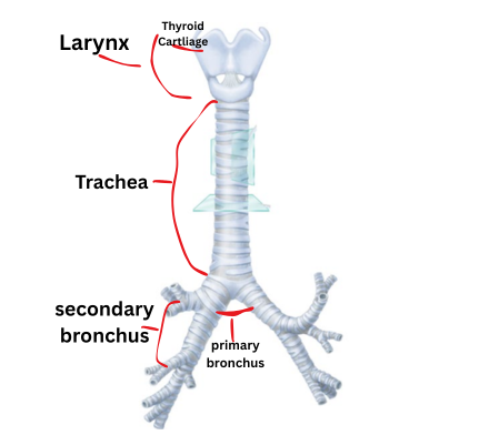
Right: shorter, wider, more vertical. Left: longer, narrower, more horizontal (angled around the heart).
38
New cards
Which bronchus is aspirated food most likely to enter?
The right primary bronchus, because it is wider, shorter, and more vertical, so objects drop straight into it.
39
New cards
Conducting division
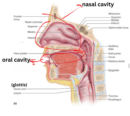
Structures that only move air to the gas-exchange area (no gas exchange). Warms, humidifies, and filters air. Nasal cavity through terminal bronchioles.
40
New cards
Respiratory division
Structures where gas exchange occurs: respiratory bronchioles, alveolar ducts, alveolar sacs, and alveoli.
41
New cards
Structures of the respiratory division
Respiratory bronchioles, alveolar ducts, alveolar sacs, alveoli.
42
New cards
Upper respiratory tract
Nose, nasal cavity, paranasal sinuses, and pharynx (nasopharynx, oropharynx, laryngopharynx).
43
New cards
Lower respiratory tract
Larynx, trachea, bronchi, bronchioles, and lungs (alveoli).
44
New cards
Epithelium: vestibule of the nostril
Stratified squamous (keratinized). Protects against abrasion and traps large particles at the entrance.
45
New cards
Epithelium: nasal cavity
Pseudostratified ciliated columnar (respiratory epithelium) with goblet cells. Traps and moves debris with mucus; warms and humidifies air.
46
New cards
Epithelium: nasopharynx
Pseudostratified ciliated columnar. Air-only passage; moves mucus and trapped debris toward the throat to be swallowed.
47
New cards
Epithelium: oropharynx
Stratified squamous. Passage for both food and air, so it needs protection from abrasion.
48
New cards
Epithelium: laryngopharynx
Stratified squamous. Food and air pass through, so it needs protection from abrasion.
49
New cards
Epithelium: alveoli
Simple squamous. Very thin, so gases diffuse quickly across it.
50
New cards
What is respiratory epithelium?
Pseudostratified ciliated columnar epithelium with goblet cells, lining most of the conducting airways (nasal cavity to bronchi). Goblet cells make mucus that traps dust and microbes, and cilia sweep it.
51
New cards
Mucociliary escalator
Cilia beat in a coordinated upward motion, moving mucus with trapped particles toward the pharynx, where it is swallowed or coughed out. It cleans the air passages.
52
New cards
Great alveolar cell (type II)
Secretes surfactant, which reduces surface tension in the alveolus and prevents it from collapsing; can also replace damaged alveolar cells.
53
New cards
Squamous alveolar cell (type I)
Thin, flat cell that makes up most of the alveolar wall; allows rapid gas exchange between the alveolus and the capillary.
54
New cards
Alveolar macrophage
Phagocytic "dust cell" that removes debris, bacteria, and particles that reach the alveoli, keeping them clean.
55
New cards
Surfactant
Lipid-protein fluid secreted by great alveolar cells that lowers surface tension and keeps alveoli from collapsing.
56
New cards
Respiratory membrane
Thin barrier for gas exchange: alveolar squamous cell, fused basement membranes, and capillary endothelial cell.
57
New cards
Direction of O2 and CO2 at the alveolus
O2 moves from the alveolus into the capillary blood. CO2 moves from the capillary blood into the alveolus (each down its concentration gradient).
58
New cards
Path of air (ventilation)
Nose/mouth -> nasal cavity -> pharynx (naso-, oro-, laryngo-) -> larynx (glottis) -> trachea -> primary bronchi -> secondary bronchi -> tertiary bronchi -> bronchioles -> terminal bronchioles -> respiratory bronchioles -> alveolar ducts -> alveolar sacs -> alveoli
59
New cards
Structures O2 crosses in external respiration
Alveolar squamous cell (type I) -> fused basement membranes -> capillary endothelial cell -> blood (red blood cell, binds hemoglobin).
60
New cards
Vessel sequence for O2 during transport
Pulmonary capillaries -> pulmonary venules -> pulmonary veins -> left atrium -> left ventricle -> aorta -> systemic arteries -> arterioles -> systemic capillaries
61
New cards
Structures O2 crosses in internal respiration
Capillary endothelial cell -> interstitial fluid -> plasma membrane of the body cell -> into the cell (down its gradient).
62
New cards
What happens to O2 in cellular respiration?
It is the final electron acceptor in the mitochondria, used to make ATP; produces CO2, H2O, and ATP.
63
New cards
CO2 in internal respiration
CO2 diffuses from cells (high CO2) across the cell membrane, interstitial fluid, and capillary endothelium into the blood.
64
New cards
Vessel sequence for CO2 during transport
Systemic capillaries -> venules -> veins -> superior/inferior vena cava -> right atrium -> right ventricle -> pulmonary trunk -> pulmonary arteries -> pulmonary arterioles -> pulmonary capillaries
65
New cards
CO2 in external respiration
CO2 diffuses from pulmonary capillary blood across the capillary endothelium, fused basement membranes, and alveolar squamous cell into the alveolus.
66
New cards
Pathway of CO2 in ventilation
Alveoli -> respiratory bronchioles -> terminal bronchioles -> bronchioles -> bronchi -> trachea -> larynx -> pharynx -> nasal/oral cavity -> out. It is the reverse of the O2 pathway.
67
New cards
How is transport different for O2 vs CO2?
O2 is mostly (about 98%) bound to hemoglobin in red blood cells, with a small amount dissolved. CO2 is mostly (about 70%) carried as bicarbonate, with the rest bound to hemoglobin or dissolved in plasma.
68
New cards
Pulmonary circuit pathway
Right ventricle -> pulmonary trunk -> pulmonary arteries -> pulmonary arterioles -> pulmonary capillaries (gas exchange) -> pulmonary venules -> pulmonary veins -> left atrium
69
New cards
Overall purpose of breathing
To supply O2 for cellular respiration (ATP production) and remove the CO2 waste, which also helps maintain blood pH.