Anatomy Tapp
 Call Kai
 Learn
Learn Practice Test
 Spaced Repetition
 Match
Match Flashcards
 Knowt Play
1/97

There's no tags or description
Looks like no tags are added yet.
Name | Mastery | Learn | Test | Matching | Spaced | Call with Kai | Chat |
|---|
No analytics yet
Send a link to your students to track their progress
98 Terms
Two types of Peripheral Nerves
Somatic and Autonomic
Somatic
Voluntary, one continuous nerve from CNS to target, move skeletal muscle & transmit sensation from body walls
Autonomic
Involuntary functions, two nerves linked by synapses, occur between CNS and target, activate organs, transmit sensation from organs, glands, and internal structures
Efferent vs Afferent
Efferent is motor and Afferent is sensory
Synapses
Electrochemical communication between nerve cells and with tissues
Autonomic Nerves (Sympathetic and Parasympathetic)
Sympathetic = Fight or Flight and Parasympathetic = Rest and Digest
Sympathetic synapse
They synapse before reaching their target, in sympathetic trunk, in prevertebral ganglia
Parasympathetic synapse
Synapse in or near the walls of their targets
Parasympathetic nerves orgin
Originate from cranial nerves or sacrum
Digestive tract, Parasympathetic
signals for secretion of fluids (e.g., saliva, pancreatic enzymes) and contraction of smooth muscle
Digestive tract, Sympathetic
stops the secretion of fluids and muscle contractionstops the secretion of fluids and muscle contraction
Cardiovascular, parasympathetic
slows heart rate and dilates arteries
Cardiovascular, sympathetic
increases heart rate and constricts arteries

What is in the image
Manubrium

What is labeled
Sternum

What is labeled
Xiphoid process

What is labeled
Costal Cartilage

Label the image
ribs, pectoralis major, retromammary space, lactiferous ducts, nipple, suspensory ligaments

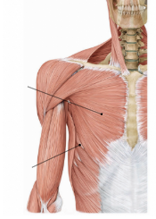
Label the two muscles
Pectoralis major, Serratus anterior

Label the two muscles
Pectoralis minor, serratus anterior
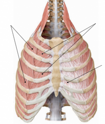
Label the muscles
External intercostal muscles, Internal intercostal muscles, External intercostal aponeurosis


Label the muscles
Endothoracic fascia, Innermost intercostal muscles


Label the muscle fibers
External intercostal muscles, Internal and innermost intercostal muscles

Intercostal neurovascular bundle definition
The intercostal neurovascular bundle is a group containing a vein, an artery, and a nerve that travels along the space between each pair of ribs


Label
Right dome of diaphragm, left dome of diaphram

Label 4 things (2 domes, 1 tendon, and 1 bone)
Left dome of diaphragm, right dome of diaphragm, central tendon, vertebra


Label
Right dome of diaphragm, Costodiaphragmatic recess

Label
The blue is expiration maximum, the pink inspiration maximum
The amount of air you can inhale and exhale
Diaphragm innervation
Phrenic nerve
Where does the phrenic nerve originate from
C3, C4, C5
Pleural cavity
space between parietal pleura (superficial) and visceral pleura (deep) containing serous fluid
Coelom
The body cavity within the trunk that contains and surrounds the internal organs
Coelomic sacs
Fluid-filled sacs within the coelom that surround and protect internal organs
Pleura (visceral and parietal)
Visceral pleura covers the lungs; parietal pleura lines the thoracic cavity
Pleural cavity (normal and collapsed lung)
Potential space between pleurae; normally minimal, but expands when the lung collapses.
Costodiaphragmatic recess
Potential space between the costal and diaphragmatic pleura where the lungs expand during deep inspiration.
Apex
Superior tip of the lung

Horizontal fissure
Separates the superior and middle lobes of the right lung

Oblique fissure
Separates the lower lobe from the upper lobe; also separates the lower lobe from the middle lobe on the right

Hilum (root)
Area where bronchi, blood vessels, nerves, and lymphatics enter and leave the lung


Label the lines and Upper and Lower lobe
Apex, Upper Lobe, Lower Lobe, Middle Lobe, Oblique Fissures, Apex, Horizontal Fissure


Label

Hilum (root) of the lungs
Area where bronchi, blood vessels, nerves, and lymphatics enter and leave the lungs.
Trachea and hilum (root) of the lungs
The trachea carries air to the bronchi; the hilum is where bronchi, vessels, nerves, and lymphatics enter and leave the lungs
Bronchopulmonary units
Functional lung regions supplied by a segmental bronchus and its accompanying blood vessels
Bronchopulmonary units
Lung regions organized by the airway branching pattern: main bronchus → lobar bronchus → tertiary/segmental bronchus → bronchioles → respiratory bronchioles

Label

Serous fluid
Attracts visceral and parietal pleurae and reduces friction between them
Diaphragm Contraction
Pulls the central tendon down, expanding the pleural cavity and creating negative pressure
Rib elevation
Lifts and expands the ribs, increasing thoracic volume
Lung filling
Lungs passively expand to fill the space created by thoracic expansion
Vertical changes during inspiration and expiration
Inspiration: diaphragm moves down, increasing thoracic volume; expiration: diaphragm moves up, decreasing thoracic volume
Rib cage movement during inspiration
The rib cage increases thoracic volume in three dimensions: vertical, anteroposterior, and transverse
Forced respiration
Breathing that uses additional muscles to actively increase or decrease thoracic volume beyond normal quiet breathing
Pneumothorax
Air enters the pleural cavity, causing the lung to collapse

Label the heart


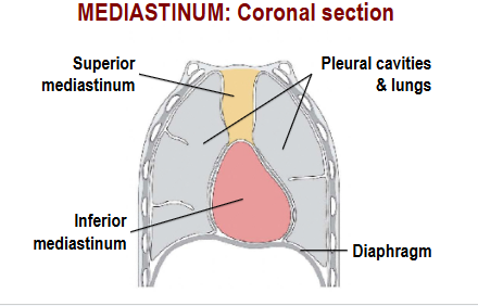
Label
Superior mediastinum, Pleural cavities and lungs, Inferior mediastinum, Diahphragm

Label


Label


Label

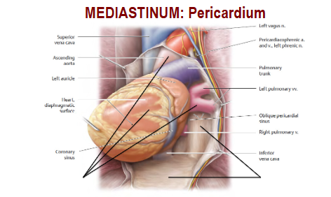
Pericardium
Double-layered sac surrounding the heart; protects the heart and reduces friction as it beats


Label

Label


Label


Label


Label


Label
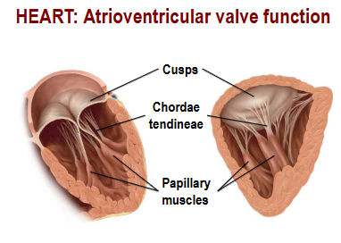
Heart: Blood flow, right atrium & ventricle
Right atrium → right ventricle - Deoxygenated blood enters through the right atrium from the venae cavae → passes through the tricuspid valve → enters the right ventricle

Pulmonary vessels
Pulmonary arteries carry deoxygenated blood from the right ventricle to the lungs; pulmonary veins return oxygenated blood to the left atrium


Label


Label
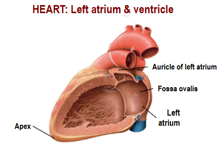

Label

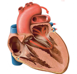
Left atrium and ventricle blood flow
Oxygenated blood enters the left atrium from the pulmonary veins → passes through the mitral valve → enters the left ventricle → leaves through the aortic valve into the aorta.

Label


Label

Heart conduction
SA node starts the heartbeat → AV node delays it → AV bundle → bundle branches → Purkinje fibers spread the signal through the ventricles.
Diastole vs Systole
Diastole: heart chambers relax and fill with blood. Systole: chambers contract and pump blood out

Label


Label

Label
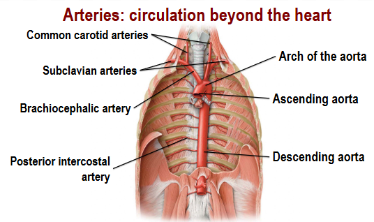
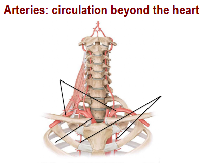
Label
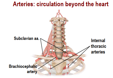
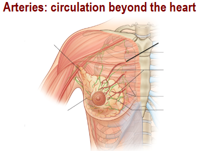
Label


Label it

Veins circulation beyond the heart
Veins return blood from body tissues back to the heart; valves prevent backflow and skeletal muscles help push blood forward
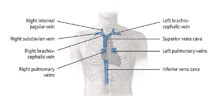

Label


Label


Label


Label

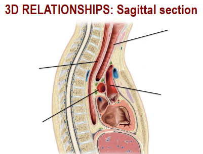
Label


Label


What is the yellow circle
Thoracic Duct

Label

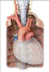
What nerves are these
Phrenic and Vagus nerves
Intercostal neurovascular bundle
Runs along the inferior border of each rib in the costal groove; arranged VAN: Vein, Artery, Nerve

Sympathetic trunk
A chain of sympathetic nerves alongside the vertebral column that carries autonomic signals to organs, blood vessels, and glands

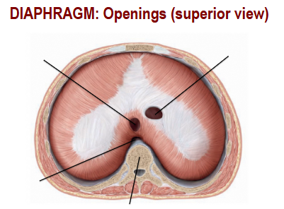
Label

Partial pneumothorax
Air enters the pleural cavity, separating the pleural layers and reducing lung expansion, causing partial lung collapse
