OTR 704 3.1 Palpation, Screening, & ROM
 Call Kai
 Learn
 Practice Test
 Spaced Repetition
 Match
 Flashcards
 Knowt Play
1/100

There's no tags or description
Looks like no tags are added yet.
101 Terms
Anatomical palpation
Method of physical examination using tactile touch with fingers and hands to assess skin, bony landmarks, and soft tissues; palpate from superficial to deep (light pressure gradually increased) and check for:
Temperature, tone, edema, mobility of skin, scar tissue
Crepitus, nodules, spasms, trigger points, tissue consistency
Functional Movement Screening (ROM Screening)
Assesses basic ability to produce volitional movement and typically begins with AROM against gravity
Occurs at onset of evaluation and throughout plan of care; informs need for further ROM assessment (PROM & goniometry)
Looks for evidence of: abnormal limb posturing, deviations in trunk alignment, compensatory movement patterns, weakness, inability to selectively control joints, discoordination of movement timing and/or multi-joint movements
How does muscle tissue influence ROM?
Often associated with decreased flexibility in concert with their respective tendons and fascia
Increasing flexibility relies on elastic properties of muscle
Length can be changed over time
Muscle strength may also impact ROM due to size, type, and length-tension relationship
How does connective tissue influence ROM?
Ligaments and joint capsules, while possessing some elastic properties, can lose their elasticity during periods of disuse and immobilization; may also be loose or lax
Connective tissue composition
Collagen: strong, flexible, inelastic structure that holds connective tissue together; enables tissue to resist mechanical deformation
Elastin: extensible; allows elongation then return to normal length
Reticulin: fine fibers
Ground substance: material between fibers functioning to decrease friction; nutrient transport
Fibrous connective tissue
Dense (regular & irregular): tendons, ligaments, joint capsules, aponeurosis, periosteum — highly organized, greater cross links creating more dense tissue
Areolar (loose): adipose, fascia associated with skin, muscle fibers, nerves — unorganized, increased space between the individual fibers
Cartilage
Rigid connective tissue composed of chondrocytes within a collagen, elastin, ground substance matrix
Types: hyaline, fibrocartilage, and elastic
Reticular connective tissue
Supports structural walls of organs
Elastic connective tissue
Composed of elastic fibers and found in blood vessels, airways, and hollow organs
How do bony structures/joint structures influence ROM?
Can limit end point range, can also stop movement at normal end points in the range
Ligaments, capsule integrity, and joint surface all influence overall ROM
How does adipose tissue influence ROM
Can act as a “wedge” between lever arms; restricts movement wherever it is found
How does skin influence ROM?
Injury or surgical procedure may alter skin, resulting in tightness & scar tissue formation (e.g., burns)
Stretching can improve ROM to varying degress
How does neural tissue influence ROM?
Develops tightness as a result of compression, chronic repetitive microtrauma, muscle imbalances, joint dysfunction, or poor posture
How do demographic/personal factors influence ROM?
Age: increased age = decreased ROM
Sex: hormonal & anatomical differences impact ROM
Pre-existing conditions & diagnoses: conditions like arthritis and SCI influence ROM
End-feel
The resistance in a joint that is felt at the end ranges of passive limits of motion
An abnormal end-feel may indicate a comorbidity or complication
Hard end-feel
Bone-to-bone; stop in movement as bony surfaces meet
Ex: elbow extension, knee extension
Soft end-feel
Soft-tissue approximation; stop in movement due to soft tissues (e.g., muscles)
Ex: elbow flexion, knee flexion, shoulder adduction
Firm end-feel
Capsular end-feel; stop in movement that feels somewhat firm or leathery but has some “give” at the end-range
Ex: shoulder & hip rotation, shoulder abduction
Active ROM (AROM)
Available ROM of a specific joint when the client is volitionally moving the body part/joint
Often the focus of evaluation
Active-assisted ROM (AAROM)
Available ROM of a specific joint when the practitioner or supportive device aids the client in moving
Client’s body part/joint is still volitionally participating
Often used in treatment
Passive ROM (PROM)
Available ROM of a specific joint when the practitioner or client passively moves the body part/joint
Often used in addition to AROM or if AROM is not possible
Goniometry
ROM assessment that provides an objective measure of ROM
Universal goniometer is most commonly used device
Passive and active insufficiency
Can occur with muscles that cross more than one joint simultaneously
Passive: reaches maximum ability to elongate; prevents full ROM
Active: simultaneous movement at all the joints, reaches its shortest length and is unable to produce effective tension/force
Optimal sufficiency
Balance between the soft tissue structures surrounding the joint; balance between the agonist and antagonist musculature (length & tension) allowing for simultaneous stabilization across muscle joints
ROM assessment precautions
Healing fractures
Muscular or tendinous injury
Significant pain or discomfort
Known or chronic heterotrophic ossification (ectopic ossification)
Hypermobility of a joint
Infection or inflammatory condition
Marked osteoporosis
ROM assessment contraindications
Subluxation or dislocation of a joint
Acute myositis ossificans or ectopic ossification
Hematoma of a joint
Known DVTs
ROM assessment client positioning
Start in anatomical/neutral position (most joints are considered “in neutral" or 0 degrees)
Maintain consistent start position with trials & re-assessment
Consider age, HPI/injury, comfort, observation abilities
Allow movement through full arc of motion
ROM general procedures
Position client appropriately and support/stabilize as needed
Explain procedure and demonstrate (as needed)
Palpate & locate bony landmarks → place goniometer
Client performs AROM to best of ability (ASK: “Is that as far as you can go?”)
Observe for any compensations
Read goniometer & record degrees achieved
***ROM screening performed prior to formal measurements
Parts of the goniometer

What is occurring if PROM > AROM?
Muscle weakness
Neuromotor deficits
Pain with volitional movement
What is occurring if AROM > PROM?
Guarding
Assessor error
What is occurring if AROM = PROM but both are below normal limits?
Soft tissue tightness
Hypertonicity
Abnormal end feels (especially abnormal hard end feels)
Effects of immobilization on connective/soft tissue
Even without injury, connective tissue is consistently remodeling, therefore if movement through ROM does not occur over time, motion will be lost due to shortening of soft tissue; general soft tissue changes include:
Reduced ground substance & increased collagen cross-links,
Contracted fiber meshwork & wound contraction
Fibrosis and replacement of tissue with collagen
Weakness
Effects of immobilization on muscle
Decreased muscle fiber size & reduced number of myofibrils
Reduced oxidative capacity & force production
Increase in fibrous & fatty tissue in the muscle
Reduced intramuscular capillary density
Atrophy can be seen in two weeks
With prolonged immobilization, the normal neural feedback system of movement is lost
Histological changes: decreased ATP, creatine phosphate, & glycogen
Effects of immobilization on articular cartilage
Changes depend on position of immobilization, duration of immobilization,and weight-bearing status of joint
Less matrix organization
Necrosis of areas of constant pressure
Increased fibrofatty tissue within the joint that becomes scar tissue
Irreversible damage occurs with continued immobilization
Effects of immobilization on periarticular connective tissue
Periarticular connective tissue = soft tissue surrounding the joint, such as ligaments, the joint capsule, fascia, tendons, and synovial membranes
Connective tissue becomes thick and fibrosis
Clinical results = decreased ROM
Effects of re-mobilization
Enhances recovery; resolves abnormal collagen cross-link formation and increases fluid content in the extracellular matrix of connective tissue
Muscle fibers: early mobilization increases hematoma absorption, tensile strength, and myofiber regeneration & orientation; minimizes adhesions
Articular cartilage: early controlled loading stimulates proteoglycan & chondrocyte production = promotes healthy function of tissue
Periarticular cartilage: early mobility decreases excessive collagen fiber cross-linking and maintains fluid content of the extracellular matrix; reduced changes of tissue becoming thick & fibrous
Shoulder flexion normal ROM
0-170°
Shoulder flexion goniometry
Axis: Center of humeral head, distal to acromion process on lateral aspect of humerus
Stationary arm: Parallel to the trunk
Movable arm: Parallel to the humerus, aligned with the sagittal plane
End-feel: Firm

Shoulder extension normal ROM
0-60°
Shoulder extension goniometry
Axis: Center of humeral head, distal to acromion process on lateral aspect of humerus
Stationary arm: Parallel to the trunk
Movable arm: Parallel to the humerus, aligned with the sagittal plane
End-feel: Firm

Shoulder abduction normal ROM
0-170°
Shoulder abduction goniometry
Axis: Below the acromion process on the posterior surface of the shoulder, in line with the joint center at the humeral head
Stationary arm: Parallel to the trunk
Movable arm: Parallel to the humerus, pointing at the lateral epicondyle of the elbow
End-feel: Firm

Shoulder internal rotation (against gravity; shoulder abducted) normal ROM
0-70°
Shoulder internal rotation (against gravity; shoulder abducted) goniometry
Axis: Olecranon process
Stationary arm: Horizontal position
Movable arm: Parallel to the forearm, aligned with the ulna
End-feel: Firm

Shoulder internal rotation (gravity-reduced; shoulder adducted) normal ROM
0-40°
Shoulder internal rotation (gravity-reduced; shoulder abducted) goniometry
Axis: Olecranon process
Stationary arm: Horizontal and pointed forward in the sagittal plane
Movable arm: Parallel to the forearm, follows the arc of movement in a horizontal plane
End-feel: Firm

Shoulder external rotation (against gravity; shoulder abducted) normal ROM
0-90°
Shoulder external rotation (against gravity; shoulder abducted) goniometry
Axis: Olecranon process
Stationary arm: Horizontal in neutral, parallel to the forearm
Movable arm: Follows forearm in vertical arc in sagittal plane
End-feel: Firm
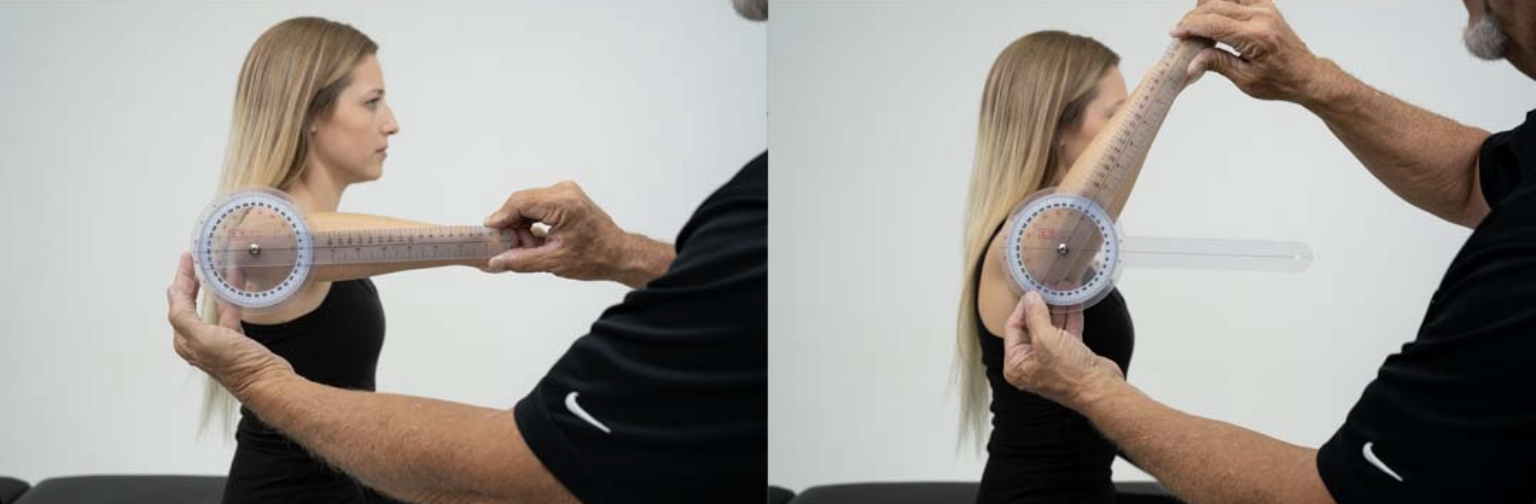
Shoulder external rotation (gravity-reduced; shoulder adducted) normal ROM
0-60°
Shoulder external rotation (gravity-reduced; shoulder adducted) goniometry
Axis: Aligned with and below the olecranon process
Stationary arm: Parallel to the forearm
Movable arm: Parallel to the forearm, follows the forearm in lateral arc towards the abdomen
End-feel: Firm

Shoulder horizontal adduction normal ROM
0-130°
Shoulder horizontal adduction goniometry
Axis: Over acromion process
Stationary arm: Parallel to the line between the left and right acromion, over the shoulder toward the neck
Movable arm: Parallel to the humerus on the superior aspect
End-feel: Firm or soft
Shoulder horizontal abduction normal ROM
0-40°
Shoulder horizontal abduction goniometry
Axis: Over acromion process
Stationary arm: Parallel to the line between the left and right acromion, over the shoulder toward the neck
Movable arm: Parallel to the humerus on the superior aspect
End-feel: Firm or soft
Elbow flexion normal ROM
0 to 135-150°
Elbow flexion goniometry
Axis: Lateral epicondyle at end of elbow crease
Stationary arm: Parallel to midline of humerus or aimed at humeral head
Movable arm: Parallel to radius
End-feel: Soft, hard, or firm

Elbow extension normal ROM
0°
Elbow extension goniometry
Axis: Lateral epicondyle at end of elbow crease
Stationary arm: Parallel to midline of humerus or aimed at humeral head
Movable arm: Parallel to radius
End-feel: Hard or firm
Forearm supination normal ROM
0 to 80-90°
Forearm supination goniometry
Axis: Ulnar border of the volar aspect of the wrist, just proximal to ulnar styloid
Stationary arm: Vertical, perpendicular to floor
Movable arm: Resting against volar aspect of wrist
End-feel: Firm

Forearm pronation normal ROM
0 to 80-90°
Forearm pronation goniometry
Axis: Ulnar border of dorsal aspect of wrist, just proximal to ulnar styloid
Stationary arm: Perpendicular to floor
Movable arm: Squarely across center of dorsum of distal end of forearm between the styloids
End-feel: Hard

Wrist flexion normal ROM
0-80°
Wrist flexion goniometry
Axis: Lateral wrist just distal to radial styloid in anatomical snuffbox
Stationary arm: Parallel to the radius
Movable arm: Parallel to 2nd metacarpal (index finger)
End-feel: Firm

Wrist extension normal ROM
0-70°
Wrist extension goniometry
Axis: Lateral wrist just distal to radial styloid in anatomical snuffbox
Stationary arm: Parallel to the radius
Movable arm: Parallel to 2nd metacarpal (index finger)
End-feel: Firm or hard

Wrist radial deviation normal ROM
0-20°
Wrist radial deviation goniometry
Axis: Dorsum of wrist at base of 3rd metacarpal in a hollow over the capitate
Stationary arm: Follows the midline of the dorsal aspect of the forearm
Movable arm: Parallel to the 3rd metacarpal aligned with the third MCP joint
End-feel: Firm

Wrist ulnar deviation normal ROM
0-30°
Wrist ulnar deviation goniometry
Axis: Dorsum of wrist at base of 3rd metacarpal in a hollow over the capitate
Stationary arm: Follows the midline of the dorsal aspect of the forearm
Movable arm: Parallel to the 3rd metacarpal aligned with the third MCP joint
End-feel: Firm or hard

MCP flexion normal ROM
0-90°
MCP flexion goniometry
Axis: Dorsal aspect of MCP joint
Stationary arm: Dorsal surface of hand following metacarpal
Movable arm: Dorsal surface of proximal phalanx
End-feel: Hard or firm
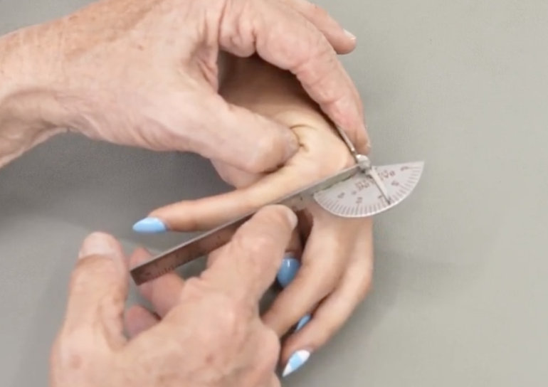
MCP extension normal ROM
0-45°
MCP extension goniometry
Axis: Lateral aspect of MCP joint
Stationary arm: Parallel to the metacarpal
Movable arm: Parallel to the proximal phalanx
End-feel: Firm

MCP abduction normal ROM
0-25°
MCP abduction goniometry
Axis: Over MCP
Stationary arm: Over corresponding metacarpal
Movable arm: Over proximal phalanx
End-feel: Firm

PIP flexion normal ROM
0-110°
PIP flexion goniometry
Axis: Lateral surface of PIP
Stationary arm: Lateral side of proximal phalanx
Movable arm: Follows middle phalanx
End-feel: Usually hard

PIP extension normal ROM
0°
PIP extension goniometry
Axis: Lateral surface of PIP
Stationary arm: Lateral side of proximal phalanx
Movable arm: Follows middle phalanx
End-feel: Usually hard

DIP flexion normal ROM
0-80°
DIP flexion goniometry
Axis: Dorsal surface of DIP
Stationary arm: Over middle phalanx
Movable arm: Over distal phalanx
End-feel: Soft or firm

1st CMC extension (radial abduction) normal ROM
0-50°
1st CMC extension (radial abduction) goniometry
Axis: Dorsal surface of CMC at the base of the thumb metacarpal, in the anatomical snuffbox
Stationary arm: Parallel to the radius
Movable arm: Aligned with the 1st metacarpal of the thumb
End-feel: Firm
1st CMC abduction (palmar abduction) normal ROM
0-50°
1st CMC abduction (palmar abduction) goniometry
Axis: Junction of thumb and index finger metacarpals, in the anatomical snuffbox
Stationary arm: Over the radius
Movable arm: Parallel to the 1st metacarpal of the thumb
End-feel: Firm
1st MCP flexion normal ROM
0-50°
1st MCP flexion goniometry
Axis: Parallel to the dorsal surface of the MCP
Stationary arm: Over the thumb metacarpal
Movable arm: Over the proximal phalanx
End-feel: Hard or firm
1st MCP extension normal ROM
0°
1st MCP extension goniometry
Axis: Parallel to the dorsal surface of the MCP
Stationary arm: Over the thumb metacarpal
Movable arm: Over the proximal phalanx
End-feel: Firm
1st IP flexion normal ROM
0 to 80-90°
1st IP flexion goniometry
Axis: Over the dorsal surface of the IP
Stationary arm: Over the proximal phalanx
Movable arm: Over the distal phalanx
End-feel: Firm
1st IP extension normal ROM
0 to 20-30°
1st IP extension goniometry
Axis: Over the dorsal surface of the IP
Stationary arm: Over the proximal phalanx
Movable arm: Over the distal phalanx
End-feel: Firm
Thumb to 5th digit opposition normal ROM
0 (if pads of thumb and 5th digit can touch)
Thumb to 5th digit opposition goniometry
Measure the distance between centers of the pads of the thumb and 5th digit
End-feel: Soft or firm

Hip flexion normal ROM
0-120°
Hip flexion goniometry
Axis: Greater trochanter
Stationary arm: Middle of lateral aspect of pelvis at greater trochanter, parallel to the trunk
Movable arm: Parallel to the long axis of the femur on the midline of the lateral aspect of the thigh
End-feel: Soft

Knee flexion normal ROM
0-135°