Horse state part B - surgery & orthopaedics
 Call Kai
 Learn
Learn Practice Test
 Spaced Repetition
 Match
Match Flashcards
 Knowt Play
1/24

Earn XP
Description and Tags
Equine surgery and orthopaedics
Name | Mastery | Learn | Test | Matching | Spaced | Call with Kai | Chat |
|---|
No analytics yet
Send a link to your students to track their progress
25 Terms
1. Principles and indications of local anaesthesia.
Local anaesthesia = numbing an area of the body using medications called local anaesthetics.
Gives temporary loss of sensation and pain.
Used for surgical procedures and diagnostic conclusion.
Reduces the required amount of anaesthetic agent and allows complicated procedures to be performed on a standing horse.
Injected around the nerve at a site where the nerve is not covered by bone.
Risks following this type of procedure, including epidural anaesthesia:
Ataxia of the hind limbs
Hind-limb motor blockage
Recumbence
Respiratory depression
Indications ✅
Surgical procedures: removal of teeth, laparoscopy and wound management.
Lameness diagnostics: pain blocking using peripheral nerve blocks, intraarticular and intrabursal injections, and ring blocks.
Contraindications ❌
Fracture confirmation
Septic inflammation, including:
Periarticular cellulitis
Hoof abscesses
Regional anaesthesia
Regional anaesthesia can be used to localize the area of pain:
➡ Apply local anaesthesia and register any improvement in lameness after 15 minutes.
Start with a palmar digital nerve block and go upwards on the leg if there is no improvement.
Regional anaesthesia is divided into two main regions:
Head
Limbs
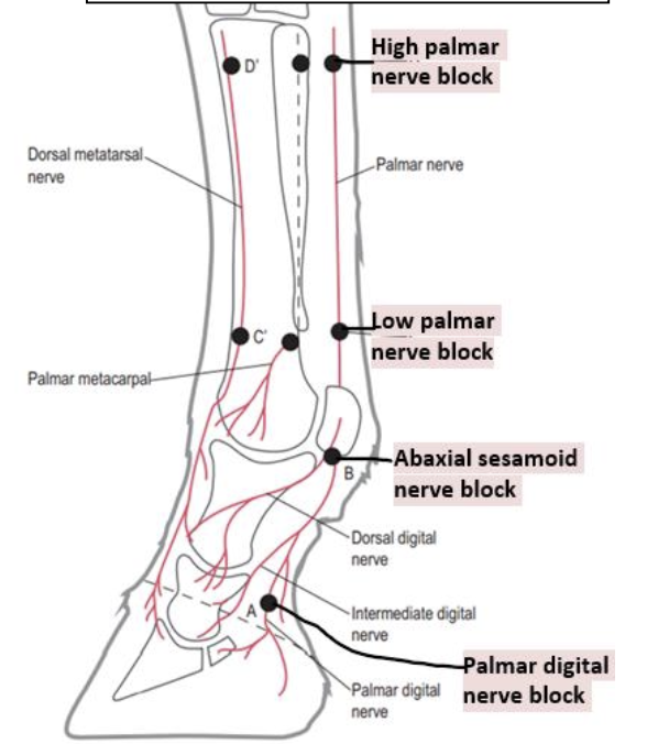
Forelimb nerve blocks 🐴
Palmar digital nerve (PDN) block – “heel block”
Semi-ring block at the pastern
Abaxial sesamoid nerve block – basisesamoid nerve block
Low 4-point block – low palmar nerve block
High 4-point block – high palmar nerve block
Lateral palmar nerve block
Hindlimb nerve blocks 🐴
Plantar digital nerve block – as in the forelimb
Abaxial sesamoid nerve block – as in the forelimb
Low 6-point nerve block – low plantar nerve block
High 6-point nerve block – high plantar nerve block
Tibial nerve block
Head nerve blocks 🐴
Maxillary nerve block
Infraorbital nerve block
Mandibular nerve block – inferior alveolar nerve block
Mental nerve block
Auricopalpebral nerve block
Diamond block – supraorbital, lacrimal, zygomatic and infratrochlear nerve blocks
Retrobulbar nerve block – muscles of the eye

Teeth
subgingival, supraperiostal, interincisival or intralesional infiltration
Epidural anaesthesia
⚠ Epidural anaesthesia is injected into the epidural space, not the subarachnoid space. Injection into the subarachnoid space is called intrathecal or spinal anaesthesia.
Used in a standing horse.
Analgesics should be warmed to body temperature before injection into the spine.
Indications
Analgesia or anaesthesia of the perineal area, including:
Perineum
Rectovestibular laceration repair
Surgeries involving the rectum
Anus
Tail
Urethra, Bladder, Vulva, Vestibule, Vagina
Relaxation of abdominal contractions in dystocia
Location
Between the 1st and 2nd coccygeal vertebrae
Or the lumbosacral space
⚠ It is forbidden to use proximal epidural anaesthesia in horses because it may cause severe hindlimb motor blockade, ataxia and recumbence.
Drugs
Adjust the dose to prevent ataxia and recumbence because the horse is in a standing position!
Local anaesthetics: lidocaine, mepivacaine
Alpha-2 agonists: xylazine, detomidine
Opioids: morphine, methadone, hydromorphone
Tramadol: analgesia
Ketamine: analgesia and systemic effects
One can use lidocaine (2%, 7-9ml), xylazine or detomidine (alpha-2-agonists) or opioids like morphine (butorphanol) or methadone.
Types of local anaesthesia 💉
Superficial, Infiltration, Perineural, Local perfusion
Intrasynovial:
Intraarticular
Intrabursal
Intrathecal
Local anaesthetic drugs and duration ⏱
Drug | Onset of effect | Duration |
|---|---|---|
Procaine | 5–10 min | — |
Lidocaine | 5 min | 30–60 min |
Mepivacaine | 10 min | 60–120 min |
Bupivacaine | 30 min | 120–140 min |
Etidocaine | 3–5 min | 5–10 hours |
Proparacaine | <1 min | 5–25 min |
Equine sedation and incomplete general anesthesia
Sedation = the depression of a patient's awareness to the environment and reduction of its responsiveness to external stimulation.
Sedatives and tranquilizers are commonly used in combinations as pre-anesthetics before general anesthesia to relax and sedate the animal. Some sedatives also provide analgesia.
💊 Drugs
Alpha2-adrenergic agonists
Xylazine, detomidine, medetomidine, romifidine → sedation + analgesia
Can be used alone or in combination with opioids.
Induce bradycardia, decreased CO, hyperglycemia, decreased bp, hypothermia, hypotension, diuresis, polyuria, can kick
Xylazine: (Rometar) lasts about 15–20 min. Dose: 1.1 mg/kg IV.
Detomidine: (Domosedan) 0.01-0.04mg/kg IV or IM, lasts about 30–60 min.
Atipamezole to reverse the effect
Phenothiazines
Acepromazine → sedative, mild tranquilizing, often in combo with alpha2 agonist or opioid, is anti-arrhythmic, and cause vasodilation.
often used in oral surgery (reduces chewing).
Contraindicated in breeding stallion (penis prolapse) or hypovolemic patients.
Opioids
Butorphanol, morphine, buprenorphine, meperidine
Analgesic → always in combo with sedative.
Benzodiazepines
Diazepam, midazolam → sedative, muscle-relaxing.
Induce ataxia → not used in standing procedures.
Foals
⭐ Common sedation in horse: Detomidine + butorphanol (domosedan og tourbugesic IV, 0,1ml av hver per 100kg)
→ Just know that you combine an alpha2 agonist + opioid.
🐴 Standing sedation
Necessary for a variety of surgical and non-surgical procedures in the standing horse.
Common drug combination for 500–600 kg horse:
💉 Detomidine (0.5 ml) + butorphanol (1 ml)
Benefits:
Smoother recovery than GA.
Sometimes cheaper than GA.
Simpler to do standing sedation in some procedures, like laryngoplasty, dental procedures.
In head surgeries (a lot of bleeding) → less bleeding in standing position than recumbence.
Decreased risk for horse compared to general anesthesia, but more risk for staff.
Indications:
- Dental procedures, stomatology
- Diagnostic imaging
- Sinus surgeries
- Urogenital
- Orthopedic
- Wound assessment
- Castration
🐴 Sedation of adult horse
Short-term sedation is achieved by administering a bolus of an alpha2 agonist, with or without an opioid.
When used in combo with an opioid:
1⃣ Alpha2 agonist FIRST → 2⃣ Opioid
The horse must first be sedated with the alpha2 agonist prior to opioid to avoid opioid-induced excitement.
A combo of opioid + alpha2 agonist is only done when:
Heavy sedation is required.
Additional analgesia is required.
🐴 Sedation and anaesthesia in foals
Sedation and anesthesia may be required in foal to allow diagnostic and therapeutic procedures.
Differences in foals compared to adult horses:
❤ Circulatory system (transition from in-utero to newborn)
💊 Metabolism of drugs
🌡 Susceptibility to hypothermia and hypoglycemia
❌ Avoid:
Drugs that lower heart rate → xylazine, detomidine
Drugs that decrease preload → acepromazine
→ May produce diminished cardiac output and tissue perfusion.Premedication if using gases.
Halothane.
Sedation of foals:
Neonatal foals become recumbent when sedated, and the foal should be supported until it assumes recumbence.
avoid drugs that lowers heart rate (xylazine and detomidine) and dec. preload (acepromazine).
In foals younger than 4 weeks one should avoid A2A and use benzodiazepines, opioids and ketamine/propofol.
Alpha2 agonist
Given to healthy foals, minimum dose.
Older foals are sedated as adult, but with higher dose.
Benzodiazepines – diazepam and midazolam
Slow administration.
Ataxia may prolong.
Don’t repeat dose or give high dose.
Not analgesic.
💉 Benzodiazepines + ketamine
Combined for painful procedures.
Light anesthesia.
Diazepam/midazolam + ketamine
Diazepam commonly used for sedation in foals because it provides tranquilization and muscle relaxation with relatively little cardiovascular depression, and is commonly combined with ketamine for induction of GA
Equine general anesthesia
General anaesthesia = controlled and reversible CNS-depression.
It gives:
Analgesia, Amnesia, Immobility, Unconsciousness, Muscle relaxation
📋 Patient preparation
Obtain medical history.
Pre-anesthetic clinical exam, with focus on cardiovascular and respiratory system.
Conditions affecting recovery or induction should be considered and implementing plans for assisting → like musculoskeletal injuries or neurological diseases.
🌾 No hay 12 hours before anesthesia.
Mouth should be flushed to remove debris.
💉 A jugular catheter should be placed if GA is intended or prolonged sedation (MENTION THIS ON STATE)
💧 Preoperatory stabilization of cardiovascular system with IV fluids is necessary for horses undergoing emergency surgery!!! (IMPORTANT ON STATE)
⚠ Horses must be sedated before induction of any anesthesia → cause excitement in horse without sedative premedication.
Why sedation before induction?
Sedation:
Improves ease of handling
Makes induction safer
Decreases dose of induction
Decreases dose of maintenance drugs
Provides analgesia
Improves quality of recovery by prolonging the time until the first attempt to stand
➡ Sedate with alpha2 agonist / Xylazine → wait 3–5 minutes after apparent effect of sedation before induction of anaesthesia.
Filip’s master mix: Xylazine for sedation, diazepam + ket for induction and xylazine + ket every 5-7 min for maintenance
💉 Total Intravenous Anesthesia – TIVA
TIVA = the use of intravenous agents for induction and maintenance of anesthesia, by the injection of a liquid anesthetic in a vein and by catheterization of vein (infusion anaesthesia).
✂ Procedures
Castration, Cryptorchidectomy, Crib biting, Ovariectomy, Wound management, Orthopedic → lag screw, implant removal
💊 Induction regimes
⚠ Always first sedate with alpha2 agonist → then induction agent
Induction agents:
Ketamine + diazepam
Ketamine + guaifenesin → muscle relaxant, analgesic
Thiopental
Propofol
⏱ Prolongation of anesthesia with injectable drugs:
Short periods:
➡ Bolus of xylazine + ketamine
Prolonged period:
➡ Thiopental + alpha2 agonist
Never use ketamine alone!!
For maintenance, do half the amount of actual dose.
Triple dip → 1L of 5% guaifenesin and adding 1-2g of ketamine and 500mg of xylazine then administered up to a rate of 1ml/kg/h, and is used for maintenance of surgeries up to 90 min.
⭐ Importance of TIVA
TIVA is frequently used in horses.
It’s cheaper than standing anesthesia and many procedures can be performed.
TIVA provides:
Smooth, excitement-free induction phase
Slow lowering of the body into sternal or lateral recumbence
Minimal cardio-pulmonary depression
Calm recovery period
Minimal ataxia
😷 Inhalation anaesthesia

Most common method of maintaining anesthesia in the hospital setting.
Requires strict monitoring and a professional anesthesiologist
Expensive and time consuming
Used for surgeries >1 h
🏥 Equipment
Endotracheal intubation
Anesthetic machine
Recovery box → soft covering, dark and quiet room
➡ Induction starts with injectable → then maintenance with gas.
💨 Gases
Isoflurane, Sevoflurane, Desflurane
High oxygen flow rate should be used during the first 10–15 min of anesthesia to carry the inhalation anesthetic into the circuit and thereby into the horse.
Flow rates:
First 15 min → 20 ml/kg/min
Remainder → 10 ml/kg/min
Foals → 40–60 ml/kg/min
✅ Pros
Minimal drug accumulation
Good monitoring
❌ Cons
Cardiovascular decrease
No analgesia
Too rapid recovery
⚠ Recovery from iso and sevo is fast and sometimes uncontrolled.
➡ Sedation decreases speed of recovery.
📊 Monitoring
❤ Heart rate: 28–44 bpm
Bradycardia → atropine to increase HR
🌡 Body temperature: 37.5–38°C
🫁 Respiratory rate: >4 breaths/min
🩸 Blood pressure: 120/70 mmHg
Hypotension → dopamine increases BP
🩸 Direct arterial blood gas
O₂
CO₂
💨 Capnography
CO₂ in expired air
❤ ECG
Heart’s electrical activity
🫁 Pulse oximetry
Measures oxygen level of blood
Attached on tongue
🚽 Urinary catheter
If procedure is >1 hour
Prevents over-distention of the bladder
⚠ Risks associated with equine anaesthesia
Horse can be dangerous to itself and the veterinarians during excitation stage.
1⃣ Complications at induction
Intracarotid injection
Administration of incorrect drug
Haematoma formation
Perivascular injections
2⃣ Intraoperative complications
🫁 Hypoxaemia
Common in all recumbent adult, full-sized horses
🩸 Hypotension
Higher risk in injectable anaesthetics
Mean arterial pressure should be >70 mmHg
💨 Hypercapnia
PaCO₂ >45
Due to depressant effects of anaesthetic drugs on respiratory function
3⃣ Post-operative complications
1.💪 Myopathy
Dorsal recumbency: gluteal and longissimus dorsi muscles are most likely to be affected due to inadequate circulation.
Prevention:
Careful positioning
Adequate padding
Short operating time
Maintain BP
2.🦴 Long bone fractures
Higher after colic surgeries
3.Neuropathy
Most commonly seen on peripheral nerves, e.g. radial nerve
Ischemic in origin
Sweeney shoulder
🏥 Complications during hospitalization period
🩸 Shock
☠ Endotoxaemia → fluid + electrolytes are important
🐴 Postoperative Ileus (POI)
🤕 Colic → some horses suffer from one or more episodes of colic after colic surgery
🩹 Wound complications
Adhesions
🩸 Thrombophlebitis
🦶 Laminitis → after acute abdomen
💩 Postoperative diarrhoea
🦠 Septic peritonitis → contamination of ingesta in peritoneal cavity
→ Atb, NSAIDs and hydration!
Equine wounds
Wound = a sudden and violent disturbance of soft tissue connections with simultaneous skin and mucosal damage due to mechanical, physical or chemical factors. fra lecture: a wound is a disruption of the anatomical and cellular continuity and integrity of the tissue.
Categorization of wounds
Cause: accidental or intentional (surgical incision)
Open wounds: Incisions (sharp cut, sharp edges), Lacerations (most common, ireegular skin edge, painful), Puncture, Avulsion (wound with tissue loss), combined, complicated
Closed wounds (not entire skin thickness): Abrasion, Contusion, Burn (chemical, friction, freeze and sunburn, 1st-3rd degree)
Location: head, thorax, abdomen, joints (heals completely different at different locations)
🔍 Wound Assessment
🐴 General assessment of horse
History (what happened)
Physical examination of patient (initial wound assessment, patient cooperative or not, restrain, sedation?)
💉 Important to know tetanus vaccination status! need tetanus toxoid administration if not vaccinated within 6 months, 13-20ml, but careful with pregnant mares (acute hepatitis/Theiler´s Ds)
Penetrating wounds above abdomen/thorax: hemorrhage should be eliminated (Hematocrit, cavity liquid content assessment)
Contraindicated to use phenotiazines (acepromazine) on horses with hemorrhagic wounds → cause vasodilation → worsening of hypotension → hemorrhagic shock
🔎 Initial assessment of wound
Influence of healing
Severity involving structures:
Nerves, Blood vessels, Ligaments, Bones, Joints
Degree of contamination (initial cleaning before palpation)
In joints, use sterile saline or contrast medium (water only in verry dirty wounds, water is hypotonic and is not sterile)
Use sterile gloves to not contaminate the wound with human skin
Remove foreign body!
⚠ Factors inhibiting healing:
Infection, Contamination, Necrosis, Movement, Loss of blood supply, Hypoxia, Loss of tissue, Tumors (sarcoid = fibroblastic tumor, wound in this area will behave differently), Cutaneous habronemiasis, Phytiosis (fungal involvement)
Wound healing depends strongly on location:
Head wounds → generally heal well because of good blood supply.
Eyelid → gentle debridement + suture
Lip/tongue → suture; severe tongue injury may require partial amputation
Nostrils → careful layered closure
Ear → suture soft tissue, not cartilage
Neck, trunk & proximal limbs → may include lacerations, punctures and burns; treat according to wound type with cleaning/debridement, analgesia ± antimicrobials/supportive treatment.
Distal limb wounds → poorer healing because of lower temperature/blood supply, contamination, movement and tension. Particularly important complications:
Tendon laceration
Extensor → partial injuries may heal conservatively; complete injuries may require suturing/support.
Flexor → repair + strong immobilization/support.
Digital tendon sheath penetration → risk of septic tenosynovitis → lavage + drainage ± systemic/local antibiotics.
Joint penetration → risk of septic arthritis/synovitis → lavage + intra-articular/systemic antibiotics + immobilization as indicated.
🩹 Methods of Wound Management
1⃣ Primary closure/healing (First intention healing)
Union of tissue by sutering wound margins
Minimal granulation tissue, desired type of healing, often very good outcome
Clean and non-infected
Sutured during golden period = 6–8 h (know on state)
Signs not to: inflammation, swelling, exudate formation
Preparation: Hemorrhage control, hair removal, clean, wound debridement (remove necrotic tissue), lavage (saline, lactated ringer´s, hydrogen peroxide)
2⃣ Secondary intention healing
Left unsutured
Heals via contraction + epithelialization
Contraction begins after the lag phase (mainly by myofibroblasts)
Done with wounds that:
Have lost tissue and/or are infected
new epithelium is visible after ca 2weeks after injury on extremities, it grows at rate 1-1.5mm/10 days
control with: counter pressure (bandage/cast), immobilisation, antimicrobials, topicals
3⃣ Delayed primary closure
Apposition after golden period
Before granulation tissue → 4–5 d
Sutured when ready for primary closure
4⃣ Delayed secondary closure
Closure after granulation tissue formation
5⃣ Grafting
Used in wounds that are too large to heal or cannot be sutured
Pedicle graft
Free graft (full-thickness, split, sheet graft, island graft (pinch, punch/biopsy punches)

🚑 Initial Wound Treatment
🎯 Goal: Decontaminate the wound as much as possible and prevent further contamination.
💧 Flush/lavage with saline
✂ Sharp debridement of gross contaminants
💊 Local antiseptics or ATBs packed into wound to prevent further contamination
🦴 Limb immobilization according to injury
🩸 Pressure bandage applied directly over bleeding area to control hemorrhages
🔄 Stages of Wound Healing
1⃣ Inflammatory phase
⏱ 2–3 days after wounding
Increased permeability
Fibrin depositis and WBC (first neutrophiles)
later in distal extremities
⬇
2⃣ Debridement phase (lag phase = debridement + inflammation phase. KNOW ON STATE)
⏱ Within 6–8 hours of injury
Macrophages (made from monocytes) move into wound
Remove debris
Induce fibroblasts and cytokines
Stimulate fibroplasia + angiogenesis
⬇
3⃣ Repair phase (fibroblastic/proliferative phase)
Formation of granulation tissue
Fibroblasts and blood vessels start to appear in the lesion by day 3 (fibroblasts are crucial for ct formation)
Epithelial cells move from the wound edges across the granulation tissue (this is what we want)
⬇
4⃣ Remodelling/maturation phase
⏱ Begins 2 weeks after wounding → 6–12 months later
wound contraction, granulation tissue and collagen production diminishment, collagen cross-linking → scar formation
🧠 Remember:
Inflammation → Debridement → Repair → Remodelling
🧼 Wound Preparation
1⃣ First treatment – Active immunization
💉 TAT = immunoserum tetanus
Dosage: 4000–6000 IU (13–20 ml)
2⃣ Anaesthesia / sedation
3⃣ Preparation of wound environment
Shave the area
Clean wound with antiseptic soap
4⃣ Surgical debridement
🩸 Living tissue bleeds → dead tissue does not bleed
Can be done:
Mechanically
Dakin's solution → less discomfort
Mechanically:
Cut new edges for wound
Excise grossly non-viable or damaged skin with scalpel
⚠ If wound contains devitalized tissue or debris → wound should be left open:
Secondary intention healing OR
Delayed primary closure
5⃣ Lavage (flushing) of wound
💧 Sterile saline or electrolyte solution
Application under pressure:
Betadine
Chlorhexidine
→ antimicrobial activity
6⃣ Wound revision
Palpation and control of wound for foreign objects
Sterile gloves and probes
X-ray examination?
Bandaging
Corticosteroid ointment?
Honey?
🩹 Wound Dressings
Wounds on dorsal and palmar surface of fetlock, tendons and ligaments
➡ Fixation bandage (cast)
Functions of wound protection/bandaging
🛡 Protective
💧 Absorption
🩸 Compression
🦴 Stabilizing
🧻 Wound Covering
Types
Biological → skin grafts
Synthetic → semi-permeable, impermeable or permeable
1⃣ Underlying materials
Bandage cotton wool / padding
2⃣ Bandage
Elastic flexible cohesive bandage
Plaster padding
Cast
3⃣ Cast – fixation bandage
Cast bandage embedded in polyurethane resin
Hard and solid after 20–30 minutes
⭐ Radiolucent! Important for X-ray controls

🧩 Skin Transplant
Used for wounds that are healing unsatisfactory or not at all.
Preparation
Clip
Shave
Disinfection
ATB ointments + bandage for 3 days
💉 Sedation / anaesthesia
Butorphanol
Detomidine
TIVA
Types
Pinch grafting
Punch grafting
Meshing grafts
Full-thickness sheet grafting
📍 Donor sites
Ventrolateral part of abdomen
Lateral shoulder area
Cranial area of chest
Side parts of neck
💧 Wound Drainage
Deep wounds → dead space → accumulation of secretion → bacterial growth → disturbed healing process
Types of drains
Passive:
Penrose
Tubular
Active:
Closed suction systems
Open suction systems
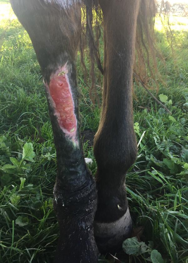
5. Second Intention Healing Wounds in Horses
Second intention healing = wounds left un-sutured to heal by second intention.
Indications:
Wounds that
Have lost tissue
Are infected
Are susceptible to infection
🔄 Two mechanisms of healing
Contraction
Epithelization
➡ Both dependent on formation of granulation tissue.
🧼 Treatment
💧 Decontaminate/flush wound by lavage saline solution
✂ Debridement:
Mechanically → cut new edges or scrape off dead tissue
Dakin's solution → less discomfort
🧩 Skin grafting if needed
🍯 Apply topical ointment → Manuka honey (also aid in debride)
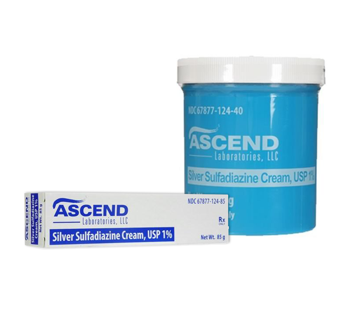
💊 Topical ATB → silver sulfadiazine
💧 Wet dressing with antiseptic → moisture + low pH
🩹 Bandage or cast
💊 NSAIDs
🔄 Stages of Second Intention Healing
1⃣ Wound Expansion
Right after a wound is made, the wound enlarges from retraction of the surrounding skin.
Wounds on the distal limb of horses may expand for 11–13 days before contraction begins.
⬇
2⃣ Granulation Tissue Formation
⏱ Appears 3–6 days after injury.
Granulation tissue provides:
🧱 Surface for migration of epithelial cells
🦠 Barrier for infection
🔄 Myofibroblasts for contraction
🧬 Fibroblasts for collagen formation
⚠ There is more granulation tissue in leg wounds VS other places on the body, because of the blood supply and lack of muscles on legs.
Factors promoting formation:
Larger body size
📍 Location → distal portion of limbs, mobile areas
🌡 Environment → moisture, warmth, low pH, low O₂ tension
🍯 Topical medication → honey, scarlet oil
⬇
3⃣ Epithelization
Epithelialization = the first signs of reparation.
Formed from the edges of the wound
Migrating epithelium moves beneath the clot that covers the wound
Can be seen at about 2 weeks after wounding
More prominent in distal part because of big skin tension
Factors increasing rate
🔥 Heat
🫁 Increased tissue oxygenation
Hyperbaric oxygen therapy
💧 Moisture
💊 Topical medication
➡ Therefore use:
🩹 Bandaging or casting
💧 Wet dressing
💊 Silver sulfadiazine
Factors decreasing rate:
🦠 Infection
Necrotic tissue
Exuberant granulation tissue
Glucocorticoids
Changing bandage too often
⬇
4⃣ Contraction Phase
Contraction = wound closure by active shrinking/reduction of the wound, by pulling the surrounding tissue centripetally.
Contraction begins after the lag phase
⚠ Wounds distal to the carpus and hock/tarsus are not capable of significant contraction
Factors inhibiting contraction:
Hypovolemic anemia
Hypoalbumin
🧠 Remember the order:
1⃣ Expansion → 2⃣ Granulation → 3⃣ Epithelization → 4⃣ Contraction
EXPAND → FILL → COVER → SHRINK
Equine hernias
Hernia = a protrusion of an organ, or part of it, through a defect in the wall of the anatomical cavity in which it normally resides.
Most are reducible, but in some cases the contents of the hernia cannot be returned to their normal location due to:
Incarceration
Strangulation
Adhesions
📚 Classification
🔹 Origin
Congenital or Acquired
🔹 Direct vs. indirect
Direct hernia → occurs through a tear
Indirect hernia → occurs through a natural passage (inguinal canal, umbilicus)
🔹 External vs. internal
External hernia → occurs through the body wall producing a visible and palpable swelling covered by skin.
Examples:
Umbilical, Inguinal/scrotal
Traumatic abdominal wall hernias, Ventral
Incisional hernias
Internal hernia → within the abdominal cavity.
hernia where abdominal viscera protrude through a ring, rent, or aperture located entirely within the abdominal cavity, example:
Epiploic foramen, Mesenteric, Diaphragmatic, Omentum, Mesocolon, Gastrosplenic ligament, Hepatogastric ligament, Hepatoduodenal ligament, Ductus deferens
⚠ Incarcerated vs. Strangulated
🟡 Incarcerated hernia
Passage of ingesta through the protruding loop of intestine is arrested.
Blood flow in its wall is maintained.
🔴 Strangulated hernia
Both irreducible + incarcerated, AND blood circulation is also arrested ➡ Results in gangrene unless speedy relief is given.
🧠 Remember:
Incarcerated = contents blocked
Strangulated = contents + blood blocked
Umbilical Hernia
More common in fillies (young female horses) and may be hereditary.
Causes:
Congenital: Present at birth, Failure of abdominal wall to close
Acquired: Develop at 3–4 weeks of age, Excessive straining to defecate/urinate, Umbilical infection
Hernial sac comprises:
Inner peritoneal layer
Outer layer of skin
Linked by connective tissue
May contain:
Small intestine, Cecum, Omentum
⚠ Incarceration and strangulation of herniated intestine is rare!
🔍 Clinical signs
Typical oval swelling in umbilical region
Intestine or omentum inside
Size varies
Tense, painful swelling → irreducible

🔎 Diagnosis
Palpation of hernia:
Can it be reduced/put back into abdomen?
Check hernial ring → size, shape, rigidity
💊 Treatment
Conservative:
Bandages, Metal clamps, Rubber rings
Cheap but dangerous
⏳ Smart to wait with surgery until foal is 6–12 months old, because it can disappear by itself.
Surgical:
GA and Dorsal recumbency
Elliptical skin incision around hernia
From here either open hernial sac but not peritoneum and push sac inside, followed by suturing the muscle or remove hernial sac by penetrating peritoneum.
If no damage to hernial sac/intestine → can be returned to abdominal cavity, but chance of re-herniation
Resection of hernial sac is the better option
Suture ring with continuous suture pattern overlapping mattress
Inguinal Hernia
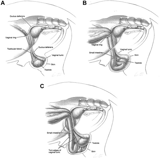
1⃣ Intravaginal (untrue scrotal) hernia in foals
Frequent problem in foals.
➡ Internal inguinal ring is very wide for passage of testes in scrotum up to 6 weeks of age.
➡ Small intestines can pass this ring and canal → enters vaginal process.
🔍 Clinical signs
Asymmetric scrotum
Possible to palpate testicles and distal intestine
🔎 Diagnosis
USG → intravaginal dislocation of intestine
Possible to reposition
💊 Treatment
Reposition intestine until inguinal ring gets smaller
Check every day
Reposition if it falls back
Check by palpation for signs of strangulation
➡ Often resolves spontaneously by 3–6 months old.
➡ Application of a truss/bandaging may hasten resolution.
2⃣ Intravaginal (untrue) hernia in adult stallions
Rare, but more dangerous due to narrow inguinal canal → may cause strangulation.
💊 Treatment
Conservative:
Manual compression of vaginal process
Surgical:
Laparotomy, Closure of inguinal canal, Resection
Unilateral castration
Inguinal surgical approach → expose herniated intestine → reduce intestine back into abdomen → assess viability → resect if non-viable → close/narrow the inguinal ring → often unilateral castration.
Diagnosis (ASKED ON STATE)
⚠ Sedate first (stallion + pain/colic can be dangerous!)
Rectal palpation: palpate the internal inguinal/vaginal ring (cranially and laterally to pelvis). Normally you identify structures of the spermatic cord at the ring; with an inguinal hernia, intestine can be palpated entering the inguinal ring/canal.
Transrectal USG → identify intestine entering the inguinal ring and assess the entrapped bowel.
External/scrotal examination + USG can show unilateral scrotal enlargement/intestine.
3⃣ Extra-vaginal (true scrotal) hernia
➡ Rupture of vaginal process
➡ Intestine is in the cavum scroti + traumatisation of serosal surface.
🔍 Clinical signs
Enlargement of scrotum
Continuous enlargement
🔎 Diagnosis
Subcutaneous localisation of intestine in scrotum
USG shows black Olympic rings filled with fluid above strangulation.
Growing enlargement of scrotum
Colic
🚨 Treatment
SURGERY IMMEDIATELY!!
Bilateral castration
Ligature of processus vaginalis
Close external inguinal ring with suture
4.Inguinal rupture (direct inguinal hernia)
is a herniation through a tear in the peritoneum and abdominal musculature close to vaginal ring which lies subcut in inguinal/scrotal region. Surgery.
Ventral Hernia
= Hernia coming through any part of the abdominal wall other than umbilicus or inguinal canal.
Causes
Mainly seen in mares after:
Perforation of abdominal cavity from kicks
Rupture of prepubic ligament
Traumatic:
Kicks
Collisions with blunt objects
Halfway-jump over straddling gates
Traumatic lateral abdominal hernias: along costal arch, ventral midline, and low flank
Incisional/postoperative:
Improper closure of incision
Tearing of sutures or sutured tissue
Postoperative wound infection
🔎 Diagnosis
Signs of:
Depression
Abdominal discomfort
Cessation of defecation → indicates intestinal obstruction
⚠ Important to differentiate from an abscess:
Palpate swelling
Rectal palpation
Ultrasound
Exploratory surgery
💊 Treatment
Surgery
➡ Reposition intestine → reconstruct and close abdominal wall.
🫁 Diaphragmatic Hernia
Can be:
Congenital
Acquired → fractured ribs, dystocia, trauma
🔍 Clinical signs
🚨 Violent intermittent colic
🫁 Respiratory distress
🔎 Diagnosis
X-ray
USG
Absence of lung sounds
Intestinal sounds in chest
Sanguineous fluid in thoracocentesis
💊 Treatment
Surgical repair
Primary technique OR Mesh technique
🧠 Quick Overview
🐴 Equine hernias
Umbilical → young foals → often resolves spontaneously
Inguinal – foal → intravaginal → intestine enters vaginal process → often resolves by 3–6 months
Inguinal – adult → intravaginal → strangulation risk → unilateral castration
Extra-vaginal → rupture of vaginal process → 🚨 immediate surgery + bilateral castration
Ventral → trauma/incision → reconstruct abdominal wall
Diaphragmatic → colic + respiratory distress → surgical repair
Stallion castration
Castration = surgical removal of the testis.
Performed at any age
Most commonly at 12–18 months old
📌 Indications
Remove masculine behaviour and management problems
Trauma, Testicular neoplasia, Inguinal herniation, Torsion of the spermatic cord, cryptorchidism
Preoperative actions; starvation for 8-10h if GA, tetanus prophylaxis, maybe ATB, NSAIDs, physical exam → descended testicles, herniation, etc.
🔪 Procedure – Three ways
Closed
➡ Amputate whole vaginal process (do not open vaginal tunic)
Open
➡ Incision through vaginal process (both layers)
Semiclosed
open tunica vaginalis, then closing it after
🐴 Standing Castration
⭐ Preferred by Zert For stallions up to 1 year old.
✅ Advantages
Popular method, Easy, Quick
Less assistance, Less space
Avoid risk of recumbency and recovery
💉 Sedation
Alpha2 agonist + opioid: Xylazine / detomidine + Butomidor
💉 Local anaesthesia
2% procaine
S.C. incision
20 ml 2% procaine intratesticular
10 ml in spermatic cord
⚠ No need for suture material → cannot be done aseptically.
➡ Only use an emasculator. Possible to ligate.
Often using open method
Recumbent Castration
💉 General Anaesthesia
Xylazine, Ketamine, Diazepam
💉 Local anaesthesia
Procaine: S.C., Intratesticular, Spermatic cord
Castration methods
Standing or recumbent, open, closed or semi-closed method. Scrotal or inguinal approach
1. Open castration
Parietal vaginal tunic is opened, and the testicle is exposed, the spermatic cord is uncovered
Procedure:
Scrotal skin incision → tunica dartos/fascia → open parietal vaginal tunic → exteriorize testicle → transect caudal ligament of epididymis → separate/perforate mesorchium → expose spermatic cord → emasculate the uncovered cord.
The spermatic cord may also be both twisted and ligated for additional hemostasis.
The scrotal incision can then be left open for drainage and secondary intention healing, which is common, or closed under appropriate sterile conditions.
⚠ Open does NOT mean that the skin must remain open.
It means the vaginal tunic was opened!!
2. Closed castration
The parietal vaginal tunic is not opened. The testicle remains enclosed within the tunic. The spermatic cord is covered.
Procedure:
Scrotal or inguinal incision → dissect fascia away from intact parietal vaginal tunic → exteriorize testicle still covered by tunic → isolate the covered spermatic cord → ligate and/or emasculate the entire cord together with the vaginal tunic → remove testicle.
In closed method you can choose from these methods what to do with the covered spermatic cord:
Emasculation
Ligature + emasculation
Henderson method → twisting/torsion
Equitwister → twisting/torsion machine
The important sentence:
3. Semi-closed castration
Testicle is initially isolated while enclosed in the parietal vaginal tunic, but the tunic is then incised and manipulated. The remaining tunic is retained with the spermatic cord during emasculation
Procedure:
Scrotal incision → exteriorize covered testicle → small incision into parietal vaginal tunic → insert thumb/fingers and manipulate/invert tunic → strip scrotal fascia from spermatic cord → extend tunic incision → emasculate spermatic cord with associated tunic.
uncovered method: open tunica vaginalis, emasculate and suture the spermatic cord, then do the same with the tunica vaginalis → semi-closed (som vi gjorde i timen)
Scrotal vs inguinal approach
Inguinal approach: an incision is made over the superficial ring, rather than directly over the scrotum.
With ligation of vessels + closing of abdominal cavity + wound closure
Can be used in horses older than 1 year
Safer method for bleeding
Insurance cover
Primary healing
❌ More expensive
❌ Retention of wound secretion
Done in dorsal recumbency
3 Types of Closed Castration:
1. Uncovered
➡ One ligature on spermatic cord
+
➡ One ligature on vaginal process
(som han gjorde i timen)
2. Covered ⭐ Zert prefers this
➡ One ligature around spermatic cord + vaginal process
⚠ More dangerous as spermatic cord might slip into abdominal cavity and bleed
3. Castration from Regio inguinalis
➡ Resection of vaginal process
Opening vaginal process
Ligation of spermatic cord
Suture of wall of vaginal process
Suture of skin
OPEN METHOD
Without ligature
Recumbent castration
Without closure and ligation
Done with emasculator or twisting
➡ Stops the bleeding
Spermatic cord can be:
Uncovered
Covered
🔧 Emasculators
Sandy
Henderson
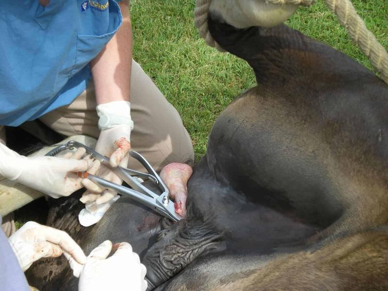
🔄 Twisting method
Henderson method + Equitwister
➡ Castration by machine and torsion of testicle
➡ Torsion of whole proc. vaginalis
➡ Closure of abdominal cavity
Mechanical twisting of the entire spermatic cord proximal to the testis (15–25 rotations) using a specialized drill-attached clamp until the cord severs.
Major post-operative complication: Bilateral evisceration of abdominal contents due to lack of a suture ligature on the open vaginal ring.
🩹 Scrotal Healing
1⃣ Secondary intention healing – MOST COMMON
Common to stretch the incision to help post-operative drainage
Ensure excess fascia is trimmed
2⃣ Primary intention healing
With absorbable sutures
If performed under sterile conditions
Requires adequate hemostasis
⬇ Decreases risk of post-operative complications
Postop care
→ 2-3 weeks of healing.
If open method one should restrict activity and of closed method one should confine horse to stall for 2 weeks and monitor closely.
Do 15 minutes of exercise twice daily, starting 24h after surgery, to prevent preputial and scrotal swelling (edema).
Tetanus prophylaxis, systemic IM procaine penicillin for 3–5 days, and IV/oral NSAIDs (e.g., flunixin or phenylbutazone) for 1–3 days.
⚠ Complications
1.🩸 Haemorrhage
➡ Grasp and re-crush with emasculator for >30 mins
Usually due to:
Improper tying before removal of emasculator
Cord was too large
Too hard exercise of horse before castration
2. Evisceration
➡ Prolapse of intestinal or omental contents → must do laparotomy
predisposing factors: Pre-existing wide superficial/deep inguinal rings, increased intra-abdominal pressure, slippage of cord ligatures, or unclosed open castration in draft breeds/older horses.
3.💧 Oedema
➡ Due to insufficient exercise after surgery or to short incision → give NSAIDs and drainage
4.🦠 Septic funiculitis
➡ Infection of remaining cord stump due to:
Contaminated emasculator
Ligature (using un-asbsorbable suture)
→ Give doxycycline and NSAIDs
Poor wound drainage
5.🦠 Clostridial infection
➡ Tetanus (give penicillin)
6.🦠 Septic peritonitis → doxycycline and NSAIDs
7.⚠ Penile damage ➡ Uncommon
8.💧 Hydrocoele ➡ Idiopathic, painless, fluid-filled enlargement of the parietal tunic following open castration→ scrotal swelling
9.🐴 Persistent masculine behaviour ➡ Caused by improper castration resulting in retention of epididymal tissue, or failure to recognize and remove a cryptorchid testicle
Colic
🧠 Quick Overview
STANDING 🐴
→ Up to 1 year
→ Alpha2 agonist + opioid + local procaine
→ Emasculator
→ ❌ No suture
→ Secondary healing
RECUMBENT – CLOSED 🛏
→ Older than 1 year
→ Ligation + closure
→ Primary healing
→ Uncovered / Covered / Regio inguinalis
RECUMBENT – OPEN 🔴
→ No ligature
→ Emasculator or twisting
→ Uncovered or covered
🐴 Stallion castration — methods
OPEN
No ligatures/closure.
Emasculator or twisting controls bleeding.
Scrotal wound left open → secondary intention.
CLOSED – 3 methods:
Uncovered
Open vaginal tunic → expose spermatic cord.
Cord: twist → emasculator → ligature/suture.
Vaginal tunic: twist → emasculator → separate ligature/suture.
🧠 Two structures → two ligatures.
Covered
Cord remains covered by vaginal tunic.
Deal with them together.
One ligature around cord + vaginal process.
🧠 One package → one ligature.
Regio inguinalis
Approach through inguinal region.
Resect vaginal process.
Ligate spermatic cord.
Close vaginal process + wound.
🧠 Memory
OPEN = no ligature, leave open
CLOSED:
Uncovered = TWO separately
Covered = ONE together
Inguinal = approach from groin
8. Diagnosis and Castration of Cryptorchids
Cryptorchidism = failure of both or one of the testes to descend into the scrotum.
Can be unilateral → usually still fertile
Can be bilateral → usually sterile
Left testicle most commonly affected
If testicle has not descended by 3–4 weeks of age → unlikely to descend
Doesn’t count as cryptorchid until 2 years old
🧬 Etiology
Hereditable:
Genetics
Improper gubernaculum function
Too large testis to pass through inguinal canal
Hormonal:
Inadequate stimulation of androgen or testosterone
⚠ These stallions should not be used for breeding.
📍 Types of Cryptorchidism
1⃣ Complete abdominal cryptorchidism
➡ Both epididymis + testis are within the abdomen
2⃣ Incomplete abdominal cryptorchidism
➡ Epididymis has descended into the inguinal canal, but testis remains in abdomen
3⃣ Inguinal cryptorchidism
➡ Epididymis + testis have descended into the inguinal canal, but NOT into scrotum
Also called “high-flankers.”

🔍 Diagnosis
History
Is the horse castrated earlier?
Rectal palpation of inguinal rings
No structures in inguinal region
➡ Cryptorchidism is completely abdominal
Vessels in inguinal region
➡ Cryptorchidism is inguinal or incompletely abdominal
External palpation
Give sedatives to relax m. cremaster → easier to palpate
Can always palpate it in case of inguinal cryptorchidism
USG
➡ Typical homogenic echogenic appearance
Hormonal assays
Testosterone concentration (hCG stimulation test)!! → Administration of hCG causes a significant rise in serum testosterone levels if functional testicular tissue is present. Measure testo before and after!
Blood: ⬆ Anti-Müllerian hormone → from Sertoli cells in cryptorchid testes, ⬇ Testosterone
Urine: Estrogen
✂ Treatment
➡ Surgical removal of BOTH testes under general anaesthesia
⚠ Always locate and remove cryptorchid testicle FIRST.
If you cannot find the testicle:
➡ Send horse to professional clinic → you will only ruin the situation for other vet.
Surgical method depends on location of testicle.
Other options:
Laparoscopic surgery
Immunological castration
💉 Immunological castration
Immunization against luteinizing hormone-releasing hormone (LHRH)
➡ Decreases serum concentration of testosterone
⚠ Variable result
🔪 Methods of Surgery
⭐ Zert's approach:
Open abdominal cavity close to the prepuce and remove both testicles from one operation wound.
1⃣ Inguinal Approach
📍 Used for:
Inguinal
Incomplete abdominal
Procedure
Dorsal recumbency → Incise under inguinal canal → Cut rectus abdominis → Locate vaginal process from gubernaculum → Incise vaginal process to locate testicle and epididymis → Find proper ligament of testis → Pull testis through vaginal ring → Suture superficial inguinal ring with absorbable suture ➡ Prevents evisceration
2⃣ Para-inguinal Approach
📍 Used if inguinal method didn't work.
Incision several cm medial to inguinal ring
Incision between raphe scroti and left leg
Incision in the aponeurosis of the external abdominal oblique muscle, parallel and several centimeters medial to the superficial inguinal ring.

3⃣ Paramedian Approach
📍 Used in abdominal cryptorchidism
Procedure
Skin
⬇
Subcutis
⬇
External fascia flava abdominis
⬇
Aponeurosis of oblique abdominal muscle → cut
⬇
Rectus abdominis → separate fibers with finger
⬇
Transverse fascia + peritoneum → perforate
⬇
✋ Introduce hand into abdomen
⬇
🥚 Locate + remove testicle
⬇
Ligature
⬇
Reposition stump of spermatic cord
⬇
Suture fascia transversalis
⭐ Fascia transversalis = main structure holding abdominal wall
4⃣ Flank Approach
10–15 cm incision
Through skin + subcutis
Paralumbar fossa of affected side
Horse can be standing or recumbent
Layers
Skin + subcutis
⬇
Cut external abdominal oblique
⬇
Split internal abdominal oblique
⬇
Split transversus abdominis
⬇
Cut peritoneum
⬇
🥚 Locate + remove testis
5⃣ Laparoscopic Approach
Minimally invasive
Requires expensive equipment
Dorsal recumbency in the Trendelenburg position (head tilted down, pelvis elevated).
Procedure
Distend abdomen with gas
Insert camera through umbilical region
Locate testicle(s) around vaginal ring
Remove testicle(s)
Dorsal recumbency in the Trendelenburg position (head tilted down, pelvis elevated).
🩹 Post-operative Care
Stall rest 7 days
Lead walking only
Exercise gradually resumed over 10–14 days
External sutures removed 7 days post-op
give NSAIDs for 5 days, ATB for 7-10 days (trimetoprim sulphonamide)
After laparoscopy
➡ Less invasive
➡ Resume activity after first 72 hours
⚠ Complications
Rare, but include:
Anaesthetic complications
🩸 Excessive haemorrhage
Evisceration
Bowel damage
🦠 Infection
Post-op swelling
Incision breakdown
Continued stallion behaviour
🧠 MOST IMPORTANT TO REMEMBER
📍 Where is the testicle?
Complete abdominal
➡ 🥚 Testis + epididymis = ABDOMEN
Incomplete abdominal
➡ 🥚 Testis = ABDOMEN
➡ Epididymis = INGUINAL CANAL
Inguinal
➡ 🥚 Testis + epididymis = INGUINAL CANAL
🔪 Match location → surgery
Inguinal/incomplete → 🔪 Inguinal approach
Can't access by inguinal → 🔪 Para-inguinal
Abdominal → 🔪 Paramedian / flank
Abdominal, minimally invasive → Laparoscopy
Committee questions:
Diagnosis — VERY HIGH YIELD
External + rectal palpation of inguinal rings/canal.
For rectal: sedation + Buscopan was specifically asked.
USG → retained testicle described in these reports as round/hyperechoic with a central vessel.
Blood: AMH.
Urine: estrogen.
⭐ hCG stimulation test → they repeatedly care about this:
test testosterone before → give hCG → test testosterone after.
Increase indicates functioning testicular tissue.Used particularly when deciding castrated gelding vs retained testicular tissue.
Hormones
Testosterone → produced by Leydig cells.
If they ask control:
Hypothalamus GnRH → pituitary LH → Leydig cells → testosterone.
⚠ Don't say GnRH comes from pituitary—the Oct 2023 report phrases this incorrectly.
Surgical approaches
Know the names:
Inguinal → especially inguinal/incomplete abdominal cryptorchid.
Parainguinal
Paramedian
Flank
Laparoscopy
⭐ Complete abdominal cryptorchid: they particularly asked about paramedian approach and the muscle layers, so know the layers from your notes.
Laparoscopy — only mention if prepared for follow-ups
October 2025 examiner immediately followed it with:
3 incisions/ports
camera
instruments
gas/insufflation
CO₂
Ligasure → hemostasis
advantages → faster recovery, fewer complications/less hemorrhage.
Equine laparotomy
Laparotomy = incision into the abdominal cavity, usually in the case of life-threatening gastrointestinal abnormalities.
Exploratory laparotomies can also be performed to address:
Reproductive issues
Lesions in other organs in the abdominal cavity
Upon incising the peritoneum, assess:
→ The color and character of peritoneal effusion,
→ The pattern/distribution of distended bowel
→ Any gross discoloration of visceral or peritoneal surfaces.
Gas/fluid filled bowel may need to be decompressed before continuing
→ Large intestine: use needle to decompress
→ Small intestine: strip content into cecum in simple obstructions or do enterotomy
Keep intestines warm and moist with Hartmann’s solution during laparotomy (warmed sterile isotonic saline)
Monitor, give ATB and IV fluids post op, also look for incisional swelling, herniation and evisceration post op.
📌 Indications
🐴 Colic → diagnose exact cause of colic when obstructing lesion requires surgery
No exact diagnosis, but enough evidence that surgery is required to save the horse's life
No response to treatment of colic (persistent severe pain)
Recurrent colic over a period of days or weeks
Obstruction
Neoplasia
🔪 Different Ways to Open the Abdominal Cavity
1⃣ Inguinal
Rectus abdominis muscle is cut
Does not alone allow a thorough exploration and decompression of the non-herniated bowel
Used in conjunction with ventral midline incision
📌 Used for:
Stallions with inguinal or scrotal hernia
Cryptorchid (inguinal and incomplete abdominal) castration
2⃣ Parainguinal
Incision located cranially to the external inguinal canal
Rectus abdominis muscle is cut
3⃣ Flank
🐴 Standing approach
📌 Most common for:
Small colon
Nondistended large colon
Uterine torsion
🔪 Incision
Made in a vertical line midway between:
Last rib ↔ Tuber coxae
➡ Starting dorsal → continuing ventral
Layers
Skin
⬇
🔪 External abdominal oblique → sharply divided
⬇
Internal abdominal oblique → bluntly divided parallel to fibers
⬇
Transverse abdominal muscle → bluntly divided parallel to fibers
⬇
🔪 Peritoneum punctured
⬇
Abdominal cavity
🧠 Muscles involved
External abdominal oblique
Internal abdominal oblique
Transverse abdominal muscle
🪡 Closure
Closure is done for 3 layers:
First two layers of muscles → appositioned and sutured with absorbable material
Skin → sutured or stapled
4⃣ Paramedian
📍 10 cm lateral to the midline
Incision on either right or left midline
Through tunica flava abdominis and rectus abdominis muscle
⚠ Careful to avoid:
Deep epigastric vessels
Superficial epigastric vessels
🪡 Closure
➡ Suture external fascia of rectus abdominis sheath
5⃣ Ventral Midline ⭐
⭐ Mostly done in linea alba in case of colic
➡ Allows complete revision of abdominal cavity
Indication: Surgical colic, urolith removal, ovariectomy, and caesarean section
📏 Incision
Small intestine → 15 cm
Large intestine → 60 cm
🔪 Approach
Initial incision made accurately in midline → Through linea alba → Extending cranially from umbilicus → Open peritoneum → Enter abdominal cavity
🔍 When opening the peritoneum, assess:
Color and character of any effusion, Distribution/pattern of distended bowel, Discoloration of visceral surfaces, Discoloration of peritoneal surfaces
🪡 Closure
⚠ Do NOT include peritoneum → causes more irritation/inflammation → adhesions
1⃣ Linea alba
Suture 1 cm from wound edge
Simple continuous suture
⬇
2⃣ Subcutaneous tissue
⬇
3⃣ Skin
Ford interlocking suture, Prolene
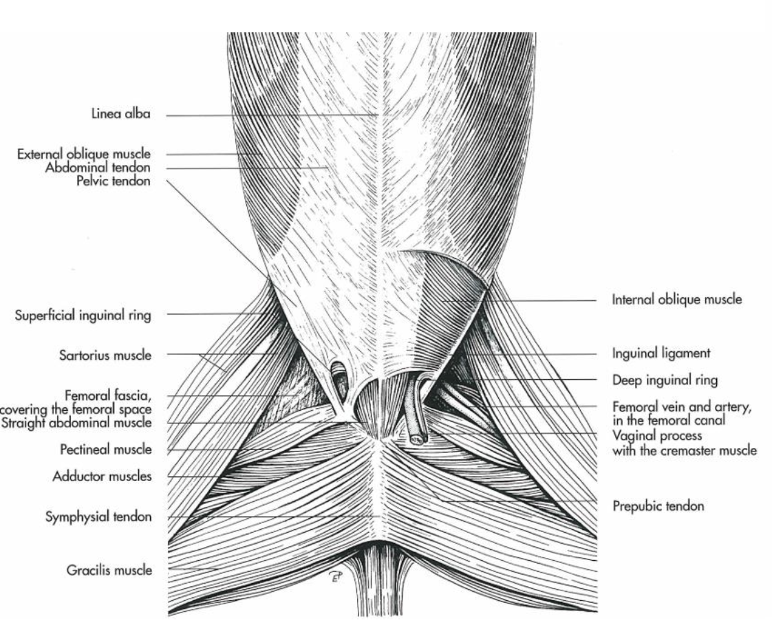
→ A wound stent or incise drape is applied, followed by removal after 12 hours and placement of an abdominal bandage
🧠 Quick Overview
Approach | 📍 Location | ⭐ Main use |
|---|---|---|
Inguinal | Inguinal region | Inguinal/scrotal hernia, cryptorchid |
Parainguinal | Cranial to external inguinal canal | Alternative inguinal access. crypto, ovariectomy, inguinal hernia. |
Flank | Between last rib + tuber coxae | Standing; ovariectomy, uterine torsion, small colon surgeries, impaction, rectal tear. |
Paramedian | 10 cm lateral to midline | Through rectus abdominis. colic, crypto, cystotomy. |
Ventral midline ⭐ | Linea alba | Colic + complete abdominal exploration |
🧠 Most important:
COLIC → VENTRAL MIDLINE → LINEA ALBA → COMPLETE EXPLORATION
⭐ Approaches + what you cut
Approach | Important layers |
|---|---|
Ventral midline / median | Linea alba → peritoneum |
Paramedian | Rectus abdominis → peritoneum |
Inguinal | Rectus abdominis |
Parainguinal | Rectus abdominis |
Flank | External oblique → internal oblique → transversus abdominis → peritoneum |
Which approach for what?
Ventral midline → most common for colic/exploratory laparotomy; report also mentions removal of uroliths.
Inguinal → cryptorchid surgery, particularly inguinal/incomplete abdominal cryptorchid.
Flank → know the approach/layers; the March 2023 report suggests Zert may ask about an important fascia flavis? here.
🚨 When does a colic need laparotomy?
Think:
Severe/persistent pain despite analgesia + diagnostic findings suggesting surgical lesion.
Can assess:
Clinical signs/pain
Rectal examination
Nasogastric reflux
USG
Abdominocentesis
Bloodwork
⭐ Lactate was specifically asked.
↑ Lactate = tissue hypoperfusion/hypoxia, especially important with strangulated/ischemic intestine.
🧠 Persistent severe pain + ↑ lactate → think ischemia/strangulation → surgery
Equine enterotomy
Enterotomy = the surgical incision into the intestinal lumen.
Commonly performed in:
Jejunum, Cecum, Small colon

📌 Indications for Enterotomy
Remove foreign bodies → commonly in right dorsal colon:
Enteroliths, Fecoliths, Bezoars
Removal of fluid and ingesta
Right and left dorsal displacement
Parasitic infestations → Ascarid larvae in small intestine
Ileum and cecum → obstruction and impaction
⭐ Pelvic flexure enterotomy → most commonly performed, for evacuation of sand or feed impactions of the large colon, enteroliths, and colonic volvulus.
Right ventral colon enterotomy → access cecum in cases of cecocolic intussusceptions
Colonic volvulus → pelvic flexure enterotomy
Small colon strangulation → pelvic flexure enterotomy to release ingesta
💉 Surgical Preparation
💊 Pre-medication
Broad-spectrum peri-operative ATB
Anti-inflammatory medication → Flunixin
🍽 Dietary
Fasted if possible
⚠ Majority of cases are emergent
🔪 Procedure
1⃣ General anaesthesia ➡ Horse is put under GA
2⃣ Midline laparotomy ➡ Abdomen opened using midline laparotomy
3⃣ Secure bowel ➡ Fix with Forceps OR Stay suture
4⃣ Incise intestinal wall
➡ Longitudinally
➡ Antimesenteric side
5⃣ Limit spillage of intraluminal fluid
Aspirate intraluminal fluid OR
Restrict flow using extraluminal digital pressure
6⃣ Remove the problem
➡ Remove luminal obstruction / fix whatever problem / do the job
7⃣ Lavage
💧 Lavage bowel with sterile saline
8⃣ Close
🪡 One- or two-layer suture technique:
Synthetic absorbable
Cushing or Lambert
Minimal inversion of serosa
First layer with Cushing’s or lambert and 2nd layers appositional + inverting
➡ Staple closure with thoracoabdominal (TA) stapler
COLON Enterotomy
📍 Common locations
⭐ Pelvic flexure
Left dorsal → cecocolic intussusception
Right dorsal colon → enteroliths
Most common sites: pelvic flexure, cecal apex, descending colon (small colon), right dorsal colon
Procedure
Incision on antimesenteric side when possible
Large colon is exteriorized
Placed on a colon tray:
On left side OR Caudally between horse's hind legs
and isolated with sterile drapes
💧 One hose → inserted into colon
💧 Another hose → continuously lavages serosal surface
How is luminal fluid and ingesta evacuated during a pelvic flexure enterotomy?
→ A full-thickness stab incision is extended longitudinally (ca. 10 cm), and water hoses are used to flush and siphon out content into a receptacle.
🪡 Closure
Thorough lavage of the enterotomy site and surrounding bowel with several liters of sterile saline BEFORE first suture layer
Two layers:
1⃣ Simple continuous
⬇
2⃣ Inverting musculoserosal layer using Cushing or Lambert
➡ Colon rinsed thoroughly with sterile saline
➡ Replaced into abdomen
Post-surgical complications:
Peritonitis (due to leakage or contamination)
Postoperative ileus / diarrhea
Abdominal adhesions
Incisional/luminal hemorrhage
SMALL INTESTINE Enterotomy
⚠ Enterotomy in SI should be avoided because closure can produce:
Adhesion formation and Narrowing (stricture) of lumen
➡ More common to do enterectomy in SI than enterotomy.
📌 Indications
A longitudinal enterotomy on the antimesenteric surface of SI may be indicated for:
Removal of obstructions
Impaction with food components
Foreign material
Ascarids → anoplocephala, cyathostomin, parascaris equorum
Empty a segment of distended bowel to facilitate reduction of a strangulation in the epiploic foramen
Suture in small intestine: A single-layer inverting pattern (Cushing or Lembert) using synthetic absorbable monofilament suture
💧 Prevent adherence of intestinal contents to serosa
Use:
Constant lavage with warm sterile saline OR
Precoat site of affection with sodium carboxymethylcellulose
🩺 Aftercare
Physical Monitoring: every 4h (adult) or 2h (foals) the first 24h
💧 Fluid therapy: Give oral fluid 24-36h post op
Feeding: small amount of food after 48 hours (gradually 24-72h)
Avoid thrombophlebitis.
Give domperidone (or lidocaine IV) for SI and metoclopramide for LI → to increase peristaltic movement
Lidocaine IV ⭐ — commonly used after equine abdominal surgery, especially for postoperative ileus (POI). It has analgesic/anti-inflammatory effects and may promote GI motility.
Neostigmine — directly increases cholinergic activity → increases intestinal contractions/motility.
💊 Analgesia → Peri- and post-operative NSAIDs 3-5 days
💊 Antimicrobial → Peri- and post-operative broad-spectrum ATB 3-5 days (penicillin, aminoglycosides)
other med: laxatives/lubricants
follow up and evaluation after 30 days
⭐ Most Common Sites of Enterotomy
📍 Cecal apex
📍 Pelvic flexure ⭐
📍 Right dorsal colon
📍 Descending colon

🧠 QUICK MEMORY
ENTEROTOMY = ENTER the intestine
GA
⬇
Midline laparotomy
⬇
Exteriorize + secure bowel
⬇
Longitudinal ANTImesenteric incision
⬇
Remove obstruction / ingesta
⬇
Lavage 💧
⬇
Cushing/Lambert closure 🪡
⬇
Rinse + replace
⭐ Large colon → enterotomy common, especially pelvic flexure
⚠ Small intestine → enterotomy avoided if possible → enterectomy more common
⭐ Most common sites:
Pelvic flexure — #1
Cecal apex — #2
Descending/small colon
Right dorsal colon has also been asked.
Cecum: incision specifically at the apex.
Indications/causes: obstruction/impaction, and examiners specifically asked about left and right dorsal displacement.
Know which displacement is more common and which is worse — this was directly asked.
Important complication/progression of displacement → large-colon volvulus.
⭐ Closure: 2–3 layers.
Simple continuous/interrupted
Then Cushing or Lembert
Also know inverted/vertical mattress — specifically requested once.
⭐ Drugs to restart intestinal motility:
Neostigmine
Physostigmine
Small intestine: previous answers emphasize that enterotomy is mainly performed on large intestine; SI may instead require enterectomy.
Enterectomy
Enterectomy = surgical removal of a portion of the intestine.
📍 Commonly:
Jejunum
Ileum
📌 Indications
Any intestine with:
Abnormal color
Decreased motility
Thickened appearance
⚠ Dead jejunum wall:
Black serosa
❌ No pulsation of vessels
Strangulation → most commonly
After correction of obstruction which caused secondary vascular damage without return of:
Normal color, Motility, Pulsation
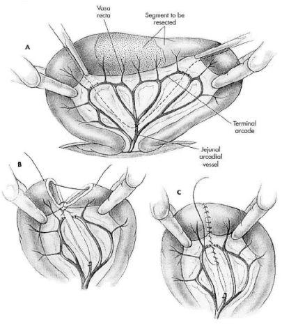
Resection of the Small Intestine
📏 Physiological limit of small intestine resection = maximum 8 metres
➡ SI is 18–21 m long
⭐ End-to-end anastomosis is the simplest and most physiologically compatible method.
🔪 Procedure
1⃣ Isolate bowel
Fluid-filled ischaemic bowel should be isolated with intestinal clamps before manipulation begins.
⬇
2⃣ Select resection sites
Suitable site proximal and distal to the strangulated bowel
⬇
3⃣ Ligate vessels
Mesenteric vessels to compromised bowel are double ligated
⬇
4⃣ Transect intestine
Distal end of gut is transected
Mesentery incised between ligatures
⬇
5⃣ Cut vessels
Each vessel is cut
⬇
6⃣ Anastomosis
Remaining healthy intestine is joined together
Long end of proximal gap of mesentery is closed when anastomosis is completed

🪡 Closure Alternatives
Use synthetic absorbable monofilament suture material.
A) ⭐ Zert!
Two-layer closure:
➡ Simple interrupted suture pattern in two layers
Mucosa
Serosa
B)
1st layer: Simple continuous
2nd layer: Cushing seromuscular suture
C)
Single layer:
➡ Interrupted Lembert sutures
Resection of the Large Intestine
⚠ Large intestinal resection is performed very rarely in horses.
Why?
1⃣ Most obstructions don't require resection
Many obstructions are caused by:
Displacement with no/minimal interference of blood supply
Simple obstruction of lumen → e.g. enteroliths
➡ Can usually be relieved by a simple enterotomy.
2⃣ Strangulation may involve too much bowel
If strangulation obstruction is present:
➡ Length of bowel involved may be so great that resection + anastomosis is not considered
➡ Euthanasia
🔗 Anastomosis
Anastomosis = surgical joining of tubular structures (arteries, veins, intestine) together so they can become continuous.
1⃣ End-to-End Anastomosis ⭐
➡ Connects two open ends of intestine together
Intestine:
━━━━✂ ✂━━━━
↓
━━━━━━🔗━━━━━━
🪡 Suture
Simple continuous → serosa
Cushing → seromuscular layer
⭐ Simplest and most physiologically compatible method.
2⃣ End-to-Side Anastomosis
➡ Connects end of intestine to a larger piece
━━━━━━┓
┃
┃
3⃣ Side-to-Side Anastomosis
➡ Connects the sides of intestine together
Used for:
➡ Jejunal anastomosis
━━━━━━━
↕
━━━━━━━

⚠ Post-operative Complications
💧 Leakage of content
🕸 Adhesions
🚫 Obstruction / stricture
🩸 Bleeding
🦠 Infection
💊 Post-op Care
NSAIDs
ATB
Stool softener
🧠 QUICK MEMORY
Enterotomy vs. Enterectomy
ENTEROTOMY
➡ ✂ Cut INTO intestine
➡ Remove obstruction/content
➡ Intestine stays
ENTERECTOMY
➡ ✂ Remove a SECTION of intestine
➡ Usually because bowel is dead/damaged
➡ Join healthy ends = ANASTOMOSIS
⭐ Most important sequence:
Strangulation
⬇
🖤 Dead/damaged intestine
⬇
✂ Resection / enterectomy
⬇
🔗 Anastomosis
⬇
⭐ Usually end-to-end
Disorders of teeth exchange and exodontics
Equine dental developmental abnormalities can involve:
🔢 Tooth number
🦷 Morphology
📍 Position in the dental arcades
🦷 Tooth Anatomy
Horses have hypsodont teeth = continue to grow their whole life → 2–3 mm each year.
Tooth divided into:
Crown → visible
Reserve crown → hidden in alveolar bone
Apex/root of tooth
Tooth consists of:
Outer → inner:
Cementum, Enamel, Dentin, Pulp
Types
Incisors, Canines, Premolars, Molars
🔢 Numbering System
Two systems:
1⃣ Based on type of teeth
PM1, M2, etc.
2⃣ Triadan system
🧮 Dental Formula
Deciduous teeth
I 3/3, P 3/3 × 2 = 24
12 incisors
12 molars
🐴 Permanent teeth
I 3/3, C 1/1, P 3 or 4/3, M 3/3 × 2 = 36–44
➡ Depends on presence of:
Canines
P1 = wolf teeth
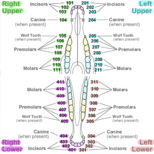
⏰ Eruption Schedule
Foals are born with 4 premolars
⚠ Critical age = 2.5 years
Replacement of 12 teeth
➡ Can impact food processing, decline performance and cause health problems
Deciduous Teeth
Foals get deciduous teeth at around 6 weeks old.
🦷 Incisors – RULE OF 8 ⭐
Central → 8 days
Intermediate → 8 weeks
Corners → 8 months
🧠 8 days → 8 weeks → 8 months
Premolars
P2, P3, P4
➡ Erupt immediately after birth or within 2 weeks
🐴 Permanent Teeth
Horse has complete set of permanent teeth at 5 years.
🦷 Incisors
All erupted at 4.5 years:
Central → 2.5 years
Middle → 3.5 years
Corner → 4.5 years
🧠 2.5 → 3.5 → 4.5
🐺 Wolf teeth – P1
1st premolar
In maxillary arcades
Only in some horses
🦷 Premolars
All erupted by 4 years:
PM2 → 2.5 years
PM3 → 3 years
PM4 → 4 years
🦷 Molars
All erupted by 3 years:
M1 → 1 year
M2 → 2 years
M3 → 3 years
➡ Have 2 infundibulum in maxillary arcade
🧠 M1 = 1, M2 = 2, M3 = 3
🦷 Canines
4 in stallions
May not be developed
Rudimentary
Erupt around 3.5–5 years
📅 Age Determination of Horse
1⃣ Eruption of teeth
Presence of deciduous teeth
2⃣ Occlusal surface of lower incisors
Infundibulum wears away over time
Gone in central by 8 years
3⃣ Shape of incisors
Flattened → until 10 years
Oval → 12–13 years
Round → 15–20 years
Triangular → >20 years
4⃣ Angle of incisors
Young → more vertical/flat
Older → greater/sharper angle
5⃣ Galvayne's groove
➡ Mark on upper corner incisors that appears and disappears in ageing horse
⚠ Congenital & Developmental Defects
1⃣ Oligodontia
Congenital absence of a tooth germ OR retention and inclusion of a tooth within jaw.
Consequences
Missing tooth →
Mesial drift
Tipping of neighboring teeth
Lack of wear of antagonist
Dental elongations
Abnormal mastication
🔎 Diagnosis ➡ X-ray
2⃣ Polyodontia / Supernumerary Teeth
➡ Too many teeth
Can be:
Retained deciduous teeth
Supplementary teeth
Often:
Mandibular premolar
Maxillary molar
Remnants called “caps.”
Consequences
Difficulty eating
Unusual facial development
Maxillary sinusitis
Nasal discharge
Facial swelling
🦷 Developmental Defects
1.Delayed tooth eruption
➡ Due to overcrowding
➡ Usually small breeds with limited jaw space
2.Retained deciduous dentition
➡ Caps retained because of entrapment between adjacent teeth
3.Maleruption / misalignment
➡ Cheek teeth → cause overgrowth of tooth erupting first
4.Dental dysplasia
Abnormal bending
Abnormal size
Double teeth
5.Diastema
➡ Abnormal spaces between teeth
➡ Most common form of periodontitis
➡ Food impaction
6.Abscess
Apical
Root
Periapical
7.Dental caries
➡ Bacterial destruction of dentin
➡ Extraction if caries result in fractures
🕳 Infundibular Caries
Very common in maxillary cheek teeth because they have 2 infundibulums:
1 rostral
1 caudal
Incisors have 1 infundibulum.
What is an infundibulum?
➡ Funnel-like structure/layer consisting of cementum
⚠ M1 is the oldest permanent tooth in the cavity and is often predisposed to caries.
Hole fills with food
⬇
🦠 Tooth can rot/become infected
⬇
🦷 Tooth becomes weakened
⬇
💥 Fracture
💊 Treatment
Grade 1 → clean + fill hole with composite
Grade 2–4 → extract tooth
🔢 Grades of Infundibular Caries
0 = Normal
1 = Hypoplasia of cementum
2 = Cementum + dentinum hypoplasia
3 = Cementum + dentinum + enamel hypoplasia
4 = Fracture of tooth 💥
🧠 0 Normal → 1 C → 2 C+D → 3 C+D+E → 4 Fracture
🦷 Exodontics
Exodontics = teeth extraction
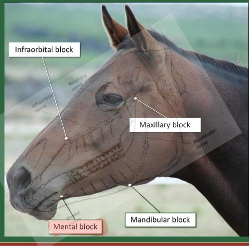
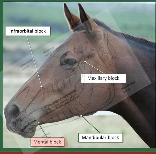
💉 Nerve Blocks
Maxillary nerve block
📍 Ventral to zygomatic arch, dorsal to facial vessel
➡ Blocks:
Maxillary teeth, Maxillary sinuses
Infraorbital nerve block
➡ Blocks:
Upper incisors, Canines, Wolf teeth, Upper Premolar 2
Mandibular nerve block
📍 Foramen mandibulae, medial side
➡ Blocks mandibular teeth
Mental nerve block
📍 Mental foramen
➡ Blocks:
Lower incisors, Canines, Wolf teeth
📌 Indications for Extraction
🐺 Wolf teeth
Extract if:
Rostrally displaced, Unerupted, Fractured, Mandibular wolf teeth
🦷 Incisors, canines & cheek teeth
Extract for:
Apical infection, Fracture, Severe periodontal disease, Tooth mobility due to trauma
🔧 Extraction Procedures
1⃣ Intraoral Extraction
Forceps extraction
Fragmentation
Segmentation
Fragment elevation/luxation
🐴 Done in standing sedation
💉 Alpha2 agonist + opioid
2⃣ Surgical Extraction
😴 Done in general anaesthesia
Methods:
Surgical buccotomy
Transbuccal extraction → minimally invasive
Trephination extraction → minimally invasive
⚠ Transbuccal Extraction
Avoid:
Facial nerve
Dorsal buccal branch
Ventral buccal branch
Parotid salivary duct
Facial artery
Facial vein
💉 Local Anaesthetics
Drug | ⏱ Effect after | ⌛ Duration |
|---|---|---|
Procaine | 5–10 min | — |
Lidocaine | 5 min | 30–60 min |
Mepivacaine | 10 min | 60–120 min |
Bupivacaine | 30 min | 120–140 min |
Articaine | 3 min | 75 min |
🧠 QUICK MEMORY
⭐ Deciduous incisors = Rule of 8
8 days → 8 weeks → 8 months
⭐ Permanent incisors
2.5 → 3.5 → 4.5 years
⭐ Molars
M1 = 1 → M2 = 2 → M3 = 3 years
⭐ Exodontics
Standing + alpha2/opioid
➡ Intraoral extraction
GA
➡ Surgical extraction
⭐ Nerve blocks
Maxillary nerve → maxillary teeth + sinus
Infraorbital → upper front teeth
Mandibular → mandibular teeth
Mental → lower front teeth
Sharp teeth
Sharp teeth = the most common dental/stomatological pathology in horses.
➡ Include minor sharp points → large unopposed overgrowths.
Etiology
Overgrowth of enamel due to:
➡ Lack of wear + continuous eruption
Causes
Missing teeth
Malerupted teeth
Fractured teeth
Overall disparity in apposition of arcades:
Parrot mouth
Diastema
Lack of normal lateral movements of mandible during mastication → due to diet
⭐ Diastema – most common cause
Diastema = abnormal space between two teeth
➡ Leads to malocclusion of opposite tooth
⚙ Pathogenesis
Insufficient/excessive attrition of teeth
⬇
Sharp points/spurs of enamel
⬇
Lack of attrition of occlusal surface
+
Continuous eruption
⬇
🦷 Overgrowth
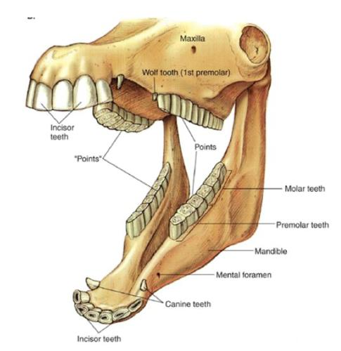
⭐ Why do sharp points form?
The maxillary arcades are set wider apart than the mandibular arcades.
This leads to areas of lack of wear during mastication.
MAXILLA
➡ Sharp points/spurs form on BUCCAL side
MANDIBLE
➡ Sharp points/spurs form on LINGUAL side
🤕 Consequences
Sharp points can cause:
➡ Abrasion and ulceration of:
Cheeks and Tongue
🩺 Clinical Signs
⚠ Many horses are asymptomatic.
Most common:
Dysphagia
Soft-tissue trauma
Quidding
Riding problems
🔎 Diagnosis
Clinical examination
💉 Sedation
Head gag
Rinse mouth prior to examination
✋ Palpation
👀 Visual inspection
🔦 Light for visualization
Occlusion probe → inspect for damage
🔧 Treatment – Rasping
➡ Rasping of teeth using:
Hand rasp or Motorized instruments
Where do you rasp?
MAXILLA → BUCCAL side
➡ Because maxillary cheek teeth form spurs on the buccal side
MANDIBLE → LINGUAL side
➡ Because mandibular cheek teeth form spurs on the lingual side
This occurs because maxillary cheek teeth are wider than mandibular cheek teeth.
⚠ Important During Rasping
❌ Do NOT rasp away too much
➡ Secondary dentine protects the pulp.
When reducing an overgrowth:
➡ Maintain the normal lateromedial angulation of the occlusal arcades.
📈 Prognosis
✅ Good prognosis for most cases.
⚠ Excessive removal of occlusal surface OR heating of tooth structure using uncooled instruments can cause:
➡ Post-procedure quidding
⏱ Can take months to years to resolve.
🧠 QUICK MEMORY
Why sharp teeth?
Continuous eruption + ↓ wear = sharp teeth
Where are the sharp points?
🐴 Upper/MAXILLA
➡ BUCCAL 🫦
➡ Cuts CHEEK
🐴 Lower/MANDIBLE
➡ LINGUAL 👅
➡ Cuts TONGUE
Treatment
🔧 RASP/FLOAT
⚠ Don't rasp too much → protect secondary dentine + pulp
Surgical diseases of nasal cavity and paranasal sinuses
👃 DISORDERS OF THE NARES
1⃣ Wry Nose “campylorrhinus lateralis”
Congenital damage
Shortening of premaxilla on one side
➡ Upper jaw + nose deviate to one sideTypically accompanied by deviation of nasal septum
➡ Problems breathingDifferent degrees, but often euthanasia is best
2⃣ Hypertrophy of Alar Folds
➡ Vibrant sound during inspiration = high blowing
Causes exercise intolerance in performing horses
🔎 Diagnosis + Treatment
Confirm noise originates from alar folds during exhalation/inhalation:
➡ Place sutures from skin at dorsal aspect of nose across openings of false nostrils
➡ Compare noise before vs. after suture placement
Positive result
➡ Correct diagnosis
➡ Indication for resection of alar folds
3⃣ Atheroma of False Nostrils
➡ Development of epidermal inclusion cysts (sebaceous cysts) in lining of false nostril.
Painless
Usually no respiratory obstruction
Mainly cosmetic significance
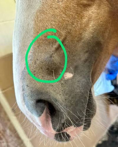
4⃣ Facial Paralysis
➡ Dysfunction of nasolabialis muscle
Causes:
Facial surgery, Dental extraction
⚠ Can cause an aerodynamic disaster for sport horses
5⃣ Trauma
Usually when nose is caught in a hook.
➡ Requires anatomical restoration
👃 DISORDERS OF THE NASAL CAVITY
🩸 Progressive Ethmoidal Hematoma – PEH
Repeated epistaxis at rest → unilateral or bilateral.
Can progress to:
Respiratory stridor
Facial deformity
⬇ Airflow through affected nostril
➡ Unknown etiology.
⚙ Pathogenesis
Expanding sub-mucosal hemorrhages develop on surface of ethmoidal turbinate labyrinth
⬇
Mucosal capsule splits
⬇
🩸 Bloody discharge
More common in horses >4 years.

🩺 Clinical Signs
⭐ Repeated hemorrhages from one nostril at rest
Dyspnoea → nasal obstruction, reduced drainage
Dirty nasal discharge → not fresh blood
Facial swelling
🔎 Diagnosis
History, Physical exam, Endoscopy, X-ray, CT
💊 Treatment
First:
➡ Chemical ablation with formalin 10% (inject/apply it on lesion to remove it)
➡ Repeat at 2–3 week intervals until resolved
If not successful:
➡ Surgical removal through frontal flap
Other Nasal Cavity Disorders
Trauma
Intra-nasal foreign bodies
Neoplasia
Polyps
Mucoid degeneration of nasal conchae
Nasal septum deviation
Hyperplasia of nasal septal mucosae
Subcutaneous emphysema
Concha necrosis and metaplasia
🦴 PARANASAL SINUSES
⭐ Five Paired Paranasal Sinuses
Frontal
Frontal sinus
Conchofrontal sinus
Caudal maxillary
Rostral maxillary
Ethmoidal
Sphenopalatine
⚠ Paranasal sinuses are not completely separated.
➡ All sinuses communicate with or drain into the middle meatus.
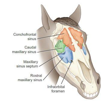
🦠 Primary Sinusitis
Pathogenesis:
Stagnation of mucus
⬇
Inhibited muco-ciliary clearance by URT viral agents
⬇
Opportunistic bacteria follow
⬇
Purulent exudate
⬇
Chronic sinusitis
⬇
Hyperplasia of lining
⬇
Narrowing of ostia + inspissation of pus
🦷 Secondary (Dental) Sinusitis
➡ Caused by dental periapical suppuration
Roots of 4th–6th maxillary teeth lie within maxillary sinuses.
Fracture or necrosis
⬇
Dental infection
⬇
🦠 Secondary sinusitis

🩺 Clinical Signs
Early:
➡ Mucoid unilateral nasal discharge
Later:
➡ Purulent + malodorous discharge
Also:
Facial swelling, Nasal obstruction
🔎 Diagnosis
Clinical signs, Percussion, Oral inspection, X-ray
⭐ CT = BEST / GOLD STANDARD – Zert
💊 Treatment
Conservative:
Systemic ATB
Volatile or steam inhalation
Light exercise
Surgical:
Catheter placement
Radical surgery
🦷 Secondary Empyema
➡ Caused by problems with first molar (M1)
⭐ Most frequent problem of rostral maxillary sinus
Treatment
Trephination (making a hole through the bone to access the sinus)
Push tooth out
Flush bulla
Make drainage into oral cavity without extraction of tooth
💧 Sinus Cysts
Unknown etiology
Common features with PEH
Often in region of drainage ostium (large sinus cyst → blocks drainage ostium → mucus can't drain → sinus expands → facial swelling/nasal obstruction)
Cysts contain yellow fluid
🩺 Clinical Signs
Nasal obstruction, Facial swelling
Rarely ocular proptosis → exophthalmos
Mucoid discharge
🔎 Diagnosis
Clinical signs, Physical exam, Endoscopy, X-ray
🔪 Treatment
➡ Fronto-nasal flap surgery
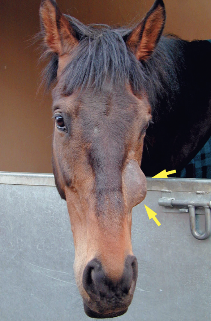
🍄 Mycotic Rhinitis & Sinusitis
Unknown etiology
May be opportunistic fungal infection secondary to other supportive conditions
🩺 Clinical Signs
Low-grade unilateral purulent discharge
Maybe epistaxis
🔎 Diagnosis
➡ Endoscopy
💊 Treatment
Topical benzimidazole
Foley balloon catheter
🧬 Neoplasia & Polyps
Neoplasia ➡ True tumors are uncommon
Polyps
➡ Pedunculated inflammatory proliferations enclosed in mucous membrane
➡ Can develop from complication of dental periapical disease
🩺 Clinical Signs
Putrid nasal discharge mixed with blood
Ocular proptosis
🔎 Diagnosis
Physical exam, Endoscopy, X-ray, CT
🔪 Treatment ➡ Fronto-nasal flap surgery
🧠 QUICK MEMORY
👃 NARES
Wry nose → congenital deviation
Alar fold hypertrophy → high blowing
Atheroma → painless cyst
Facial paralysis → nasolabialis dysfunction
Trauma → anatomical restoration
🩸 PEH
Older horse + repeated unilateral epistaxis AT REST
➡ Think progressive ethmoidal hematoma
Treatment:
Formalin 10% → if unsuccessful → frontal flap
🦷 SECONDARY SINUSITIS
Dental disease → unilateral → purulent + malodorous discharge
⭐ CT = GOLD STANDARD
💧 SINUS CYST
Facial swelling + obstruction + yellow fluid
➡ Fronto-nasal flap
🍄 MYCOTIC
Unilateral purulent discharge ± epistaxis
➡ Endoscopy → topical benzimidazole

Surgical Diseases of the Pharynx, Larynx & Guttural Pouch
PHARYNX
Pharynx = soft tissue passage for food and air.
Connected with:
Nares, Middle ears, Oral cavity, Larynx, Esophagus
Supported by striated muscle for:
Swallowing and Phonation
1⃣ Dorsal Displacement of the Soft Palate – DDSP
🏇 Only a problem in racing horses while exercising.
Likely due to abnormal contraction of muscles of soft palate → causing them to be flaccid.
⚙ What happens?
Soft palate displaces dorsally
⬇
“Sits” on the epiglottis
⬇
🚫 Obstructs opening to trachea
⬇
Difficulty getting air during exercise
🩺 Clinical Signs
Loud expiration noises
Unable to maintain speed while running
🔎 Diagnosis
Clinical signs, History, Endoscopy at rest
⭐ Endoscopy during exercise = GOLD STANDARD
🔪 Treatment
Laryngeal advancement – tie forward
Move larynx rostrally + dorsally
Laser palatoplasty
⚠ Epiglottic Entrapment – Don't Mix with DDSP
➡ Enlargement/entrapment of epiglottis in younger horses
Treatment
➡ Gentle resection of epiglottic folds by laser
Other Pharynx Diseases:
Choanal atresia/stenosis
Palatal defects
Instability of soft palate
Dynamic collapse of pharyngeal walls
Intra-palatal cysts
Pharyngeal cysts
Neoplasia
Extra-mural distortion by:
Strangles, Abscess, Neoplasia
LARYNX
Larynx = symmetrical, tube-shaped musculo-cartilaginous organ connecting:
Pharynx → Larynx → Trachea
2⃣ Laryngeal Hemiplegia= Recurrent Laryngeal Neuropathy
Progressive functional loss of laryngeal nerves
⬇
Larynx collapses on affected side during inspiration
⬇
Permanent dysfunction of muscles
⬇
🚫 Partial obstruction of airways
⭐ Typical
Higher prevalence in males
Long-neck, large breeds . long vagus recurrent nerve, trauma or wear on the nerve
Usually LEFT side
🩺 Clinical Signs
Inspiratory noise during exercise (high pitched whistling/roaring sound)
Exercise intolerance
Usually asymptomatic at rest
🔎 Diagnosis
Palpation
➡ Atrophy of intrinsic laryngeal musculature
Other:
Arytenoid depression test
“Grunt-to-the-stick” test
Endoscopy
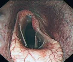
🔪 Treatment
⭐ Prosthetic laryngoplasty
➡ Suture between cricoid + arytenoid
➡ Mimics action of CAD muscle
Other:
Nerve/muscle grafting
Permanent tracheostomy
Cordectomy → Zert does not like
3⃣ Fourth Branchial Arch Defect – 4-BAD= Laryngeal Dysplasia
Syndrome of irreparable congenital defects due to failure of development of some/all derivatives of the 4th branchial arch.
Can be:
Unilateral or Bilateral
❌ Defective Structures
4 structures:
1⃣ Wings of thyroid cartilage
2⃣ Crico-thyroid articulation
3⃣ Cricothyroideus muscles
4⃣ Crico-pharyngeal sphincter muscles
🩺 Clinical Signs
Abnormal respiratory sounds during exercise, Eructation, Nasal discharge, Coughing, Recurrent colic
4-BAD→ missing/abnormal laryngeal structures → abnormal respiratory sounds + swallowing problems.
🔎 Diagnosis
Palpation, Dynamic endoscopy, Ultrasonography
Palpation:
➡ Unusually wide gap between:
Caudal margin of thyroid and Rostral edge of cricoid
Normally, these structures overlap.
❌ Treatment ➡ No way to reconstruct the absent structures

GUTTURAL POUCHES = Auditory Tube Diverticulum (ATD)
2 air-filled, balloon-like structures
📍 Located between:
Base of cranium → dorsally
Pharynx + esophagus → ventrally
Each pouch:
Is in contact with the other
Separated by thin areolar tissue → septum
Connected to the nasopharynx
4⃣ Guttural Pouch Tympany
⚙ Pathogenesis
Guttural pouch ostium acts as non-return valve
➡ Air can enter
➡ ❌ Air cannot leave
⬇
🎈 Guttural pouch expands

Etiology
Foals a few days after birth
Congenital malfunction of pharyngeal opening of pouch
🩺 Clinical Signs
🎈 Tympanic swelling of parotid region
Mucopurulent discharge
Bacterial infection → strangles
Dysphagia, Dyspnea
🔪 Treatment
Improve airflow from guttural pouch:
➡ Opening into larynx OR
➡ Perforation of septum between the pouches
5⃣ Diverticulitis of Guttural Pouch
➡ Inflammation of ATD mucous membranes
Etiology
Strangles
Chronic diverticulitis → chronic catarrhal inflammation
Empyema
Chondroids
6⃣ Chronic Empyema & Chondroids
⚙ Pathogenesis
Failure of drainage
⬇
Mucus/pus accumulates in pouch
⬇
Pus becomes stagnant
⬇
Formation of solid concretions = CHONDROIDS
⚠ Risk of Streptococcus equi
🩺 Clinical Signs
Bilateral purulent nasal discharge, Swelling of parotid region
🔎 Diagnosis
Clinical signs, Lateral X-ray
💊 Treatment
➡ Liquify chondroids by repeated lavage
➡ Drainage via Foley balloon catheter

7⃣ Guttural Pouch Mycosis 🍄
➡ Invasive fungal plaque on mucosal wall of ATD.
🚨 Clinical Signs
⭐ Spontaneous epistaxis AT REST (plaqe erodes internal carotid artery lining the guttural wall)
Minor hemorrhages
Dysphagia
Pharyngeal paralysis
➡ Ingesta in nasal discharge
Cranial nerve defects:
Laryngeal hemiplegia
Facial palsy
Horner's syndrome
Abnormal head posture
🔎 Diagnosis
Clinical signs
Endoscopy ⚠ can be dangerous

💊 Treatment
Conservative:
➡ Antimycotic drugs
Surgical:
➡ Occlusion of carotid artery branches
Ligature OR Transarterial coil embolization (must also do surgery, will be a lot of bleeding. the potensial risk of severe hemorrhage is the risk, not the fungi itself)
Other Larynx Diseases
Epiglottal hypoplasia
Laryngeal granuloma
Neoplasia
Sub-epiglottal cysts
Epiglottal entrapment → younger horses → laser
Arytenoid chondropathy
Axial deviation of ary-epiglottal folds
🧠 QUICK DIFFERENTIATION
🏇 DDSP
Soft palate → over epiglottis
➡ EXPIRATORY noise
➡ Exercise
➡ Dynamic endoscopy
➡ Tie-forward
🗣 Laryngeal hemiplegia
Usually LEFT arytenoid doesn't function
➡ INSPIRATORY noise
➡ Exercise
➡ Endoscopy
➡ Prosthetic laryngoplasty
🎈 Guttural pouch tympany
Foal + air trapped
➡ Parotid swelling
➡ Create drainage/airflow
Empyema/chondroids
Pus → stagnant → solid chondroids
➡ Purulent discharge
➡ Lavage + Foley catheter
🍄 Guttural pouch mycosis
Fungal plaque
➡ 🚨 Spontaneous epistaxis at rest
➡ Cranial nerve problems
➡ Occlude carotid artery branches
Diseases of the eyelid and conjunctiva
👁 Importance of the Eyelids
Protect the eye
Produce and distribute tears
Aid tear drainage
Help control the amount of light entering the eye
👁 DISEASES OF THE EYELID
Congenital Disorders
1⃣ Entropion
Entropion = inward rolling of the eyelid margin.
⭐ Common in foals.
Etiology
Foals:
Dehydration and Congenital disorders
Adults:
Trauma
⚙ Pathogenesis
Eyelid edge turns inward → Eyelashes rub against eye surface → Irritation of conjunctiva + cornea
Long term:
Scarring, Abnormal coloring, Slow-healing sores
🩺 Clinical Signs
💧 Epiphora = excessive tearing
😣 Blepharospasm = blinking
Conjunctivitis, Keratitis
💊 Treatment
Temporary repair:
Vertical mattress suture, Subconjunctival injections
Surgery: rarely required.
⚠ Not recommended until adult age → avoid over-correction / iatrogenic ectropion.
➡ Blepharoplasty = removal of skin below eye
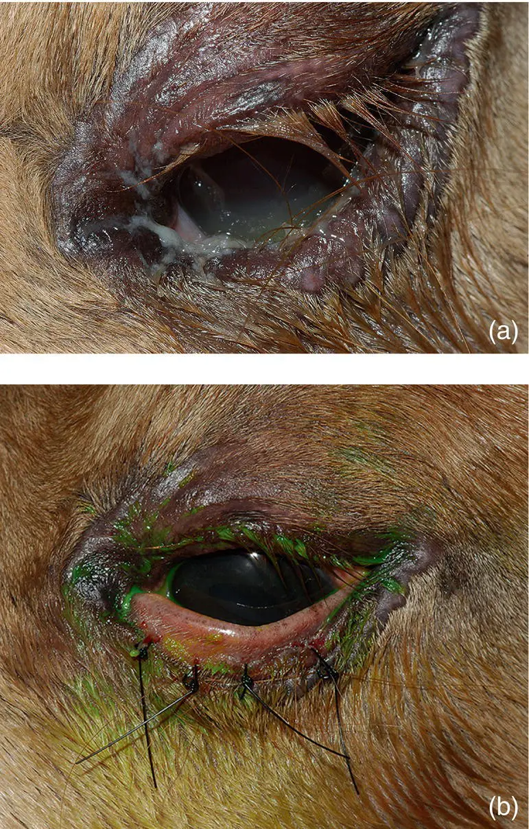
2⃣ Ankyloblepharon
➡ Failure of eyelids to open after birth
3⃣ Dermoid
➡ Focal congenital masses of displaced skin/tissue
May include hair.
🔪 May require surgical removal if causing irritation.
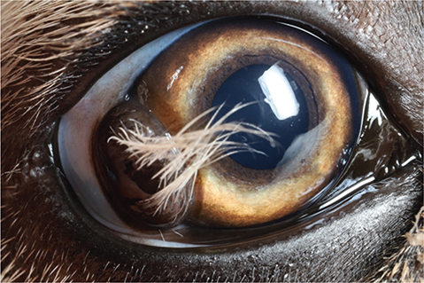
4⃣ Agenesis & Coloboma
Coloboma (dysgenesis)
➡ Full-thickness absence of portions of normal eyelid
Agenesis
➡ Complete absence of eyelid
If ocular disease is present:
➡ Reconstructive blepharoplasty
🐴 ACQUIRED EYELID DISORDERS
5⃣ Trauma / Laceration
⭐ Common because of the horse's prominent lateral eye position.
⚠ All eyelid injuries should be treated ASAP to avoid further damage to eye.
🩺 Clinical Signs & Diagnosis
Usually obvious: Edema, Bleeding
💊 Conservative Treatment
Topical compress
Topical ATB
Systemic NSAIDs
🔪 Surgical Treatment
Temporary tarsorrhaphy
Re-apposition of eyelid margin
🚨 NEVER RESECT / CUT OFF AN EYELID LACERATION!
Preserve as much eyelid tissue as possible.
6⃣ Ectropion
Ectropion = eversion of eyelid margin.
Usually due to complication of eyelid trauma.
⚙ Pathogenesis
Eyelid turns outward → Conjunctiva exposed → Irritants + secondary bacterial infections → Long-term/recurrent conjunctivitis + scarring
🔪 Treatment
➡ Surgical V and Y blepharoplasty:

7⃣ Blepharitis
Blepharitis = inflammation of the eyelids.
🦠 Infectious Causes
Bacteria:
Moraxella equi
Parasites:
Oncocerciasis, Habronemiasis
❌ Non-infectious Causes
Dermatological condition. Photosensitization, Abscess, Neoplasia
🩺 Clinical Signs
Blepharospasm. Hyperemia, Swelling, Exudation, Alopecia, Pruritus, Epiphora
🔎 Diagnosis
History, Clinical signs
💊 Treatment
➡ Depends on etiology and condition
8⃣ Neoplasia
Most common
Squamous cell carcinoma (SCC)

Sarcoid
Melanoma
Etiology / Risk Factors
Depends on:
Age, Breed
Lack of ocular pigmentation
Genetics
☀ UV radiation
Infectious agents
Chronic irritants
🔎 Diagnosis
Histopathological examination, Biopsy
💊 Treatment
BCG (Bacillus Calmette-Guérin) → injection used to dissolve sarcoid
⭐ Gamma-radiation = best
⚠ Resection could destroy the eyelid margin.
9⃣ Distichiasis
➡ Double set of eyelashes
Extra hair/cilia grow from Meibomian glands.
Treatment
➡ Surgical electrolysis
🔟 Trichiasis
➡ Inward direction of eyelashes
Eyelashes contact cornea
⬇
Irritation
1⃣1⃣ Prominence of Nictitating Membrane= Third Eyelid
Etiology
Altered globe position or size, Inflammation
Neoplasia → SCC
Tetanus
Horner's syndrome
Prolapse of orbital fat pad
Conjunctival amyloidosis
🔎 Diagnosis
Clinical signs, Biopsy, Cytology
💊 Treatment
Depends on underlying cause:
Surgical, Antitoxins
CONJUNCTIVITIS
The conjunctiva covers:
Inner eyelids
Third eyelid
Sclera
Conjunctivitis = inflammation of the conjunctiva.
⭐ Common in foals due to:
Reduced tear production
Low corneal sensitivity compared to adults
🦠 Etiology
Primary Irritants: Environmental, Allergic, Chemical
Foreign bodies
Infectious: Moraxella equi
Parasites: Thelazia, Habronema musca, Onchocerca cervicalis
Secondary ⭐ More Common
Secondary to:
Keratitis
Disorders of tear production/draining
Eyelid disease
Uveitis
Endophthalmitis
Trauma
Ulceration
Neoplasia → SCC
Systemic disease
Respiratory viral infections
Equine influenza, Equine herpesvirus, Rhinovirus
Other infections
Strangles, Equine viral arteritis, Actinobacillus spp., Corynebacterium spp.
🩺 Clinical Signs of Conjunctivitis
Swelling, Edema,💧 Epiphora
Discharge:
💧 Serous → viral
🟡 Purulent → bacterial
🔴 Hyperemia
🔎 Diagnosis
Physical examination
Scraping + culture
💧 Schirmer tear test
Occasionally biopsy
💊 Treatment
➡ Depends on etiological agent.
💧 Flushing of conjunctival sac
Ophthalmic preparation of ATBs
Granulomatous lesions:
Antihelmintic therapy
Anti-inflammatory therapy
🪱 Mechanical removal of parasites
🧠 QUICK MEMORY
Eyelid position
Entropion = IN ↩ → eyelashes damage cornea
Ectropion = OUT ↪ → conjunctiva exposed
Eyelashes
Distichiasis = extra/double eyelashes
Trichiasis = eyelashes directed inward
Trauma
🚨 NEVER cut off eyelid laceration → re-apposition
Conjunctivitis
🔴 Inflamed conjunctiva
💧 Serous = viral
🟡 Purulent = bacterial
Useful terminology
Epiphora = excessive tears 💧
Blepharospasm = excessive blinking/squeezing eye 😣
Blepharitis = inflammation of eyelid
Conjunctivitis = inflammation of conjunctiva
Corneal diseases
👁 Anatomy of the Cornea
Outside → inside:
💧 Tear film
⬇
Corneal epithelium
⬇
Corneal stroma
⬇
Descemet’s membrane
⬇
Endothelium
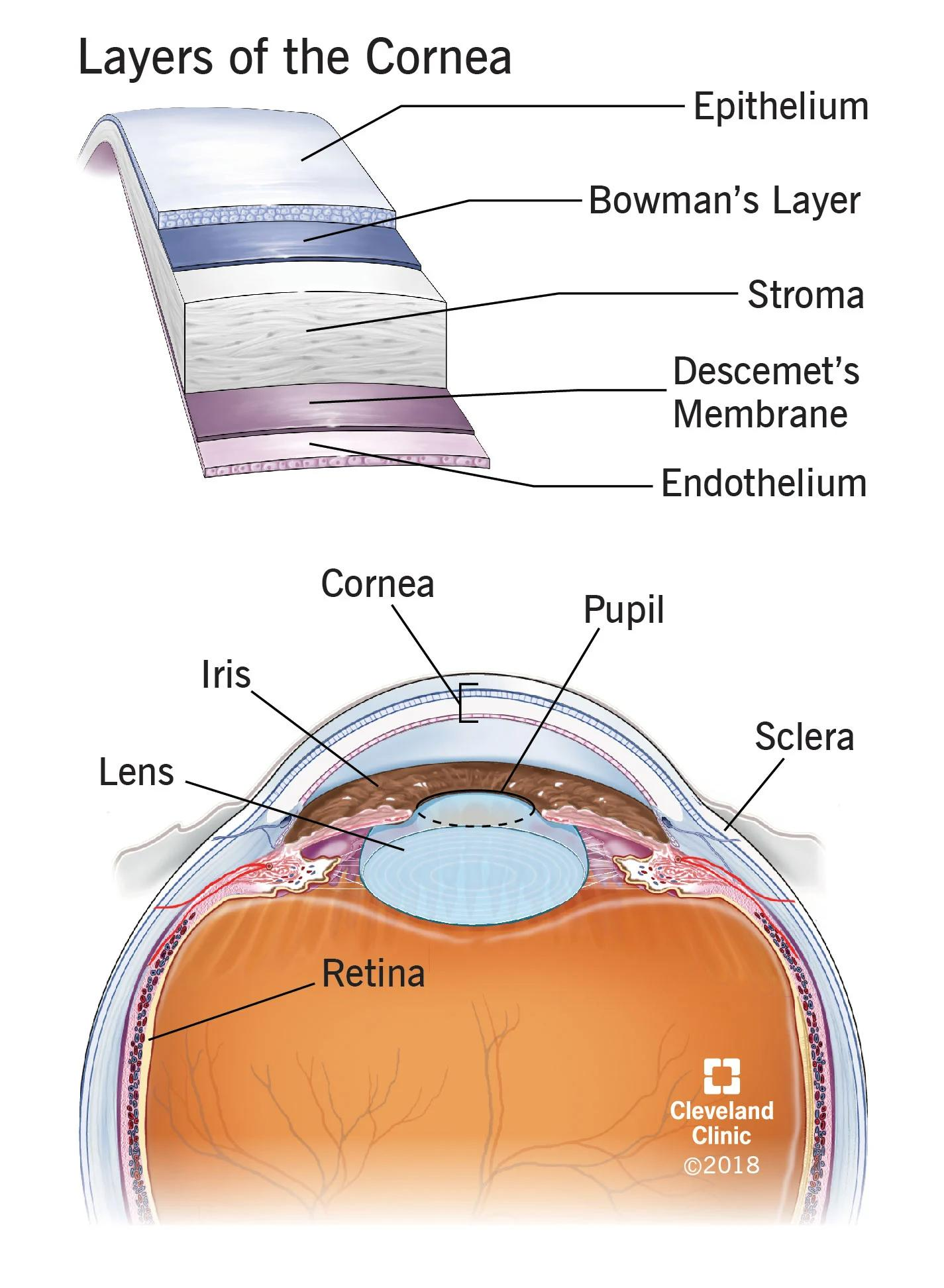
Acquired corneal diseases are common and can be:
Traumatic / non-traumatic
Inflammatory / non-inflammatory
Ulcerative / non-ulcerative
1⃣ Corneal Ulcer / Traumatic Keratitis
Corneal ulcer = loss of corneal tissue.
The cornea is widely exposed to the environment → prone to disease and ulceration.
📏 Classification according to depth
Superficial
Deep
Perforating
🔍 Etiology
⭐ TRAUMA
Also:
Disorders of adnexa:
Ectopic cilia
Entropion / ectropion
Eyelid laceration
Foreign bodies
Infectious → Equine herpesvirus 2
🩺 Clinical Signs
💧 Epiphora, Ocular discharge, Miosis, Blepharospasm, Photophobia,⬆ Vascularization / erythema
Loss of transparency → opaque cornea
➡ Damage of corneal epithelium causes absorption of water from precorneal tear film.
🔎 Diagnosis – Fluorescein Stain ⭐
🟢 Superficial wounds → stain GREEN
🟢 Deeper wounds → halo appearance

💊 Treatment
Superficial / deep epithelial wounds:
Topical ATB, NSAIDs, Topical 1% atropine, EDTA → protease inhibitor
Full-thickness lacerations:
🔪 Surgery: Conjunctival flap, Corneal graft, Remove loose flaps of cornea
🚨 CORTICOSTEROIDS CONTRAINDICATED WITH CORNEAL ULCERS
➡ Corticosteroids speed up activity of proteases
➡ Worsen the ulcer
2⃣ Keratomalacia / Melting Ulcer 🚨
Keratomalacia = liquefactive necrosis of the corneal stroma.
🚨 OCULAR EMERGENCY
🦠 Etiology
Proteases produced by:
Bacteria:
Pseudomonas, Streptococcus
Fungi
Inflammatory cells
⚙ What happens?
Proteases
⬇
Break down corneal stroma
⬇
🫠 Cornea begins to “melt”
🩺 Clinical Signs
Greyish, edematous, gelatinous corneal opacity
Blue color
Blepharospasm
Serous → purulent ocular discharge
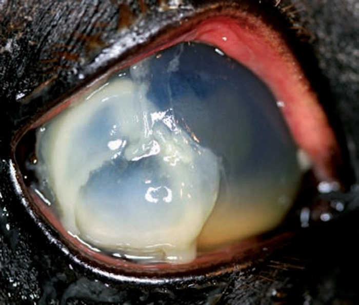
🔎 Diagnosis
➡ Fluorescein stain
💊 Treatment
Topical ATB, Antifungal, NSAIDs
⭐ Topical anti-proteases:
EDTA, Acetylcysteine
Treatment every 4–6 hours
3⃣ Immune-Mediated Keratitis – IMK
Common non-infectious eye disease in horses.
Often affects one eye, but can affect both.
⚠ Serious → can threaten horse's sight.
Four Types – Based on Depth
1⃣ Epithelial / superficial
Inflammation of eyelids
Congestion of conjunctival blood vessels
Swelling of conjunctiva
2⃣ Midstromal
3⃣ Deep stromal
4⃣ Endothelial
🚨 Most dangerous and damaging type
➡ Affects innermost layer → endothelium

💊 Treatment
⚠ Important to distinguish from other causes of keratitis, especially infectious keratitis.
Topical NSAIDs
Topical cyclosporine A
❌ No corticosteroids!
4⃣ Dry Eye Syndrome= Keratoconjunctivitis Sicca – KCS
KCS = aqueous deficiency of the precorneal tear film, causing progressive inflammatory changes of the cornea + conjunctiva.
Two types: tear-deficient dry eye due to insufficient aqueous secretion, and evaporative dry eye often linked to meibomian gland dysfunction or eyelid abnormalities
🔍 Etiology
Tear deficiency, Evaporative disorders, Eyelid problems, Meibomianitis, Exophthalmos
Can result from head trauma, damage to the facial nerve controlling tear production, or secondary systemic/local inflammation
🩺 Clinical Signs
Ocular pain, Dull cornea, Corneal edema, Keratitis, Mucopurulent ocular discharge
🔎 Diagnosis
Schirmer tear test → <10 mm/min
Rose Bengal stain → Remains red if tissue is devitalized
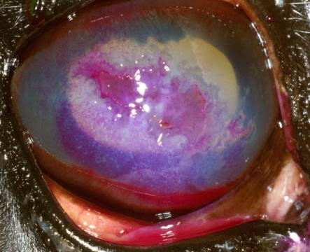
💊 Treatment
Replace precorneal tear film:
Synthetic mucins, Artificial tears, Topical cyclosporine A
5⃣ Corneal Stromal Abscess
Corneal stromal abscess = pus-filled sore in the connective tissue of the cornea.
🦠 Etiology
Healing ulcer / corneal defect
⬇
Bacteria or fungi become trapped inside
⬇
Stromal abscess
⚠ Can cause secondary uveitis.
It typically develops when a superficial corneal microtrauma or ulcer heals over, sealing bacteria or fungi (frequently fungal) inside the avascular stroma where the immune system and topical drugs struggle to reach.
🩺 Clinical Signs
White/yellow material in connective tissue, Corneal inflammation + swelling, Formation of blood vessels
Ocular pain

💊 Treatment
Topical + systemic ATB
Antifungal
Painkiller, NSAIDs
🔪 Surgery may be required to:
Remove abscess, promote corneal healing
⚠ Prognosis = guarded
🦠 Infection in Corneal Ulcers
Most ulcers are presumed to have secondary bacterial and fungal infection.
⭐ Take swabs BEFORE any topical treatment
➡ Microbiology
If fungal infection confirmed:
➡ Antifungal treatment 6–8 weeks
6⃣ Protozoal – Besnoitia benetti
Coccidia → Besnoitia benetti
FH: Cats
IH: Horse (mostly donkeys, rare in horse)
Vector: Flies
🔄 Life Cycle
🐱 Cat sheds oocysts
⬇
🐴 Horse ingests from grazing/water
⬇
Formation of tissue cysts in skin/nostrils
⬇
Edema + hyperkeratosis + alopecia
⬇
Can also infect eyes/sclera
⬇
⚪ Small dots/cysts on surface
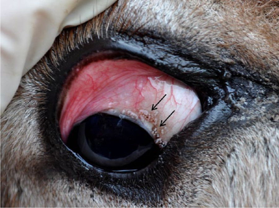
🩺 Clinical Signs
Fever, Nasal discharge, Ocular discharge
Salivation, Stiff gait, Orchitis
Subcutaneous edema
Multifocal pinpoint parasitic cysts:
Nostrils, Ears, Face, Body
crusty hard skin lesions
👁 Scleral pearls → tiny cysts in eyes
🔎 Diagnosis
Skin biopsy
Endoscopy of nasal cavity
ELISA confirmation
💊 Treatment
⚠ Problematic → no drug stops formation of cysts in organs/skin.
Early infection:
➡ Try trimethoprim (stop cyst forming, but not kill parasite)
Otherwise:
➡ Mostly symptomatic treatment
7⃣ Equine Herpesvirus 2
🩺 Clinical Signs
Superficial punctate keratitis
Blepharospasm
Lacrimation
🔎 Diagnosis
Clinical signs
Response to antiviral drugs:
Idoxuridine, Acyclovir
8⃣ Parasitic – Onchocerca cervicalis
Large nematode inhabiting:
Subcutaneous tissue, Tendons, Tendinous ligaments
➡ Microfilariae in skin.
⚙ Pathogenesis
Migration + death of Onchocerca cervicalis microfilariae
⬇
Corneal + conjunctival tissues
⬇
👁 Keratitis / keratoconjunctivitis
🩺 Clinical Signs
Pain, Hyperemia
Corneal opacity
🔎 Diagnosis
Clinical signs
Demonstration of microfilariae in biopsy
💊 Treatment
Ivermectin
Corticosteroids
🧠 QUICK DIFFERENTIATION
Corneal ulcer
Loss of corneal tissue
➡ Trauma most common
➡ Fluorescein +
➡ ATB + NSAID + atropine
🚫 NO corticosteroids
🫠 Melting ulcer
Corneal stroma literally breaking down
➡ Proteases
➡ Gelatinous/blue-grey cornea
🚨 Emergency
➡ EDTA + acetylcysteine
🛡 Immune-mediated keratitis
Non-infectious immune disease
➡ Different depths
➡ Cyclosporine A
💧 KCS
Not enough tears
➡ Schirmer <10 mm/min
➡ Artificial tears + cyclosporine
Stromal abscess
Bacteria/fungi trapped INSIDE healing cornea
➡ White/yellow material
➡ Can cause secondary uveitis
➡ Guarded prognosis
⭐ One important rule
CORNEAL ULCER + CORTICOSTEROIDS = ❌
Corticosteroids
➡ ⬆ Protease activity
➡ ⬆ Corneal breakdown
➡ Ulcer gets worse
Uveitits
The uvea = middle layer of the eye, beneath the sclera. It consists of: IRIS, CILIARY BODY, CHOROID
Uveitis = inflammation of the uvea.
🚨 Most common cause of blindness in horses.
Can be:
Acute, Chronic, Recurrent
🔍 Etiology
1⃣ Trauma
2⃣ Corneal Disease
Keratitis, Corneal ulcers
3⃣ Equine Recurrent Uveitis – ERU ⭐
➡ Autoimmune disease ⭐ Most frequent!
4⃣ Infectious Causes
🦠 Bacteria
Leptospira interrogans serovars, Rhodococcus equi, Streptococcus, Brucella, Salmonella
🦠 Viruses
Equine herpesvirus, Equine influenza virus
🪱 Parasites
Onchocerca cervicalis, Thelazia, Toxoplasma, Strongylus
5⃣ Reflex Uveitis
➡ Mediated by axon effect in trigeminal nerve
For example:
Corneal injury
⬇
Trigeminal nerve stimulation
⬇
Inflammatory response inside eye
⬇
Uveitis
6⃣ Neoplasia
⭐ Melanoma = most common
Treatment
Sector iridectomy
Enucleation
7⃣ Systemic Infections
Endotoxemia
Septicemia
🩺 Clinical Signs
🔥 Acute Uveitis
😣 Pain
💧 Lacrimation
Blepharospasm
☀ Photophobia
⭐ MIOSIS
Impaired vision
Corneal edema
⭐ Reduced intraocular pressure – IOP ↓
🧠 Classic Acute Uveitis
PAINFUL + SMALL PUPIL + LOW IOP
🌫 Chronic Uveitis
➡ Cataract / cloudy eye ⭐ Typical for chronic uveitis.
🔎 Diagnosis
Clinical signs
💧 Schirmer tear test → Increased tear production
👁 IOP → ↓ Decreased pressure
Collection of:
Aqueous humor
Vitreous humor
➡ To identify etiological agent
🚨 Treatment
START TREATMENT IMMEDIATELY AND AGGRESSIVELY!
Untreated inflammation → Damage inside eye → Retinal detachment → Vision loss
1⃣ Mydriatics
Mydriatic = dilates the pupil
Atropine → long acting
⭐ Tropicamide → short acting, preferred
Why dilate the pupil?
Uveitis → Miosis → Iris/lens irritation → Risk of adhesions + lens damage → Cataract
➡ Therefore give mydriatic → dilate pupil
2⃣ Topical NSAIDs
Example: Diclofenac
Reduces:
Pain, Redness, Swelling
3⃣ Steroids
Prednisolone, Dexamethasone
⚠ Remember from corneal ulcers:
UVEITIS → steroids can be used
BUT
CORNEAL ULCER → ❌ topical corticosteroids
➡ Always check the cornea first.
4⃣ Antibiotics
Doxycycline, Gentamicin
🔄 EQUINE RECURRENT UVEITIS – ERU= Autoimmune Disease
An autoimmune condition where:
Horse's immune system
⬇
Attacks its own ocular tissues
⬇
Repeated episodes of uveitis
⬇
Progressive damage to uveal tract
⬇
👁❌ Can completely destroy vision
ERU occurs after an initial episode of uveitis.
⚠ But not every horse with uveitis develops recurrent uveitis.
🔍 Possible Triggers of ERU
Specific conditions/agents associated with ERU include:
🦠 Bacteria
Especially Leptospira
🪱 Parasitic worms
🦠 Viruses
Equine influenza
🦷 Tooth root abscess
🐴 Hoof abscess
👁 Eye injuries
Inflammation can also be stimulated by dead/dying parasite larvae that have migrated into the eye.
🔪 Treatment of Recurrent Uveitis
1⃣ Vitrectomy
Vitrectomy = aspiration/removal of material from the vitreous body
➡ Removes: Cells, Antigens, Organisms from corpus vitreum
2⃣ Cyclosporine A Implant
➡ Cyclosporine A implant:
Intravitreally / sub-sclerally ➡ Suppresses immune response
Your notes also mention:
Cephalosporin implant
⚠ Prognosis
Guarded → poor
🚨 Complications
Glaucoma
Cataracts
Cloudiness of cornea
Retinal damage/detachment
👁 Blindness
🧠 UVEITIS – QUICK MEMORY
Acute:
😣 PAIN
⚫ MIOSIS
⬇ LOW IOP
💧 TEARS
Chronic:
🌫 CATARACT / CLOUDY EYE
Treatment:
DILATE + CONTROL INFLAMMATION + TREAT CAUSE
➡ Mydriatic
➡ NSAID
➡ Steroid
➡ ATB when indicated
🪱 EYE PARASITES
1⃣ Thelazia lacrimalis
➡ Nematode transmitted by non-biting dipteran flies that lick conjunctival secretions.
🔄 Life Cycle
🪰 Fly carrying larvae → Fly feeds on horse's conjunctival secretions → Larvae migrate fly → eye → Mature into adult worms → Female sheds L1 into lacrimal secretions / tears → Flies ingest L1 → L1 → L3 inside fly → Fly infects another horse
🩺 Clinical Signs
💧 Excessive lacrimation, Conjunctivitis
🔎 Diagnosis
👀 Rapidly moving worms visible in eye
L1 detected in sediment of flushing fluid
💊 Treatment
Local anesthesia of eye
⭐ Mechanical extraction of worms
SC ivermectin
Protect horses against dipteran flies
2⃣ Onchocerca cervicalis
➡ Nematode normally residing in ligamentous tissue.
Can cause parasitic keratitis due to migration through nasolacrimal duct.
⚙ Pathogenesis
Microfilariae migrate → Eye/corneal tissues → Inflammation + irritation → Ulceration → Granuloma formation
💊 Treatment
Ivermectin, Topical corticosteroids
If granuloma irritates cornea:
➡ 🔪 Surgical removal
3⃣ Habronema musca
➡ Nematode
➡ Equine stomach worm
Hosts
FH = Equids
Stomach, Skin, Eyes, Genitalia, Lungs
IH = Musca flies 🪰
🔄 Life Cycle
🐴 Horse feces containing eggs/larvae → Flies ingest them → Develop → L3 → Fly feeds on ocular/nasal discharge → Deposits L3 on horse → Larvae normally migrate to stomach → Mature into adult worms
But larvae deposited around wounds/eyes can cause cutaneous/ocular habronemiasis.
🩺 Clinical Signs
Cutaneous / ocular form
Conjunctivitis, Thickening of eyelid
Granuloma formation in skin, Severe itching, Self-inflicted injuries
💊 Treatment
Ivermectin, Topical corticosteroids
🧠 PARASITE QUICK MEMORY
🪰 THELAZIA
FLY → EYE → visible worms
➡ Lacrimation + conjunctivitis
➡ Pull worms out + ivermectin
🪱 ONCHOCERCA
Ligaments → microfilariae → eye
➡ Keratitis + granulomas
➡ Ivermectin + corticosteroids
🪰 HABRONEMA
Stomach worm + fly
➡ Fly deposits larvae around eye/wounds
➡ Granulomatous lesions + itching
➡ Ivermectin + corticosteroids
⭐ EXAM ESSENTIALS
Uveitis = inflammation of iris + ciliary body + choroid
Acute uveitis:
➡ Pain + miosis + ↓ IOP
Chronic uveitis:
➡ Cataract/cloudy eye
ERU:
➡ Autoimmune + recurrent episodes
➡ Most important cause of blindness
ERU treatment:
➡ Vitrectomy / cyclosporine implant
Prognosis:
➡ Guarded to poor
Equine hoof diseases
HOOF ANATOMY
Outer Structures:
Hoof Wall
Covers the front and sides of the coffin bone.
Parts:
Toe = front
Quarters = sides
Heel
➡ Continually growing keratinous material
➡ No nerves or blood vessels
3 Layers of the Hoof Wall
1⃣ Outer layer → Periople
2⃣ Middle layer → Bulk of wall
3⃣ Inner layer → Laminae
⭐ Laminae attach the coffin bone to the inside of the hoof wall and bear much of the weight.
Coronary Band
Located at the top of hoof where hairline meets hoof.
➡ Primary growth + nutritional source for hoof wall.
⚖ Weight-Bearing Structures:
Sole
Protects internal structures of hoof.
Designed to bear internal weight transferred through border of sole, rather than weight directly from ground.
White Line
➡ Junction between hoof wall + sole
Frog 🐸
Tough V-shaped structure pointing down from heels.
➡ Protects digital cushion beneath it.

🦴 Inner Structures:
Digital Cushion
Located below coffin bone toward back of hoof. Cartilaginous cushion and a major shock absorber
Coffin Bone (“hovbeinet”)
= Pedal bone / distal phalanx
➡ Largest bone in hoof, Helps shape hoof wall, Surrounded by tissues forming the laminae
Navicular Bone
= Distal sesamoid bone (Small bone behind coffin bone)
➡ Helps stabilize coffin bone and is associated with deep digital flexor tendon apparatus
🔥 1. LAMINITIS (“Forfangenhet”)
Definition
Laminitis = inflammation of the laminae.
⭐ Zert definition: “Laminitis is a disconnection of the epidermal and dermal lamella, connected with failure of the suspensory apparatus.”
🔬 Lamellae
Laminae = sensitive tissues connecting hoof wall → coffin bone.
Lamellae = villi-like structures.
Two components:
Dermal lamellae
Originate from corium. Sensitive
Epidermal lamellae
Originate from the deepest layers of the hoof wall. Insensitive
They normally:
➡ Interlock with each other
➡ Suspend coffin bone inside hoof capsule
🔍 Etiology – MULTIFACTORIAL
⭐ Most common cause is obesity
🦠 Systemic infections
Enterocolitis, Endotoxemia, Septicemia
Pleuropneumonia, Metritis, Retention
🍞 Overfeeding carbohydrates
➡ Dysbacteriosis, Endotoxicosis
🌱 Lush grass (rapidly growing spring grass with high amount of sugars and starch)
➡ Hindgut fermentation, Lactic acid production
🦵 Mechanical causes
Contralateral limb overload, Chronic overload, Hard work on hard surface, Poor hoof trimming
🧬 Endocrinopathy / Metabolic
Insulin resistance, Diabetes mellitus, Hyperlipemia, Cushing, Hypothyroidism
⚙ Pathogenesis
Exact mechanism largely unknown.
Believed to involve:
Hypoperfusion → Ischemia → Necrosis of lamellae → Dermal–epidermal junction becomes edematous + weakened → Loss of interlaminar bond → Failure of suspensory apparatus → Coffin bone displacement
🔄 ROTATION vs SINKING ⭐⭐⭐
🔄 Rotation
Damage extends over large area
⬇
Suspensory support of distal phalanx lost
⬇
Body weight + pull of DDFT + dorsal soft-tissue pressure
⬇
Mechanical separation of distal phalanx from hoof wall
⬇
🦴 Coffin bone rotates away from dorsal hoof wall
➡ Palmar rotation
Heel remains suspended by laminar attachment.
🧠 Think:
TOE attachment fails → DDFT pulls → bone ROTATES
⬇ Sinking
More severe.
ALL suspensory support of laminae lost → Entire distal phalanx drops distally inside hoof → Without obvious rotation.
🧠 Difference
ROTATION 🔄
➡ Part of support remains and Coffin bone tilts
SINKING ⬇
➡ All support fails and Whole coffin bone drops → much worse prognosis.
🌍 Systemic Changes
Cardiovascular: Tachycardia, Hypertension
Endocrine:↑ Catecholamines, ↑ Cortisol
Renal: Glomerulonephritis
Liver damage
🩺 Clinical Signs of Laminitis
Systemic
↑ Heart rate, Tachypnea, Fever
Hoof
💥 Pounding digital pulse, 🔥 Warm hoof wall
Lameness, Pain, Reluctance to move
⭐ Typical Laminitis Stance:
Forelimbs stretched forward + Hindlimbs underneath body (Horse tries to remove weight from painful forefeet/toes)
🔎 Diagnosis
Clinical signs
⭐ X-ray
X-ray:
Degree of coffin bone rotation and Hoof wall thickness

🚨 ACUTE LAMINITIS = EMERGENCY
1. Cryotherapy
➡ Hoof in ice bath / ice packs
⭐ Especially important early.
💊 2. Stop Pain ➡ NSAIDs
🎯 3. Remove Initiating Cause
Examples:
Remove from pasture, ATB for infection, Anti-endotoxin medication
🩸 4. Reduce Vasoconstriction / Hypertension
Acepromazine, Potassium chloride, Topical nitroglycerin
🦶 5. Mechanical Support
Deep soft bedding, Pre-shaped frog pads
🚨 AVOID:
❌ Perineural analgesia
❌ Forced exercise
🕐 CHRONIC LAMINITIS
General
Limit carbohydrates, Potassium chloride, Biotin
✂ Corrective Foot Trimming
Remove excess horn, Trim heels
👞 Therapeutic Shoeing
Wide-webbed / seated-out bar shoe of thick steel
➡ Increase support, Minimize pressure on sole
📊 LAMINITIS GRADING
Grade | Clinical sign |
|---|---|
1 | Shifting weight |
2 | Typical stiff, stilted laminitis gait |
3 | Reluctance to walk + typical gait |
4 | Recumbency |
🧠 1 shift → 2 stiff → 3 reluctant → 4 down
❄ PREVENTION
Subacute / Preclinical Stage
Example:
Horse with colic + suspected endotoxemia
➡ Put hooves in ICE ❄
Also Support frog, Elevate heel
➡ Decreases pulling force of DDFT
General Prevention
➡ Correct feeding (Especially control glucose/carbohydrate intake)
📈 Prognosis
🟢 Good
No rotation by 10 days
Recovered by approximately day 60 (2months)
🔴 Unfavorable
Acute phase >10 days
Recumbency, High BP, Secondary infection, Displacement of distal phalanx
⚫ Hopeless
Sinking of distal phalanx >20 mm founder distance
Exungulation of hoof
💔 2. HOOF CRACKS
Deep cracks often accompanied by localized infection of the dermis.
Can be:
Partial or Complete
Etiology
Overgrown hoof wall, Poor-quality horn, Poor foot balance, Tearing, Breed predisposition
Clinical Signs
Lameness, Visible cracks

Diagnosis
Clinical signs, Hoof tester
Uni-axial analgesia → determine clinical significance
Treatment
Oil, Biotin, Methionine, Adequate immobilization
Partial hoof-wall resection
Prevention
➡ Regular good foot care
➡ Oils, biotin etc.
⚪ 3. WHITE LINE DISEASE= Horny Capsule Disease
Dermal + epidermal layers become separated at toe → Visible at white line → Dermal region exposed →🦠 Ascending infection
Etiology
Focal hemorrhage
Seroma
Chronic untreated bacterial infection
Mycotic infection of corium
Chronic pododermatitis
Pathogenesis
Persistent infection → Cavity formation → Damaged laminae fail to produce keratin → Abnormal/empty hoof wall region
Clinical Signs
Brown, crumbly horn-like material along white line
Non-keratinized void extending from white line
Lameness
Diagnosis
Clinical signs
🔨 Percussion → hollow sound
X-ray → gas shadow in soft tissue


Treatment
Regular cleansing
Wide-webbed flat shoes
Severe cases → remove affected horn
Dress exposed laminae
Allow newly formed horn to grow normally
Antifungal foot soaks
📌 4. PENETRATING WOUNDS OF THE SOLE
Puncture wound through sole or frog
➡ Can involve dermis or deeper structures.
Etiology
➡ Standing/kicking against sharp objects
⚠ Secondary infection common.
🚨 Most Dangerous Area:
⭐ Middle → palmar/plantar third of frog + sulci
Why? Deep penetration here can damage:
DDFT, Navicular bursa, Ligaments, Digital flexor tendon sheath
🩺 Clinical Signs
Acute onset lameness
May improve after foreign body removal
Non-weight-bearing lameness with secondary infection
Purulent discharge
Systemic signs of infection
Occasionally stringhalt-like gait
🔎 Diagnosis
Observation
Foreign body may still be embedded
Hoof tester
Synoviocentesis + fluid analysis
Plain radiography
Contrast radiography
Positive contrast arthrography
Sonography
💊 Treatment
ATB, Tetanus antitoxin, Cleaning, Drainage, Antiseptic drainage
🚨 Emergency surgical debridement when indicated.
If Synovial Penetration:
Navicular bursa
➡ Navicular bursoscopy
➡ Focal debridement
DIP joint
➡ Arthroscopic lavage
Aftercare ➡ Daily antiseptic dressing under sterile bandage
🦴 5. NAVICULAR SYNDROME = Palmar Heel Pain / Podotrochlosis
⭐ One of the most common causes of chronic forelimb lameness in athletic horses.
Chronic degenerative condition involving the navicular bone and associated structures.

⚙ Pathological Changes
1⃣ Medullary Changes
➡ Focal loss of medullary architecture
➡ Subsequent synovial invagination
2⃣ Sclerosis
➡ Medullary sclerosis + Damage to fibrocartilage on flexor surface
3⃣ DDFT Damage
Damaged navicular flexor surface → DDFT repeatedly contacts it → Traumatic fibrillation of DDFT → Adhesions between tendon + bone
4⃣ Enthesiophytes
➡ Bone spurs (osteophytes) at proximal + distal borders of navicular bone
🔍 Etiology
Unknown → multifactorial / complex
Important:
Distal limb conformation
Degree/type of athletic loading
Genetic/hereditary predisposition
Thoroughbred
Quarter Horse
Warmblood
🩺 Clinical Signs
Early ➡ Intermittent, progressive lameness
REST → improves
WORK → returns
⬇
Eventually becomes consistent.
⭐ Usually bilateral
Horse tries to avoid loading heels:
➡ Short stride, Choppy gait, Pointing hoof
🔎 Diagnosis
Clinical
History
Hoof tester
Sensitive over center of frog
Flexion test
Hoof shape:
Contracted heel
Atrophy of frog
💉 Nerve Blocks
Palmar digital nerve block or Navicular bursa block → ⭐ Lameness disappears/improves
🩻 X-ray
May show:
Enthesiophytes on wings of navicular bone
Cystic lesions
Calcification of DDFT

💊 Treatment of Navicular Syndrome
👞 1. Shoeing
⭐ Foot must be kept balanced.
Options:
Rolled toe, Egg-bar shoe, Adequate heel support
➡ Goal = improve mechanics and reduce stress on heel/navicular apparatus.
🔪 2. Surgery
Desmotomy of suspensory ligament ➡ Alters loading of navicular bone
Palmar digital neurotomy ➡ Removes pain sensation from affected region
💊 3. Anti-inflammatory Treatment
Aspirin, Banamine, PBZ, Polysulphated glycosaminoglycans
🧠 QUICK MEMORY
🔥 LAMINITIS
Laminae fail → coffin bone loses suspension
Two major consequences:
🔄 ROTATION
= coffin bone tilts
⬇ SINKING
= whole coffin bone drops
Classic signs:
➡ 🔥 Hot feet
➡ 💥 Pounding digital pulse
➡ 😣 Severe pain
➡ 🐴 Forelegs forward
🚨 Acute = ICE + NSAIDs + treat cause + support hoof
⚪ WHITE LINE DISEASE
White line separates → Infection moves upward → Brown crumbly horn + cavity
📌 PENETRATING WOUND
Especially dangerous through:
⭐ Middle/palmar frog
Frog → DDFT → Navicular bursa/navicular region → DIP region → Deep penetration = 🚨 possible synovial infection
🦴 NAVICULAR SYNDROME
Think:
ATHLETIC HORSE + CHRONIC BILATERAL FORELIMB LAMENESS + HEEL PAIN
➡ Short/choppy stride
➡ Worse with work
➡ Palmar digital block improves lameness
➡ Balance hoof + support heel
⭐ EXAM DIFFERENTIATION
Laminitis
➡ Laminae
➡ Acute painful feet
➡ Rotation/sinking of coffin bone
White line disease
➡ Separation at white line
➡ Brown crumbly horn + hollow cavity
Penetrating sole wound
➡ Foreign body through sole/frog
➡ Risk DDFT/navicular bursa/DIP infection
Navicular syndrome
➡ Chronic palmar heel pain
➡ Athletic horse
➡ Usually bilateral forelimbs
Equine fractures
A fracture = break in the continuity of a bone.
On X-ray: Discontinuity of cortex, Radiolucent fracture line
🔍 Etiology
⭐ Common in racing horses
Typical:
Stress fractures of carpal joint and fetlock joint
Other causes:
Direct external trauma, Kicks, Collisions
📚 Classification
1⃣ Open / Closed
Closed fracture
➡ Skin intact
Open fracture
➡ Skin communicates with fracture → High infection risk
2⃣ Simple / Complicated
Simple
➡ Single uncomplicated fracture
Complicated
➡ Associated damage to soft tissues, joints, vessels, infection etc.
3⃣ According to Fracture Line / Location
Transverse, Oblique, Longitudinal, Spiral, Comminuted, Diaphyseal, Metaphyseal
Physeal → Salter-Harris
Articular
Long-bone fractures, Short-bone fractures
4⃣ Fragment Position
Dislocation / displacement
Compression
🩺 Clinical Signs
Pain, Swelling, Lameness
Abnormal mobility
Loss of function
Often non-weight-bearing
Crepitus
🔎 Diagnosis
⭐ X-ray
Minimum 2 planes
Often multiple oblique views
Also:
CT, Scintigraphy, Ultrasonography
🦴 FRACTURE HEALING
There are two major types:
1⃣ PRIMARY BONE HEALING = Direct healing
Requires:
➡ Rigid stabilization ± compression of bone ends
The bone ends are held in very close/direct contact.
Important consequence → Little/no callus formation
Osteoblasts fill and remodel the fracture directly.
Divided into:
Contact healing and Gap healing
⭐ Best especially in intra-articular fractures
Why? Because large callus formation inside/around a joint would interfere with joint function.
2⃣ SECONDARY BONE HEALING= Indirect healing
Spontaneous fracture healing when there is no completely rigid fixation.
➡ Callus formation occurs.
There are 3 phases:
🔥 Phase 1 – Inflammatory Phase
Starts immediately.
Fracture → Bleeding → Hematoma formation → Inflammatory cells invade
Clinical → Pain and Swelling
🛠 Phase 2 – Repair Phase
Begins within a few days, before inflammation completely subsides.
Lasts weeks.
Important cells:
Chondroblasts, Fibroblasts, Osteoblasts
Sequence
1⃣ Granulation tissue forms → Soft callus → 2⃣ Cartilage formation → 3⃣ Fibrous tissue formation → 4⃣ Calcium deposited → 🦴 Hard callus
🔄 Phase 3 – Remodeling
Final phase.
Woven bone → replaced by → Lamellar bone
At the same time:
➡ Excess callus is resorbed
Result:
➡ Bone gradually regains more normal structure and strength.
🧠 PRIMARY vs SECONDARY HEALING
PRIMARY
Rigid fixation + compression
➡ Bone ends directly together, Minimal callus
SECONDARY
Some movement / indirect contact
➡ Hematoma, Soft callus, Hard callus, Remodeling
🚑 INITIAL TREATMENT OF FRACTURES
Main goals:
Minimize damage to soft tissues
Minimize further damage to bone ends
Allow horse to regain control of limb
Provide pain relief
Limit soft-tissue swelling
🚨 If Skin is Penetrated
Open fracture:
Clean wound
Water-soluble ATB ointment
Sterile dressing
Then immobilize
🔧 STABILIZATION METHODS
External Fixation
Splint
Robert Jones bandage
Cast
Transfixation cast
Pinless fixation
Internal Fixation
Lag screws
Plates
Ligatures
Major displaced fractures usually require internal fixation.
🔩 LAG SCREWS
⭐ Give contact healing / primary healing
Principle
Screw compresses the fracture fragments together.
➡ Pressure across fracture line
➡ Rigid contact
➡ Minimal movement
➡ 🚫 Avoid callus formation
Used especially in:
Transverse fractures
Oblique fractures
Metacarpal/tarsal fractures
Technique:
Small stab incision → Drill gliding hole through near cortex → Measure → Smaller drill bit into far cortex → Insert screw → Fragments compressed together

🧠 Why does a lag screw compress?
The screw threads engage only the far fragment.
When the screw is tightened:
Far fragment is pulled toward Near fragment ➡ Fracture ends are compressed.
🧱 PLATES
Used:
To stabilize fractures
Especially long bones
Types:
DCP ➡ Dynamic compression plate
LCP ⭐ ➡ Locking compression plate
LC ➡ Low-contact plate
⭐ LCP – Most Used
The screw head locks into the plate.
➡ Screw cannot move within plate
➡ Very stable fixation
➡ Better healing
Used especially in simple fractures.

🦾 TRANSFIXATION CAST
Principle:
➡ Pins/nails pass through bone
Cast → pin → bone → pin → cast
This transfers weight through pins and cast rather than through the fractured distal limb.
➡ Reduces compression/loading of fracture fragments.
👞 SHOEING
For some hoof-bone fractures:
➡ Bar shoe
Especially:
Pedal/coffin bone fractures
Navicular bone fractures
Plus:
➡ Box rest

🩺 AFTERCARE
External support
Bandage, Splint, Cast
Antibiotics, Analgesia
Box rest
🚫 Never use corticosteroids with intra-articular fractures
⚠ COMPLICATIONS OF FRACTURE HEALING
🦠 Infection
Especially:
Open fractures
Internal fixation
🦴 “Fracture Disease”
Due to prolonged immobilization:
Muscle atrophy
Soft-tissue adhesions
Joint stiffness
Osteoporosis
❌ Healing Problems
Delayed union
➡ Fracture heals too slowly
Malunion
➡ Fracture heals in wrong position
Non-union
➡ Fracture fails to unite
Foals
Prolonged immobilization can cause:
Laxity of supporting soft tissues
Growth disturbance
Angular limb deformities in contralateral limb
Adult Horses
🚨 Contralateral limb laminitis
Horse avoids loading fractured limb → Overloads opposite limb → Laminitis
✅ TREATABLE FRACTURES
🏇 Stress Fractures
Mostly:
Fetlock, Carpus
Racing horses
Treatment:
➡ Primary healing
➡ Lag screw fixation
🧩 Chip Fractures
Locations:
Fetlock, Carpus
Treatment:
➡ Arthroscopic removal
🦴 Proximal Phalanx Fractures
Sagittal/frontal plane fractures:
➡ Lag screws
🦴 Proximal Sesamoid Bone Fractures
Apical / mid-body fractures
➡ Lag screws
or
➡ Circumferential wiring
🦵 Condylar Fractures
Distal 3rd metacarpal/metatarsal bone:
➡ Lag screws
🦴 Third Metacarpal/Tarsal Bone
Transverse / oblique fractures:
➡ Compression plate
💪 Olecranon Fractures
➡ Compression plate
Long-Bone Fractures
Generally treated mainly in young foals.
➡ Compression plates can be used.
In adult horses:
➡ Prognosis often poor
➡ Euthanasia frequently advised for severe long-bone fractures.
🚫 Femoral Head Fracture
➡ Euthanasia
🧠 QUICK FRACTURE-TREATMENT TABLE
Fracture | Typical Treatment |
|---|---|
Stress fracture – fetlock/carpus | Lag screw |
Chip fracture – carpus/fetlock | Arthroscopic removal |
Proximal phalanx sagittal/frontal | Lag screw |
Proximal sesamoid apical/mid-body | Lag screw / wiring |
Condylar MCIII/MTIII | Lag screw |
Transverse/oblique MCIII/MTIII | Compression plate |
Olecranon | Compression plate |
Pedal/navicular bone | Bar shoe + box rest |
Severe adult long-bone fracture | Often euthanasia |
Femoral head | Euthanasia |
⭐ EXAM ESSENTIALS
Fracture diagnosis
➡ X-ray in 2 planes + oblique views
Primary healing
➡ Rigid fixation
➡ Minimal/no callus
➡ Best for intra-articular fractures
Secondary healing
➡ Inflammation → repair/callus → remodeling
Lag screw
➡ Compresses fragments
LCP
➡ Screw locks into plate → high stability
Transfixation cast
➡ Pins transfer load from bone into cast
Adult long-bone fractures
➡ Often poor prognosis/euthanasia
Major complication in adults
➡ Contralateral limb laminitis
Developmental Anomalies of Bones
1⃣ ANGULAR LIMB DEFORMITY – ALD
ALD = skeletal defect where a portion of the limb is bent inward or outward at an abnormal angle from the midline of the body.
⭐ Relatively common in newborn and young foals.
Most commonly affects:
Carpus, Sometimes tarsus, Fetlock
↔ VALGUS vs VARUS ⭐
When viewed FROM THE FRONT:
VALGUS: OUTWARD bending
Can be normal immediately after birth → foal may grow out of it.
VARUS: INWARD bending (⚠ Usually more severe)
🔍 Etiology
Most foals are born with some degree of limb deviation.
Often due to: Ligament laxity, Muscle weakness
Usually corrects itself as foal exercises.
Perinatal Factors:
Premature birth, Twin pregnancy, Placentitis, Perinatal soft-tissue trauma, Flaccidity/laxity of soft tissues surrounding joints, Poorly calcified bone
Developmental Factors:
Unbalanced nutrition, Excessive exercise, Trauma, Inappropriate growth, Hypothyroid hormone deficiency
🩺 Clinical Signs
Presence of valgus or varus
Swelling, Lameness
Excessive hoof wear on:
Medial side or Lateral side
🔎 Diagnosis
Clinical signs, Degree of deviation
⭐ X-ray
💊 TREATMENT OF ALD
Conservative Treatment:
For young foals with mild deviation:
Stall rest, Splints, Cast placement, Corrective hoof trimming, Glue-on shoes → Dalric type, Inside/outside hoof extensions
VALGUS
➡ Trim lateral side and Preserve medial side
VARUS
➡ Opposite:
Trim medial side and Preserve lateral side
🔪 Surgical Treatment:
Used for:
Older foals
Severe deviations
No response to conservative treatment
The principle is to alter growth on the two sides of the growth plate.
VALGUS
➡ Enhance growth on lateral side
Needle into growth plate:
4 sites
Approximately 1.5 cm
Foals a few weeks old
VARUS
⚠ Correction should start within 2 weeks.
Hoof extension on lateral side
Enhance growth on medial side
Slow growth on lateral side with screw
🔧 More Invasive Techniques
Periosteal stripping of radius
Transphyseal bridging
Transphyseal screw placement
Corrective osteotomy or ostectomy 🇺🇸 = “American way” (❌ Zert doesn't like this)
🧠 PRINCIPLE OF GROWTH CORRECTION
The limb is bent because the two sides are not growing equally.
To straighten it:
OPTION 1:
➡ Enhance growth on the short/slow side
OR
OPTION 2:
➡ Slow growth on the long/fast side with a screw/bridge
⬇
Foal continues growing
⬇
🦵 Limb gradually straightens.
📈 Prognosis
Without treatment ➡ Severe deformity → poor recovery
Early detection + appropriate treatment/surgery ➡ ⭐ Favorable
🦴 PHYSEAL FUSION AGE IN HORSE
Important because treatment must occur while the growth plate is still active.
Bone | Physis | Fusion |
|---|---|---|
Humerus | Proximal | 18–30 months |
Humerus | Distal | 15–21 months |
Radius | Proximal | 15–21 months |
Radius | Distal | 6–9 months |
Metacarpus | Distal | 8–12 months |
Phalanx 1 | Proximal | 6–9 months |
Femur | Distal | 24–30 months |
Tibia | Proximal | 25–30 months |
Tibia | Distal | 18–24 months |
🧠 Young foal = more growth remaining = more opportunity to correct ALD.
2⃣ PHYSITIS
What is the Physis?
Growth plate / physis = cartilage disc separating epiphysis from metaphysis
➡ Responsible for longitudinal growth of long bones.
Physitis = inflammation + swelling of a growth plate, occurs most commonly in fast-growing, well-muscled foals and weanlings between 3 and 6 months of age
The condition affects the ends of long bones near major joints—most frequently above the knee (distal radius), fetlock (distal cannon bone), or hock (distal tibia)
Common Locations:
Nursing foals:
➡ Often P1
After weaning:
➡ Often radius
Also:
Radius, Metacarpus, Metatarsus
🔍 Etiology
Trauma, Overfeeding, Obesity
Mineral imbalances:
Vitamin D
Calcium
Phosphorus
Genetic predisposition
Salter-Harris fractures
⚙ Pathogenesis
➡ Disturbance of endochondral ossification
Normally:
Growth-plate cartilage
⬇
Gradually replaced by bone
In physitis:
❌ Process is disturbed
⬇
Growth plate becomes swollen/inflamed
🩺 Clinical Signs
Swelling and warm at physis (often joint), Variable lameness, Choppy gait

🔎 Diagnosis
⭐ X-ray
Calcium : phosphorus → approximately 1:1
↑ Alkaline phosphatase
💊 Treatment
Main goal:
⭐ REDUCE EXCESSIVE GROWTH RATE + CORRECT NUTRITION
Reduce food intake
Reduce body weight
Reduce growth rate
Limit exercise
Box rest
Soft bedding/surface
Correct dietary imbalances
Keep off pasture temporarily
3⃣ PHYSEAL FRACTURES = Salter-Harris Fractures
Fractures involving the growth plate.
Typically occur in foals.
Common cause:
➡ Mare stepping on foal in stable.
⚠ Important because damage to the growth plate can disturb future bone growth.
🦴 SALTER-HARRIS CLASSIFICATION ⭐⭐⭐
Type I
➡ Through physis only
Type II ⭐ MOST COMMON
➡ Through physis + metaphysis, “Above the growth plate”
Type III
➡ Through physis + epiphysis ➡ Extends toward/into joint
Type IV
➡ Through all three: Metaphysis → Physis → Epiphysis
Type V
➡ Compression/crushing of physis
🔧 Treatment of Physeal Fractures
Important to use a locking technique.
⭐ Locking compression plate – LCP
Especially for long bones
Lag screws
Cast
➡ Metal plates and screws stabilize the fracture.
Goal
Stable fixation, Correct alignment, Protect growth plate, Minimize future growth deformity
⚠ Why Are Physeal Fractures Important?
Growth plate damaged → Growth may stop/become unequal → One side grows differently → Angular limb deformity / growth disturbance
4⃣ DEFECTS OF THE SPINE
Uncommon in horses.
Scoliosis
➡ S-shaped / lateral curvature of spine
Lordosis
= Sway-back ➡ Downward curvature of spine
Kyphosis
➡ Upward curvature of spine
Synostosis
➡ Fusion of one vertebra with the next vertebra
5⃣ DIGIT MALFORMATION 🦶
➡ 2nd or 4th splint bone develops into a complete lower limb and foot.
Normally the splint bones are reduced structures.
With this developmental anomaly:
Splint bone → Develops excessively → Formation of an additional digit/foo

6⃣ RICKETS ☀🦴
⭐ Rare in horses.
Etiology
➡ Nutritional deficiency of vitamin D
⚙ Consequence
Vitamin D deficiency → Abnormal bone mineralization → Metaphyseal flaring → Bowing of extremities
⭐ EXAM ESSENTIALS
ALD
➡ Newborn/young foals
➡ Carpus most common
➡ Valgus = outward
➡ Varus = inward
Treatment of ALD
➡ Mild/young = conservative
➡ Severe/older = manipulate physeal growth
Physitis
➡ Inflammation/swelling of physis
➡ Disturbed endochondral ossification
Salter-Harris
➡ Fracture involving growth plate
➡ Type II most common in your notes
Main danger of physeal damage
➡ Growth disturbance → angular limb deformity
Spine
➡ Scoliosis / lordosis / kyphosis / synostosis
Rickets
➡ Vitamin D deficiency → metaphyseal flaring + bowed limbs
Tendinosis and Tendon Ruptures
ANATOMY – TENDONS & LIGAMENTS
Tendons (sener) = bands of dense connective tissue connecting muscle to bone/cartilage (produce/transmits movement)
Ligaments (leddbånd) = connect bone to bone (provides stability)
FLEXOR SIDE – palmar/plantar
Superficial digital flexor tendon (SDFT)
Deep digital flexor tendon (DDFT)
Check ligaments
Suspensory ligament
EXTENSOR SIDE – cranial/dorsal
Common digital extensor
Lateral digital extensor
Long digital extensor

1⃣ SUPERFICIAL DIGITAL FLEXOR TENDON – SDFT
🐴 Forelimb
Origin: Medial humeral epicondyle
→ Runs caudally/palmarly down forelimb
Insertion: Middle / 2nd phalanx (P2)
Function
Flexes proximal and middle phalangeal joints and Stabilizes metacarpophalangeal (fetlock) joint
🐴 Hindlimb
Origin: Proximal tibia
Insertion: Middle / 2nd phalanx (P2)
Functions:
Extends the digit, Assists in extending the hock, and Flexes the stifle
2⃣ DEEP DIGITAL FLEXOR TENDON – DDFT
Main function:
➡ Flexes distal phalanx/digit
Forelimb: Medial humeral epicondyle → Runs caudally/palmarly → Distal / 3rd phalanx – P3
Hindlimb: Deep digital flexor muscle → Passes distal to tarsus → Distal / 3rd phalanx – P3
CHECK LIGAMENTS
Both SDFT and DDFT have accessory/check ligaments connecting them to bone.
Proximal Check Ligament: Belongs to SDFT → Attaches to radius
Distal Check Ligament: Belongs to DDFT → Attaches to MCIII
3⃣ EXTENSOR TENDONS
Extensors are not primarily responsible for producing extension.
➡ Their important role is to limit/reduce excessive flexion.
Therefore:
⭐ Extensor injuries are generally less critical than flexor/suspensory injuries.
Common Digital Extensor Tendon
Lateral humeral epicondyle → Runs cranially → P3
Lateral Digital Extensor Tendon – Forelimb
Lateral humeral epicondyle → P1
Long Digital Extensor Tendon – Hindlimb
Proximal tarsal region → P3
Lateral Digital Extensor – Hindlimb
Proximal tarsal region → Fuses with extensor apparatus → Associated with P3
🏗 STAY / SUSPENSORY APPARATUS ⭐⭐⭐
The suspensory apparatus:
➡ Supports the pastern (kronleddet) + fetlock (kodeleddet)
➡ Prevents excessive extension/hyperextension of the fetlock

Main components:
Suspensory ligament
DDFT
SDFT
Proximal sesamoid bones
Extensor branches
Sesamoid ligaments:
Straight
Oblique
Cruciate
Short sesamoid ligaments
🔗 SUSPENSORY LIGAMENT “Gaffelbåndet/Fesseltreget”
Origin: Proximal MCIII/MTIII → Runs down palmar/plantar surface of cannon bone → Divides into:
➡ Medial branch
➡ Lateral branch
→ Passes around/attaches to proximal sesamoid bones
→ Extensor branches continue dorsally into the extensor apparatus.
⭐ Main function:
➡ Prevent excessive fetlock extension
⚠ Severe disruption of the entire suspensory apparatus can be catastrophic and may require euthanasia.

BACK OF LEG = structures preventing fetlock from collapsing downward.
4⃣ TENDINOSIS / TENDINITIS
Tendinosis = Strain injury acquired from excessive training.
Tendinitis = Inflammatory reaction to the strain injury.
Severity ranges from: Mild inflammation → Fiber disruption / tendon rupture
🏇 Occurrence
Very common, especially in:
➡ Racing horses
Most commonly affected:
🥇 SDFT ⭐⭐⭐
Also:
DDFT
Accessory ligament of DDFT – ALDDFT (distal check ligament)
Suspensory ligament (gaffelbåndet)
More common in forelimb than hindlimb
🔍 Etiology
Overextension
Poor conditioning
Fatigue
Poor racetrack conditions
Persistent training despite inflammation
⚙ PATHOGENESIS
Especially affects digital flexor tendons.
In racehorses:
⭐ SDFT most commonly affected.
Primary lesion: Excessive strain → Central rupture of tendon fibers → Hemorrhage → Edema → Inflammation
The most frequently injured tendons/ligaments are on ➡ Palmar/plantar aspect of distal limb
🩺 CLINICAL SIGNS
🔥 Acute → Classic inflammation: Severe lameness, Heat, Pain, Swelling
⏳ Chronic → Lameness during hard work, Fibrosis, Thickened tendon, Adhesions in peritendinous area
🔎 DIAGNOSIS
⭐ ULTRASOUND – USG
Can show:
Fluid/effusion around tendon
Fiber disruption
⭐ Central core lesion
Also: MRI
Clinical examination:
Pain, Heat, Swelling, Lameness
Palpation of affected structure
💊 TREATMENT OF TENDINOSIS/TENDINITIS
⚠ Very long treatment
➡ Up to 18 months (1,5y)
🧊 Acute Phase First ~3 days
Ice packs / prolonged cooling
NSAIDs
Purpose: reduce swelling, inflammation and pain
🚶 Rehabilitation: ⭐ Controlled movement
➡ Gradually increasing controlled exercise, 🚫 No uncontrolled pasture exercise.
🩸 REGENERATIVE TREATMENT
Platelet-rich plasma– PRP ⭐⭐⭐ Zert's favorite!
Injected intratendinously under USG guidance.
PRP contains growth factors.
➡ Intended to improve tendon healing and quality of repair.
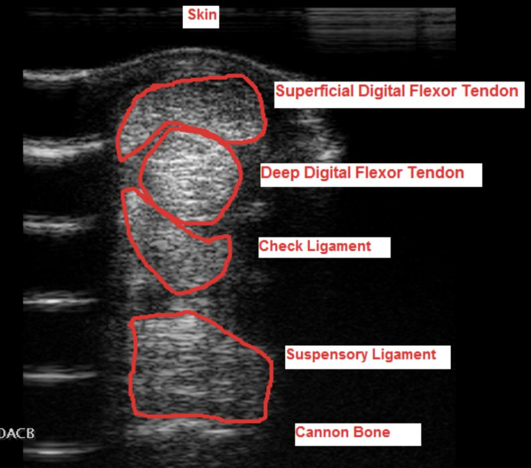
Other Growth Factors
Insulin-like growth factor – IGF
Stem Cells
Usually obtained from Bone marrow
❌ Impractical, Zert doesn't like it
Laser Therapy
Can be used as part of treatment/rehabilitation.
🧬 TENDON REPAIR
A major problem with tendon healing:
Normal tendon → Injury → Scar formation → Healed tendon becomes stiffer → Functionally inferior to original tendon → Predisposed to reinjury
⭐ Important concept: A healed tendon is not the same as the original tendon!
🕸 SCAFFOLD
Example: Urinary bladder matrix
Basically a graft/scaffold placed on/in the damaged tendon.
Think of it as: Skeleton/framework/template for healing cells
Normal scar tissue has: Less elasticity
Scaffold aims to: Improve organization/quality of repaired tissue.
🔪 TENDON SURGERY
1⃣ Proximal Check Ligament Desmotomy (Desmotomy = cutting a ligament)
Scarred tendon → Tendon becomes stiff / less elastic → Cut proximal accessory/check ligament → Allows more movement/extension → Compensates for reduced tendon elasticity
🧠 Cut check ligament → give tendon more freedom
2⃣ Annular Ligament Desmotomy
➡ Transection of proximal or distal annular ligament
Why?
Scar formation → Tendon becomes thick → Annular ligament restricts tendon expansion/movement → Cut annular ligament → More space for tendon
3⃣ Fasciotomy
➡ Cutting/opening fascia to reduce restriction and improve healing.
Locations:
Forelimb ➡ Carpal region
Hindlimb ➡ Plantar tarsal region
5⃣ TENDON RUPTURE / TRANSECTION
Traumatic rupture can involve the suspensory apparatus, with or without fractures of both proximal sesamoid bones.
➡ Loss of support of fetlock.
Common:
🏇 Racing injury
Often:
➡ Not completely ruptured
➡ Some connection remains between proximal and distal portions.
🔍 Etiology
Racing injury, Excessive weight bearing/loading, Kicks, Wire cuts
🩺 Clinical Signs
Severe acute lameness after racing/exercise, Severe pain
Abnormal limb position depending on structure damaged
⭐ The position of the limb can tell you which tendon is ruptured.
💥 SDFT RUPTURE
➡ Hyperextension of fetlock
➡ Heel drop
Why?
SDFT normally helps support the fetlock.
Rupture → Less support → Fetlock extends excessively → Heel drops
🧠 SDFT → Sinking heel
💥 DDFT RUPTURE
➡ Overextension
➡ Elevation of toe
DDFT normally flexes/supports distal digit.
Rupture → Loss of flexor pull on P3 → Toe becomes elevated
🧠 DDFT → toe UP
💥 SUSPENSORY LIGAMENT RUPTURE
Overextension of fetlock + Subluxation of pastern region
Because:
⭐ Suspensory ligament is a major anti-hyperextension structure.
🚨 WHOLE SUSPENSORY APPARATUS RUPTURE
Catastrophic.
➡ Complete loss of fetlock support
Horse may:
⚠ Step on the palmar/plantar surface of the fetlock
The limb appears essentially “broken down.”
➡ Very poor prognosis
➡ Euthanasia often indicated
💥 EXTENSOR TENDON RUPTURE
➡ Dorsal knuckling of fetlock
Why?
Loss of extensor function → Horse cannot adequately oppose flexion → Knuckling
⭐ Usually less catastrophic than flexor/suspensory rupture.
💥 PERONEUS TERTIUS RUPTURE
➡ Stifle joint is flexed
This structure is part of the hindlimb reciprocal apparatus.
🧠 RUPTURE = LOOK AT THE LIMB POSITION
Ruptured structure | Typical appearance |
|---|---|
SDFT | Hyperextended fetlock + heel drop |
DDFT | Overextension + toe elevation |
Suspensory ligament | Hyperextension + pastern subluxation |
Whole suspensory apparatus | Fetlock collapses → horse may step on palmar surface |
Extensor tendon | Dorsal knuckling |
Peroneus tertius | Abnormal reciprocal apparatus; stifle-related abnormality |
🔎 DIAGNOSIS OF RUPTURE
Palpation
Look for:
Swelling, Heat, Pain, Loss of normal tendon continuity
Always Compare with contralateral limb
🩻 X-ray
Minimum 4 views:
Dorsopalmar
Lateromedial
Oblique
Opposite oblique
Important to identify:
➡ Proximal sesamoid bone fractures
💊 TREATMENT OF RUPTURE
Depends on severity.
If NOT Completely Ruptured:
Conservative
Immediate stabilization to prevent further damage:
Cast + splint
Fixation/support bandage
Wound care
Goal:
➡ Prevent continued hyperextension
➡ Protect remaining tendon fibers
🔪 Debridement ➡ Remove damaged/devitalized tissue
BUT:
⭐ Save as much remaining viable tendon as possible
🚫 Do not suture tendon
🦴 Surgical – Severe Suspensory Apparatus Injury
If the whole suspensory apparatus is disrupted:
➡ Arthrodesis
Arthrodesis = Surgical immobilization of a joint by fusion of adjacent bones
Joint surfaces → Surgically fused → No joint movement → Provides structural stability
⚠ Expensive.
⚠ EUTHANASIA
Indicated in catastrophic injuries where functional support cannot realistically be restored.
Especially:
➡ Complete catastrophic suspensory apparatus failure.
⭐ EXAM ESSENTIALS
Tendon
➡ Muscle → bone → movement
Ligament
➡ Bone → bone → stability
Most common tendon injured in racehorses
➡ ⭐ SDFT
Most common location
➡ Palmar/plantar distal limb
Primary lesion
➡ Central rupture of tendon fibers + hemorrhage + edema
Best diagnostic method
➡ ⭐ USG → central core lesion
Healing
➡ Long, up to 18 months
➡ Healed tendon is stiffer and functionally inferior
➡ High reinjury risk
Zert's favorite regenerative treatment
➡ ⭐ PRP under USG guidance
Proximal check ligament desmotomy
➡ Increases functional elasticity/movement of scarred tendon unit
Complete suspensory apparatus rupture
➡ Catastrophic loss of fetlock support → arthrodesis or euthanasia
Developmental Anomalies of Tendons
There are 2 main types:
1⃣ FLEXURAL LAXITY
➡ Tendons/supporting structures are too long / weak / flaccid
➡ Joint hyperextends
2⃣ FLEXURAL DEFORMITY
➡ Flexor structures are too short / contracted
➡ Joint remains flexed / knuckles
🧠 MOST IMPORTANT DIFFERENCE
LAXITY | FLEXURAL DEFORMITY | |
|---|---|---|
Tendon | Too long / weak | Too short / tight |
Joint | Hyperextension | Flexion |
Limb appearance | Drops backward/down | Knuckles forward |
Think | ⬇ LOOSE | ↪ TIGHT |
🧠 LAX = loose and long
🧠 FLEXURAL = flexed because tendon is short
1⃣ FLEXURAL LAXITY= Weak / Flaccid Tendons
Laxity = deformity where joints are hyperextended because of loss/weakness of supporting structures.
Weak/long flexor structures
⬇
Cannot adequately support joints
⬇
Joint hyperextends
⬇
Fetlock drops toward ground
🔍 Etiology
Quite frequent in premature foals.
Causes:
Prematurity ⭐, Intrauterine infection, Infection/disease of mare
Older horses:
Can occur due to:
Previous injuries/diseases, Nutritional deficiencies, Lack of exercise, Normal aging
🩺 Clinical Signs
General
➡ Overextension / hyperextension
DDFT laxity
➡ Toe elevation
Because the DDFT is not providing normal flexor tension.
Severe laxity
Fetlock drops further and further:
Normal
⬇
Hyperextension
⬇
Fetlock approaches ground
⬇
Foal may walk on palmar/plantar surface of phalanges
This causes:
Skin abrasions, Pastern trauma, Fetlock trauma
🔎 Diagnosis
X-ray ⭐
Especially used to check:
➡ Skeletal maturity
Premature foal may show:
Insufficient mineralization, Delayed long-bone development

💊 Treatment
Most cases are treated conservatively.
Box stay/rest
Moderate controlled exercise
Light bandaging
👞 Shoeing
If the foal has difficulty standing:
➡ Heel extensions
Purpose:
Heel extension → Provides more support → Allows full contact between solear surface and ground
🚫 Usually Contraindicated
Cast
Surgery
Because many cases improve naturally as the foal becomes stronger.
Severe insufficient mineralization ➡ Euthanasia may be necessary.
🛡 Prevention
Foals
➡ Proper care of pregnant mare
➡ Prevent premature delivery
⚠ A severely premature/immature foal should not be encouraged to stand prematurely, particularly if skeletal mineralization is inadequate.
Older Horses
Treat tendon injuries promptly, Good nutrition, Sufficient exercise
2⃣ FLEXURAL DEFORMITY= Tendon/soft-tissue shortening
Flexural deformity = limb deviates from normal vertical alignment because structures on the flexor side are too short/tight or because of abnormalities of the extensor apparatus.
Can be congential or acquired
🐣 CONGENITAL FLEXURAL DEFORMITIES
Two important forms:
1. Shortening of flexor tendons
➡ Digital and/or carpal joints remain flexed
2. Elongation/rupture of common digital extensor tendon
➡ Cannot adequately extend digit
3⃣ CONGENITAL FLEXOR TENDON SHORTENING
Mainly affects:
➡ Forelimbs
Flexor tendons are too short → Pull joints into flexion → Foal cannot straighten limbs
🔍 Etiology
Often unknown.
Possible:
Infection of mare, Intrauterine malpositioning, Other developmental factors
🩺 Clinical Signs
Unable to stand normally
Unable to nurse if severe
Flexion of carpal joints
Contracted appearance
🔎 Diagnosis
Clinical signs
X-ray
💊 Treatment
Conservative
Stretch limbs
Bandages
Cast
💉 Oxytetracycline ⭐
According to your course notes:
➡ Oxytetracycline binds calcium
➡ Produces relaxation/lengthening of contracted flexor structures
Used in:
⭐ Congenital flexural deformity
🔪 Surgery
Severe/persistent cases ➡ Surgical release/cutting of contracted tendon or accessory ligament depending on the structure involved.
4⃣ RUPTURE / ELONGATION OF COMMON DIGITAL EXTENSOR TENDON
Occasionally seen in foals.
Usually affects:
➡ Dorsal extensor tendon
➡ Mainly around carpal region
Tendon is:
Elongated or Ruptured
🔍 Etiology
Often unknown.
Can be associated with Large foals
⚙ What Happens?
Extensor normally helps prevent excessive flexion.
Extensor elongated/ruptured
⬇
Cannot extend digit properly
⬇
Digit flexes excessively
⬇
🐴 Over-knuckling
🩺 Clinical Signs
Inability to extend digit
Difficulty stepping normally on hoof
Over-knuckling
Swelling of tendon sheath
🔎 Diagnosis
Clinical signs
⭐ USG
💊 Treatment
Cast / splint for prolonged support
Controlled standing
➡ Standing helps improve extensor function.
Prognosis
⭐ Good
🚫 IMPORTANT
Oxytetracycline is CONTRAINDICATED!
Why remember this?
The problem is already:
Extensor tendon = too long / elongated
You do NOT want treatment aimed at further relaxing/lengthening soft tissues.
🧠
Short flexors → oxytetracycline ✅
Elongated extensor → oxytetracycline ❌
🌱 ACQUIRED FLEXURAL DEFORMITIES
Two major forms:
EARLY – suckling foal
➡ Club foot
LATE – weaned foal
➡ Flexural deformity of metacarpophalangeal joint
5⃣ CLUB FOOT
Early – Suckling Foal
Also called:
🩰 Ballerina conformation
The foal walks on its toe tip.
Main problem:
⭐ DDFT is functionally too short/tight relative to the bones
⚙ Pathogenesis
Rapid bone growth
+
DDFT doesn't lengthen enough
⬇
DDFT becomes relatively too short
⬇
Pulls strongly on P3
⬇
Changes/rotates coffin-bone orientation
⬇
Heel becomes high
⬇
Foal stands on toe
🩰 = BALLERINA
🔍 Etiology
Overfeeding
Excess energy, Excess protein
Bone grows faster than tendon
Mineral imbalance:
P, Ca
Sudden changes in feed intake
Secondary hyperparathyroidism
Exercise
Trauma, Overload, Deep bedding, “Grass hoof”
🌱 What is “Grass Hoof”?
Foal repeatedly stands in the same position while grazing.
For example:
➡ Same limb repeatedly placed forward/backward
⬇
Unequal loading/growth
⬇
Can contribute to abnormal hoof conformation.
🩺 Clinical Signs – Club Foot
High heels
Stands on toes
Sole does not contact ground normally
Over-knuckling
Upright/steep hoof
🧠 Club foot = HIGH HEEL + TOE TIP
🔎 Diagnosis
Clinical appearance
X-ray
Sole elevation
💊 Treatment
👞 Hoof Correction / Shoeing
Plastic shoe
Instant shoe
Dallmer shoe
Used approximately:
➡ 2–3 months
Purpose:
➡ Improve hoof-ground relationship/support during correction.

🔪 Surgical Treatment
Accessory Ligament of DDFT Desmotomy ⭐
➡ Transect accessory/check ligament of DDFT
Elongation Tenotomy
Can be performed:
Middle metacarpal region
and/orTendon sheath region
➡ Used to release severe contracture.
🛡 Prevention of Club Foot
Remove from pasture temporarily if needed
Reduce excessive energy intake
Reduce excessive protein intake
Correct mineral balance
Harder ground
Correct hoof trimming
6⃣ LATE FLEXURAL DEFORMITY
Metacarpophalangeal / Fetlock Joint
Occurs in older foals after weaning.
Associated with shortening/contracture of multiple supporting structures:
Suspensory ligament
Distal sesamoid ligaments
DDFT
Accessory ligament of DDFT
SDFT
➡ Essentially the whole supporting/flexor apparatus becomes too tight/short.
🔍 Etiology
Poor ossification
Overfeeding
Sudden nutritional changes
Mineral imbalance
Overload
Trauma
🩺 Clinical Signs
Steep fetlock conformation
Normally fetlock/pastern:
➡ approximately 135° according to your notes
With deformity:
➡ Becomes very steep / almost straight
Also:
➡ Over-knuckling
🔎 Diagnosis
Primarily:
➡ Clinical signs

💊 Treatment
👞 Heel Support
➡ Elevate heel with wedge
Heel elevation
⬇
Changes tension/loading of DDFT
⬇
Helps correction/elongation of flexor unit
🔪 Surgery
Transect accessory ligaments of:
DDFT
SDFT
➡ Reduces restriction from contracted flexor structures.
🧠 THE WHOLE QUESTION IN ONE PICTURE
                    TENDON ANOMALIES
                          │
             ┌────────────┴────────────┐
             │                         │
          LAXITY                 FLEXURAL DEFORMITY
        TOO LONG                     TOO SHORT
        / WEAK                        / TIGHT
             │                         │
             ↓                         ↓
       HYPEREXTENSION               FLEXION
             │                      / KNUCKLING
             │
       Fetlock DOWN
       Toe may UP
                               ┌─────────┴─────────┐
                               │                   │
                          CONGENITAL            ACQUIRED
                               │                   │
                       Short flexors       ┌───────┴───────┐
                       → can't stand       │               │
                       → oxytetra        EARLY           LATE
                                         │               │
                                      CLUB FOOT      FETLOCK
                                         │          DEFORMITY
                                      DDFT short     whole apparatus
                                         │            tight
                                      🩰 TOE         knuckling
🧠 SUPER-QUICK DIFFERENTIATION ⭐⭐⭐
Condition | Main problem | Appearance |
|---|---|---|
Flexural laxity | Flexors weak/long | ⬇️ Fetlock hyperextends |
Congenital flexor shortening | Flexors too short | ↪ Limb remains flexed |
Extensor elongation/rupture | Extensor too long/damaged | ↪ Knuckling |
Club foot | DDFT relatively too short | 🩰 High heel + toe-tip |
Late fetlock deformity | Supporting/flexor apparatus too short | ↪ Steep fetlock + knuckling |
⭐ EXAM ESSENTIALS
Two types of developmental tendon anomalies:
1. Laxity = LENGTHENING
➡ Weak/long structures
➡ Hyperextension
➡ Severe = fetlock on ground
2. Flexural deformity = SHORTENING
➡ Tight/short structures
➡ Flexion/knuckling
Premature foal + fetlock dropping down
➡ Flexural laxity
Newborn + flexed limbs + cannot stand
➡ Congenital flexural deformity
➡ Stretch/bandage/cast
➡ Oxytetracycline
Newborn + extensor rupture
➡ Knuckling
➡ Cast/splint
➡ ❌ NO oxytetracycline
➡ Good prognosis
Suckling foal + high heel + walks on toe
➡ Club foot
➡ DDFT too short
➡ 🩰 Ballerina conformation
Weaned foal + very steep/straight fetlock
➡ Late flexural deformity
➡ Multiple flexor/suspensory structures contracted
Osteoarthritis
Osteoarthritis (OA) = progressive destruction of articular cartilage, accompanied by changes in:
🦴 Bone
Osteophyte formation, Degeneration/remodeling
Soft tissues
Capsular fibrosis
⭐ Very frequent problem in horses, especially in high-motion joints.
1⃣ CLASSIFICATION
PRIMARY OA
Develops mainly due to:
➡ Chronic repetitive trauma
➡ Repetitive overloading
Risk factors depend on:
Breed, Age, Sex, Conformation
Think:
🏇 Repeated loading → Small cartilage damage → More loading → Progressive OA
SECONDARY OA
OA develops as a consequence of another joint disease.
Examples:
Traumatic arthritis
Articular fracture
Osteochondrosis
Septic arthritis
🧠 Difference
PRIMARY
➡ Repetitive wear/overload itself starts OA
SECONDARY
➡ Another joint disease starts OA
2⃣ PATHOGENESIS ⭐⭐⭐
OA involves BOTH:
🔨 Mechanical destruction
🔥 Chemical/inflammatory destruction
These processes reinforce each other.
🔨 MECHANICAL DAMAGE:
Repeated abnormal loading/instability → cartilage damaged → Cartilage becomes less able to tolerate loading → More mechanical damage
🔥 CHEMICAL / INFLAMMATORY DAMAGE
Damaged joint tissues activate inflammatory pathways.
Important substances include:
Cytokines (TNF, Interleukins) ➡ Promote synovitis and cartilage catabolism.
Enzymes (Collagenase, Lysosomal enzymes, Proteoglycan-degrading enzymes) ➡ Break down components of cartilage matrix.
Inflammatory Mediators (Prostaglandins, Leukotrienes, Histamine, Nitric oxide
Other Factors: Free radicals, Antibodies
🔄 OA VICIOUS CYCLE ⭐⭐ ⭐
This is the main sequence to understand:
Mechanical injury / overload
⬇
💥 Cartilage damage
⬇
🔥 Inflammatory mediators released
⬇
Synovitis + capsulitis
⬇
Enzymes/cytokines damage cartilage
⬇
Loss of proteoglycans ⭐
⬇
Cartilage becomes soft and weak
⬇
More susceptible to mechanical damage
⬇
💥 MORE CARTILAGE DAMAGE
⬇
🦴 Subchondral bone changes
⬇
OA progresses
🔄 And the cycle continues.
🧠 WHY ARE PROTEOGLYCANS IMPORTANT?
Proteoglycans help cartilage:
💧 Retain water + Resist compression
OA
⬇
Proteoglycans lost
⬇
Cartilage becomes soft
⬇
Cannot tolerate normal forces
⬇
More mechanical damage
⭐ Initial important change = breakdown/loss of proteoglycans!!
3⃣ PATHOLOGY
OA affects:
BONE + CARTILAGE + SOFT TISSUES
🦴 BONE
Changes include:
Osteophytes, Remodeling, Subchondral sclerosis, Subchondral lysis, Fractures
Osteophyte = new bone formation around joint margins (“Bone spurs”)
CARTILAGE
Normal cartilage:
➡ Smooth and Firm
OA cartilage becomes:
Soft, Yellow, Surface fibrillation, Eroded, Disrupted/lost
🧵 What is Fibrillation?
Normal cartilage:
──────── smooth
OA:
≋≋≋≋ frayed/rough
➡ Surface starts splitting into small fibers/fissures.
🧱 SOFT TISSUES / SYNOVIUM
Congestion
Thickening
Mononuclear inflammatory infiltration
Villous hypertrophy
Capsular edema
Eventually capsular fibrosis
4⃣ CLINICAL SIGNS
Can include:
Lameness, Pain on palpation, Positive joint flexion test
Joint thickening, Reduced range of movement
Crepitus
⚠ Many horses can be relatively asymptomatic.
➡ Radiographic OA does not necessarily mean the horse is obviously painful.
5⃣ DIAGNOSIS
💉 Local Analgesia ⭐
Local/intra-articular diagnostic analgesia can be used to identify the painful region/joint.
Block joint
⬇
Lameness disappears/improves
⬇
➡ Joint is implicated as pain source
🩻 RADIOGRAPHY
Can show:
Osteophytes, Lysis
Narrowed joint space, Subchondral bone changes/remodeling
⚠ Important:
X-ray changes occur relatively LATE.
So:
Early OA
➡ Cartilage damage may already exist but X-ray may still look relatively normal.

☢ SCINTIGRAPHY
➡ Increased radionuclide uptake
Indicates:
➡ Increased bone turnover/activity
🧲 MRI
Good for detecting:
Subchondral bone abnormalities
Cartilage abnormalities
Soft-tissue abnormalities
📷 ARTHROSCOPY
Allows direct visualization inside joint.
Can identify:
Cartilage softening, Fibrillation
Cartilage loss, Osteophytosis / osteophytes
⭐ Advantage:
➡ Actually see the articular cartilage directly.
6⃣ TREATMENT
⚠ Important principle:
Articular cartilage has extremely limited regenerative capacity.
➡ Once significantly damaged, it does not simply regenerate back to normal cartilage.
Therefore treatment aims to:
Reduce inflammation
Reduce pain
Slow further destruction
Improve joint environment
Correct instability/loading
Maintain function
🟢 CONSERVATIVE TREATMENT
Light/controlled exercise
Correct overweight
Pain management
Minimize micro-instability of joint
🧠 Complete inactivity isn't necessarily the goal:
Controlled movement → maintain joint function
💉 INTRA-ARTICULAR MEDICATION
1⃣ CORTICOSTEROIDS
Strong anti-inflammatory drugs.
Used when Joint is inflamed
But according to Zert:
⚠ Not always ideal because some inflammation is part of normal healing.
Avoid/inappropriate especially when there are:
Severe lesions or Subchondral fractures
💊 Methylprednisolone Acetate
Has relatively large crystals and can be more damaging to cartilage.
➡ Therefore, use mainly in LOW-MOTION JOINTS.
Examples:
Pastern, Tarsal joints
🧠 Methylpred = LOW motion
💊 Betamethasone + Triamcinolone
Used in HIGH-MOTION JOINTS.
Examples: Coffin, Carpal, Fetlock
🧠 Beta/Triam = HIGH motion
💧 2⃣ HYALURONIC ACID – HA ⭐
Important component of normal synovial fluid/cartilage environment.
Typically used:
➡ Together with corticosteroids in mild synovitis
Can also be used:
IV after arthroscopy
Prophylactically
⭐ Zert believes in this
💉 3⃣ POLYSULFATED GLYCOSAMINOGLYCANS – PSGAGs
⭐ First choice in severe chronic arthritis
Aim → Support/protect cartilage matrix and joint environment.
🩸 4⃣ PLATELET-RICH PLASMA – PRP
⭐ Very good
Contains platelets and growth factors.
➡ Used to modify inflammation and promote a more favorable healing environment.
🧬 5⃣ IRAP (Interleukin-1 Receptor Antagonist Protein)
IL-1: binds receptor on synovial/joint cells → promotes inflammation and cartilage destruction
IRAP blocks IL-1 receptor → IL-1 cannot exert as much effect → Less inflammatory signaling
🔪 6⃣ SURGICAL ARTHRODESIS
Arthrodesis = surgical fusion of a joint
Joint surfaces are fused → Joint no longer moves → Painful movement eliminated / stability restored
Used in selected cases such as:
Intra-articular fractures, OCD, Ligament instability
Most useful where the loss of motion can be tolerated!
🏊 7⃣ PHYSICAL THERAPY
Examples:
Swimming, Underwater treadmill
Shock-wave therapy
Purpose:
➡ Maintain controlled movement/function while limiting excessive joint loading.
🧠 OA TREATMENT OVERVIEW
                    OSTEOARTHRITIS
                         │
        ┌────────────────┼────────────────┐
        │                │                │
   CONSERVATIVE     MEDICATION         SURGERY
        │                │                │
 Light exercise      Steroids         Arthrodesis
 Weight control         HA
 Pain control         PSGAG
 Stability             PRP
                       IRAP
        │
        ↓
 PHYSIOTHERAPY
 Swimming
 Underwater treadmill
 Shock wave
🧠 QUICK DIFFERENTIATION OF DRUGS
Treatment | Remember |
|---|---|
Methylprednisolone | Corticosteroid → LOW-motion joints |
Betamethasone | Corticosteroid → HIGH-motion joints |
Triamcinolone | Corticosteroid → HIGH-motion joints |
Hyaluronic acid | Mild synovitis, often + corticosteroid ⭐ Zert likes |
PSGAG | Severe chronic arthritis |
PRP | Growth factors / biologic treatment |
IRAP | 🚫 Blocks IL-1 receptor |
🧠 OA IN ONE SEQUENCE ⭐⭐⭐
OVERLOAD / JOINT DISEASE
⬇
💥 CARTILAGE DAMAGE
⬇
🔥 INFLAMMATION
⬇
TNF + IL + prostaglandins + enzymes
⬇
PROTEOGLYCAN LOSS
⬇
🟡 CARTILAGE SOFTENS
⬇
FIBRILLATION + EROSION
⬇
🦴 SUBCHONDRAL BONE CHANGES
⬇
🦴 OSTEOPHYTES
⬇
PROGRESSIVE OA
🔄 Mechanical damage and inflammation continue to promote each other.
⭐ EXAM ESSENTIALS
Definition
OA = progressive cartilage destruction + bone and soft-tissue changes.
Primary OA
➡ Chronic repetitive trauma / overload
Secondary OA
➡ Consequence of another joint disease
Pathogenesis
➡ Mechanical + inflammatory/chemical destruction
Important early cartilage change
➡ Loss of proteoglycans → cartilage softens
Bone
➡ Osteophytes + remodeling + sclerosis/lysis
Cartilage
➡ Soft → fibrillation → erosion → loss
Clinical signs
➡ Lameness + reduced ROM + joint thickening + crepitus
⚠ But OA can be asymptomatic.
Diagnosis
💉 Local analgesia → localize pain
🩻 X-ray → relatively late changes
🧲 MRI → cartilage/subchondral bone
📷 Arthroscopy → directly visualize cartilage
Treatment
➡ Controlled exercise + weight management + pain control
➡ IA medication / biologics
➡ Correct instability
➡ Arthrodesis in selected cases
Developmental Anomalies of Joints
Joint disorders can involve:
Synovial membrane, Surrounding tendons, Subchondral bone, Articular cartilage, Bursae, Synovial fluid
Main conditions:
1⃣ Septic arthritis – Joint ill in foals
2⃣ Osteochondrosis dissecans – OCD
3⃣ Subchondral bone cysts
4⃣ Spavin
1⃣ 🦠 SEPTIC ARTHRITIS/Joint ill
Septic arthritis = infection and inflammation of a synovial joint.
🚨 EMERGENCY
Can rapidly cause:
➡ Cartilage destruction, Secondary osteoarthritis, Permanent joint damage
Particularly important in foals.
🔍 Etiology
Main cause:
🦠 Bacterial infection
Important bacteria:
Coliforms, Staphylococcus aureus, Streptococcus, Salmonella, Rhodococcus, Actinobacillus
🐴 ADULT HORSES
In adults, infection usually enters the joint directly.
Common causes:
💥 Penetrating wound into joint, or extension from nearby infection
Examples:
Kick wound near hock, Kick wound near elbow
Can also be:
💉 Iatrogenic, following
Arthrocentesis
Intra-articular injection
Surgery
FOALS ⭐⭐⭐
In foals, infection commonly reaches joints through:
🩸 HEMATOGENOUS SPREAD
Bacteria enter bloodstream → Travel through circulation → Reach synovial structures → Septic arthritis
Therefore:
➡ Multiple joints may be involved (septic polyarthritis)
🚪 Where Does Infection Start in Foals?
Possible primary sources:
Umbilicus / urachus → Blood → Joint
Pneumonia
Enteritis
Maternal placentitis ➡ Infection associated with pregnancy/perinatal period
⭐ Insufficient colostrum: Failure of passive transfer → Insufficient maternal antibodies → Foal highly susceptible to septicemia → Bacteria spread hematogenously → Joint ill
🩺 Clinical Signs
Local signs: Severe lameness, Joint swelling, Heat, Pain, Joint effusion
Systemic signs may include: 🌡 Pyrexia/fever
Especially in foals:
➡ May have systemic septicemia.
🔎 DIAGNOSIS
1.💉 SYNOVIAL FLUID ANALYSIS ⭐⭐⭐
One of the most important diagnostic methods.
Normal synovial fluid:
➡ Clear
➡ Viscous
➡ Low cellularity
Septic joint:
↑ Volume
Turbid/cloudy
Clots
↓ Viscosity
↑ Cell count
Leukocytosis / high neutrophils
↑ Protein
🧠 Memory: SEPTIC FLUID = CLOUDY + WATERY + CELLS + PROTEIN
2.🧫 Microbiology
Culture synovial fluid.
⚠ Approximately 50% may be culture-negative
Possible reason:
➡ Neutrophils may have killed/removed bacteria
➡ Previous antibiotic treatment may also reduce recovery
So:
❌ Negative culture does NOT necessarily exclude septic arthritis.
3.🩻 X-RAY
⚠ Almost useless EARLY
Significant radiographic bone changes may not appear until approximately ➡ 7–10 days after infection
But septic joint is an emergency. 🚨 You cannot wait 7–10 days.
4.📡 ULTRASOUND
Useful.
Normal synovial fluid:
➡ Anechoic / black
Septic joint:
➡ Fibrin/clots/debris can become echogenic/hyperechoic
5.📷 ARTHROSCOPY ⭐
Very useful for:
Diagnosis (➡ Direct visualization of joint)
AND
Treatment ➡ Lavage, Remove fibrin/debris, Evaluate cartilage
⭐ Diagnosis + treatment at the same time
💊 TREATMENT – SEPTIC ARTHRITIS
🚨 Treat aggressively and immediately.
Main goals:
REMOVE INFECTION + REMOVE INFLAMMATORY MATERIAL + ANTIBIOTICS
💦 1. Joint Drainage & Lavage ⭐⭐⭐
Use several liters of sterile fluid.
Example:
➡ Hartmann's solution
Often performed during:
➡ Arthroscopy
Purpose: Bacteria + Inflammatory mediators + Fibrin + Debris → 💦 WASH THEM OUT
💉 2. Antibiotics
Prolonged and intensive treatment.
Can be:
Systemic, Intra-articular
Common aminoglycosides:
Gentamicin, Amikacin
Can be combined with: ➡ Procaine benzylpenicillin IM
💊 3. NSAIDs
Example:
➡ Flunixin
Purpose:
Reduce inflammation, Reduce pain
🔪 4. Synovectomy / Debridement
➡ Remove fibrin clots and abnormal synovial material.
🧠 SEPTIC ARTHRITIS IN ONE LINE
BACTERIA → JOINT → SYNOVITIS → CARTILAGE DAMAGE → OA
Treatment:
💦 LAVAGE + ANTIBIOTICS + NSAIDs + REMOVE FIBRIN/DEBRIS
2⃣ OSTEOCHONDROSIS – OC
First, understand the difference:
Osteochondrosis = underlying developmental disorder
OCD = Osteochondrosis dissecans ➡ Clinical manifestation where abnormal cartilage develops fissures/flaps/fragments at the articular surface.
🦴 NORMAL ENDOCHONDRAL OSSIFICATION
Growing foal:
Cartilage → matures → is replaced by → BONE
This is:
⭐ Endochondral ossification
❌ OSTEOCHONDROSIS = disturbance of endochondral ossification
Parts of:
Physeal growth cartilage
Articular-epiphyseal cartilage
fail to convert normally into bone.
This can contribute to:
Angular deformities, Valgus deformities, Osteochondrosis dissecans – OCD
3⃣ OSTEOCHONDROSIS DISSECANS – OCD ⭐⭐⭐
OCD is the clinically important articular form of osteochondrosis.
➡ Abnormal cartilage remains at the joint surface
➡ May fissure
➡ May form a flap
➡ May become an osteochondral fragment
🔍 Etiology
⭐ Multifactorial
Important factors:
Rapid Growth: Growth spurts followed by growth setbacks.
Mineral Imbalance: High phosphorus, Copper deficiency, High zinc
Mechanical Trauma: Site-specific loading/trauma.
Hormonal Factors: Growth hormone, Insulin, Thyroid hormones, Sex hormones ➡ More common in males
⚙ PATHOGENESIS OF OCD ⭐⭐⭐
This is the important sequence:
Normal cartilage should be converted into bone
BUT:
❌ Endochondral ossification fails → Deep cartilage is not converted to bone → Cartilage becomes abnormally thick → Deep chondrocytes become farther from nutrition → Poor nutrition to deep cartilage → Chondrocytes die → Cartilage necrosis → Fissures form → Fissure extends toward articular surface → CARTILAGE FLAP → May partially ossify → OSTEOCHONDRAL FRAGMENT
AGE
Clinical signs/lesions arise from:
➡️ Birth to approximately 2–3 years
Epiphyseal osseous development is not complete until approximately:
➡ 9–10 months
Therefore in young horses:
⭐ Some early lesions may resolve spontaneously during development.
🩺 Clinical Signs – OCD
Lameness, Stiffness
Positive flexion test
Joint effusion
Joint effusion is especially noticeable in:
Stifle, Hock
🔎 Diagnosis
🩻 X-ray ⭐
➡ Subchondral defects
➡ Osteochondral fragments
📷 Arthroscopy
➡ Direct visualization
Also:
MRI, CT
💊 Treatment – OCD
Conservative
For selected young/mild lesions:
Rest, Correct dietary intake, Correct mineral/nutritional imbalance
🔪 Surgical
➡ Arthroscopic debridement
Remove:
Separated osteochondral fragments, Abnormal cartilage, Abnormal subchondral bone
🩸 PRP
➡ Platelet-rich plasma. Used to improve the healing environment.
💧 Hyaluronic Acid
⭐ Best. Used to improve joint environment and manage synovitis.
4⃣ SUBCHONDRAL BONE CYSTS = Osseous Cyst-Like Lesions
Cyst-like lesions occur in the subchondral bone, directly beneath articular cartilage.
Reported locations include:
Medial femorotibial joint (medial femoral condyle of stifle joint)⭐, Carpus, Pastern, Coffin joint, Fetlock, Elbow, Shoulder, Hock
🔍 Etiology + Pathogenesis
Can be associated with:
Osteochondrosis
➡ Abnormal cartilage/endochondral ossification persists as localized defect within subchondral bone
OR
Trauma
➡ Localized trauma to:
Articular cartilage, Underlying subchondral bone
→ Cyst-like lesion develops (fluid filled cavity in bone just beneath articular cartilage)
🩺 Clinical Signs
Lameness localized to joint, Joint effusion
🔎 Diagnosis
Flexion test
🩻 X-ray, CT, MRI

💊 Treatment
Conservative: Rest
Surgical / Interventional:
Corticosteroid injection into cyst
Debridement
Grafts
Cartilage resurfacing
5⃣ SPAVIN
Degenerative, Non-Septic Arthritis
⭐ Seen in LOW-MOTION JOINTS
Frequently affects:
➡ Distal tarsal/hock joints
Mostly seen in:
➡ Adult horses
📌 What Is Spavin?
Spavin = chronic degenerative non-septic arthritis of low-motion hock joints.
Joint degeneration → Pain + inflammation → Progressive degeneration → Bone proliferation
⬇
Eventually:
🦴 ANKYLOSIS / FUSION

🔗 ANKYLOSIS
Ankylosis = pathological fusion/stiffening of a joint.
In spavin:
Joint gradually degenerates → Joint space disappears → Bones fuse → Joint no longer moves
Because these are low-motion joints, complete fusion may eventually reduce pain from movement.
🔍 Etiology
Overloading, Conformation problems
🩺 Clinical Signs
Pain, Lameness, Stiffness
💊 Treatment
Tildren IV ➡ Tiludronate
Purpose: Reduces osteolysis / excessive bone resorption
🧠 THE FOUR CONDITIONS – DON'T MIX THEM UP ⭐⭐⭐
Condition | Main problem | Typical patient | Key clue |
|---|---|---|---|
🦠 Septic arthritis | Bacterial joint infection | Foal or adult | Hot, swollen painful joint |
🦴 OCD | Failed endochondral ossification | Young horse | Thick cartilage → flap/fragment |
🕳 Subchondral cyst | Cyst-like lesion beneath cartilage | Young horse | Subchondral defect |
🦵 Spavin | Degenerative non-septic arthritis | Adult horse | Low-motion hock → eventual fusion |
⭐ EXAM ESSENTIALS
SEPTIC ARTHRITIS
➡ Bacterial infection = emergency
➡ Adults: penetrating wound/iatrogenic/local spread
➡ Foals: hematogenous, often due to failure of passive transfer/septicemia
➡ Multiple joints possible
➡ Synovial fluid: turbid, ↓ viscosity, ↑ cells, ↑ protein
➡ Culture can be negative
➡ X-ray changes delayed 7–10 days
➡ USG → fibrin/debris
➡ Arthroscopy = diagnosis + treatment
➡ Lavage + antibiotics + NSAID
OCD
➡ Failure of endochondral ossification
➡ Rapid growth + minerals + trauma + hormones
➡ Thick cartilage → necrosis → fissure → flap → fragment
➡ Young horses
➡ Some early lesions can resolve
➡ X-ray + arthroscopy
➡ Rest/diet or surgical debridement
SUBCHONDRAL CYST
➡ Lesion beneath cartilage
➡ OC or trauma
➡ Lameness + effusion
➡ X-ray/CT/MRI
➡ Rest or cyst treatment/debridement/grafts
SPAVIN
➡ Degenerative, non-septic arthritis
➡ Adult horse
➡ Low-motion tarsal joints
➡ Pain + lameness + stiffness
➡ Final result = ankylosis/fusion
➡ Tildren IV → ↓ osteolysis