physio LAB: skeletal system
 Call Kai
 Learn
Learn Practice Test
 Spaced Repetition
 Match
Match Flashcards
 Knowt Play
1/162

There's no tags or description
Looks like no tags are added yet.
Name | Mastery | Learn | Test | Matching | Spaced | Call with Kai | Chat |
|---|
No analytics yet
Send a link to your students to track their progress
163 Terms
206
The adult human skeleton is generally composed of how many bones
Coronal suture
a

Sagittal suture
b

Lambdoid suture
c

Frontal fontanelle
d

Occipital frontanelle
e
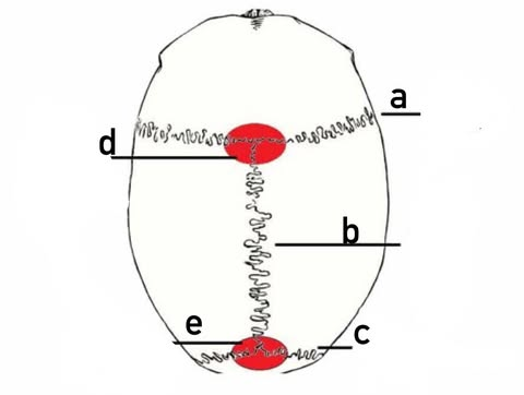
skull
protects the brain
thoracic cage
protects the heart and lungs.
Bones
act as levers
joints
serve as points of motion
skeletal muscle
provide the force for movement.
Red bone marrow
produces red blood cells, white blood cells, and platelets through haematopoiesis
haematopoiesis
Red bone marrow produces red blood cells, white blood cells, and platelets through ___
Yellow bone marrow
stores fat as an energy reserve.
fat
Yellow bone marrow stores __as an energy reserve.
axial, appendicular skeleton
what are the two major divisions of skeletal system
Axial skeleton
this division includes the Skull, vertebral column, ribs, and sternum
Axial skeleton
Forms the central axis of the body; primarily supports and protects the brain, spinal cord, heart, and lungs
Appendicular skeleton
this division icnldues the Pectoral girdles, upper limbs, pelvic girdles, and lower limbs
Appendicular skeleton
Attaches the limbs to the axial skeleton and enables movement and locomotion.
Anatomical planes
are imaginary flat surfaces used to describe sections of the body and the direction of movement.
Sagittal plane
Left and right portions
Sagittal plane
A vertical cut from front to back
midsagittal plane
creates equal left and right halves.
Frontal or coronal plane
Anterior (front) and posterior (back) portions
Frontal or coronal plane
A vertical cut from side to side.
Transverse plane
Superior (upper) and inferior (lower) portions
Transverse plane
A horizontal cut across the body
Oblique plane
Sections made at an angle
Oblique plane
A diagonal cut that is not parallel to the standard planes.
Skull
Protects the brain; supports and shapes the face; houses structures associated with vision, hearing, smell, taste, and breathing
Mandible
Forms the lower jaw, holds the lower teeth, and enables chewing and speaking
Cervical vertebrae
Support the head, protect the spinal cord, and allow neck movement.
Thoracic vertebrae
Articulate with the ribs, support the upper trunk, and protect the spinal cord.
Lumbar vertebrae
Support much of the body's weight and allow bending and twisting of the lower back.
Sacrum
Connects the vertebral column to the pelvis and transfers body weight to the pelvic girdle.
Coccyx
Provides attachment for pelvic muscles and ligaments and contributes to support while sitting.
Sternum (breastbone)
Anchors the ribs anteriorly and helps protect the heart and lungs
Ribs
Form the thoracic cage, protect the heart and lungs, and assist the mechanics of breathing
lordosis, kyphosis
what are the types or curvatures
Lordosis
refers to an inward curvature; the cervical and lumbar regions normally show this type of curves
Kyphosis
refers to an outward curvature; the thoracic and sacral regions normally show this type of curves.
True ribs
ribs 1-7 are called __
True ribs
Attach directly to the sternum through their own costal cartilages
False ribs
ribs 8-10 are called what
False ribs
Attach indirectly through the costal cartilage of the rib above
Floating ribs
ribs 11-12 are called what
Floating ribs
Have no anterior attachment to the sternum; they attach only to the vertebral column posteriorly, allowing greater flexibility.
Clavicle (collarbone)
Connects the upper limb to the trunk and helps maintain the position of the shoulder
Scapula (shoulder blade)
Provides broad attachment sites for shoulder muscles and contributes to arm movemen
Humerus
Forms the upper arm and contributes to the shoulder and elbow joints.
Radius
Forms the thumb-side of the forearm and participates in forearm rotation and wrist movement.
Ulna
Forms the little-finger side of the forearm and provides stability at the elbow.
Carpals
Form the wrist and permit flexible wrist movements.
Metacarpals
Form the palm and support hand movement and gripping
Hand phalanges
Form the fingers and allow grasping and fine movement
Pelvic girdle
Supports the trunk, transfers body weight to the lower limbs, and protects pelvic organs.
Femur
Forms the thigh, bears body weight, and enables movement at the hip and knee.
Patella
Protects the front of the knee and improves the leverage of the quadriceps during leg extension.
Tibia
Bears most of the lower leg's weight and contributes to the knee and ankle joints.
Fibula
Stabilises the lower leg and ankle and provides sites for muscle attachment.
Tarsals
Form the ankle and heel and help bear weight during standing and walking
Metatarsals
Form the middle of the foot and contribute to balance and movement.
Foot phalange
Form the toes and help maintain balance and push the body forward during walking
skull
is the bony framework of the head
skull
It protects the brain, supports and shapes the face, houses the organs of special sense, and provides attachment sites for muscles used in chewing, facial expression, and head movement
skull
The __is generally composed of 22 bones: 8 cranial bones and 14 facial bones.
22
The skull is generally composed of __bones: 8 cranial bones and 14 facial bones.
8
The skull is generally composed of 22 bones: __cranial bones and 14 facial bones.
14
The skull is generally composed of 22 bones: 8 cranial bones and __facial bones.
cranium
The __, or neurocranium, surrounds the brain, meninges, and associated blood vessels.
neurocranium
The cranium, or __, surrounds the brain, meninges, and associated blood vessels.
cranium
the roof of __is called the calvaria, while its base supports the brain and connects with the facial skeleton and vertebral column
calvaria
the roof of cranium is called the ___, while its base supports the brain and connects with the facial skeleton and vertebral column
Frontal
1; Forms the forehead, roof of the orbits, and anterior cranial cavity.
Parietal
2; Form most of the roof and upper sides of the skull
Temporal
2; Form the lower sides and part of the cranial base; house structures for hearing and balance.
Occipital
1; Forms the posterior skull and cranial base and contains the foramen magnum.
foramen magnum
occipital Forms the posterior skull and cranial base and contains the ___
Sphenoid
1; Forms part of the cranial base and orbits and articulates with many skull bones
Ethmoid
1; Contributes to the nasal cavity, nasal septum, and medial orbits; transmits olfactory nerve fibres
facial skeleton
The___, or viscerocranium, supports the soft tissues of the face and forms the orbits and the nasal and oral cavities.
viscerocranium
The facial skeleton, or ___, supports the soft tissues of the face and forms the orbits and the nasal and oral cavities.
Zygomatic
2; Form the cheekbones and part of the lateral walls and floors of the orbits.
Lacrimal
2; Form part of the medial orbital walls and contribute to the tear-drainage passage.
Nasal
2; Form the bony bridge of the nose
Inferior nasal conchae
2; Increase nasal surface area to help warm, humidify, and filter inhaled air.
Palatine
2; Form the posterior hard palate and portions of the nasal cavity and orbit.
Maxillae
2; Form the upper jaw, hold the upper teeth, and contribute to the hard palate, nose, and orbits.
Vomer
1; Forms the inferior and posterior portion of the nasal septum
Mandible
1; Forms the lower jaw and is the main movable bone of the skull.
Sutures
are strong, nearly immovable fibrous joints that connect the bones of the skull.
Coronal suture
type of suture the connects Frontal bone and the two parietal bones.
Sagittal suture
type of suture the connects Right and left parietal bones.
Lambdoid suture
type of suture the connects Occipital bone and the two parietal bones.
Fontanelles
Soft membranous spaces between some skull bones in infants.
Fontanelles
They permit moulding of the skull during birth and provide space for rapid brain growth.
landmark
may be a point, opening, projection, depression, or region used to describe anatomy and locate nearby structures.
Frontal eminence
Rounded prominence on each side of the frontal bone above the superciliary arch
Frontal eminence
contributes to the contour of the forehead and marks an early centre of ossification.