Anatomy 6511 Middle Mediastinum and Heart 낱말 카드
 Call Kai
 Learn
 Practice Test
 Spaced Repetition
 Match
 Flashcards
 Knowt Play
1/209

There's no tags or description
Looks like no tags are added yet.
210 Terms
Mediastinum
What term describes the central region between the right and left pulmonary cavities?

T1-T4
What spinal cord levels define the superior mediastinum?
T5-T12
What spinal cord levels define the inferior mediastinum?
Transverse plane passing through the sternal angle and the T4/T5 intervertebral disc
What defines the superior extent of the middle mediastinum?

Diaphragm
What defines the inferior extent of the middle mediastinum?

Pericardium, heart, phrenic nerves, pericardiophrenic vessels, and roots of the great vessels
What are the major contents of the middle mediastinum?
Pericardium
What term describes the fibroserous sac that encloses the heart and reflects at the root of the great vessels?
Fibrous pericardium
What is the superficial layer of the pericardium?

Serous pericardium
What is the deep layer of the pericardium?
Parietal pericardium
What is the outer layer of the serous pericardium?

Visceral pericardium
What is the inner layer of the serous pericardium?

Fibrous pericardium
Which layer of the pericardium is made of tough, inelastic connective tissue?
Central tendon of the diaphragm
What is the fibrous pericardium attached to?
Pericardium and heart
What does the fibrous pericardium control the position of?
Limits filling capacity of the heart (prevents overfilling)
What is the primary function of the fibrous pericardium?
Mesothelial cells
What type of cells line the serous pericardium that produce a lubricating fluid to reduce friction when the heart moves?
Creates a smooth, frictionless environment for the heart to pump
What is the primary function of the serous pericardium (mesothelial cells aid in this)?
Parietal serous and visceral serous pericardium
Between which two layers of the pericardium is the pericardial cavity?

Cardiac tamponade
What occurs when excess fluid/blood enters the pericardial cavity and restricts the volume of the heart?
Pericardiocentesis
What procedure is used to treat cardiac tamponade?
Below the xiphoid process (subxiphoid)
During a pericardiocentesis procedure, where is the needle inserted?
Reflections
What term describes the area of transition from parietal to visceral pericardium?

Around blood vessels entering and exiting the heart (great vessels of heart)
Where are reflections of the pericardium found around the heart?
Oblique sinus
What term describes the area of serous pericardium that is detached from the heart to allow for the entrance of venous vessels?
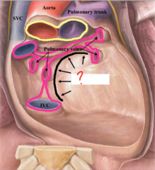
Between the line of reflection for veins and encircling arteries
Where is the transverse sinus (pericardial reflection) found?

Transverse sinus; oblique sinus
What sinus can you pass a finger through completely? Which sinus can you not and is a dead end?
Fibrous and parietal serous
Which layers of the pericardium are innervated by the phrenic nerve?

Fibrous and parietal serous
Which layers of the pericardium can transmit pain information?
Both innervated by phrenic nerve which is somatic so transmits somatosensory information
Why does the fiberous and parietal serous pericardium transmit pain information?
Visceral serous
Which layer of the pericardium is innervated by visceral afferent fibers from the cardiac plexus?
Left
Does the apex of the heart point to the right or left?
Apex of the heart
What is labeled here on the heart?

Left ventricle
Which chamber of the heart forms the apex?
Sternocostal (anterior), Left pulmonary, Right pulmonary, Diaphragmatic, Base
What are the external surfaces of the heart?

Sternocostal (anterior)
Which external surface of the heart is formed by right ventricle and right auricle?

Left pulmonary and diaphragmatic
Which external surfaces of the heart are formed by the left ventricle?

Right pulmonary
Which external surface of the heart is formed by the right atrium?

Base
Which external surface of the heart is formed by the left atrium?
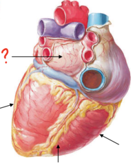
Superior vena cava (SVC), Inferior vena cava (IVC), Ascending aorta, Pulmonary trunk, Pulmonary veins
What are the great vessels of the heart?

Right brachiocephalic, Left brachiocephalic, Azygos
What veins join together to form the superior vena cava (SCV)?
Upper limbs, Thoracic wall, Head/Neck
Which body regions drain into the superior vena cava?
Abdominopelvic cavity and Lower limbs
Which body regions drain into the inferior vena cava?
Ascending aorta
What is the first part of the aorta called?
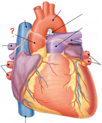
Oxygenated
Does the ascending aorta carry oxygenated or deoxygenated blood?
Arteries
Does the pulmonary trunk divide into pulmonary arteries or veins?
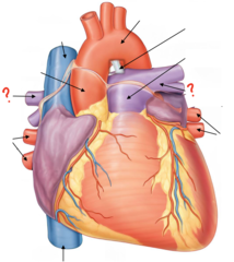
Deoxygenated; Heart to the Lungs
The pulmonary arteries carry (oxygenated/deoxygenated) blood from the (heart/lungs) to the (heart/lungs).
Oxygenated; Lungs to the heart
The pulmonary veins carry (oxygenated/deoxygenated) blood from the (heart/lungs) to the (heart/lungs).
Right pulmonary veins
What is labeled 1?

Right pulmonary artery
What is labeled 2?

Ascending aorta
What is labeled 3?

Superior vena cava
What is labeled 4?

Aortic arch
What is labeled 5?

Ligamentum arteriosum
What is labeled 6?
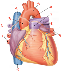
Pulmonary trunk
What is labeled 7?

Left pulmonary artery
What is labeled 8?

Left pulmonary veins
What is labeled 9?

Inferior vena cava
What is labeled 10?

Right auricle
What term describes the expanded pocket of the right atrium?
Pectinate muscle; Crista terminalis
The right auricle is made of a rough portion made of _____ muscle and a smooth portion called the _____.
Allows for additional heart volume
What is the function of the right auricle?
SVC, IVC, Coronary sinus
What are the openings in the right atrium?
Dilated venous system that drains heart
What is the function of the coronary sinus?
Right atrium
Which chamber of the heart is the coronary sinus located?
Fossa ovalis
What term describes the depression at the interatrial septum?
Between right atrium and right ventricle
Where is the right atrioventricular orifice located?
Tricuspid valve
What valve is located at the right atrioventricular orifice?
Anterior, Posterior, Septal
What are the cusps of the tricuspid valve?
True
True or False: Papillary muscles are named relative to the valve cusp in which they attach.
Papillary muscles; Chordae tendinae
Tricuspid and bicuspid valve cusps are connected to _____ muscles via _____.
Interatrial septum
What is labeled 1?
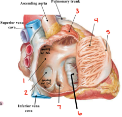
Fossa ovalis
What is labeled 2?

Right auricle
What is labeled 3?

Pectinate muscle
What is labeled 4?
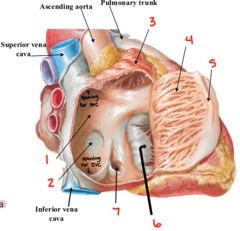
Crista terminalis
What is labeled 5?

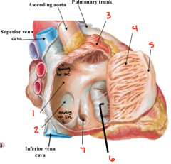
Right atrioventricular orifice
What is labeled 6?

Orifice of coronary sinus
What is labeled 7?
Muscular wall that divides left and right ventricles
What is the interventricular septum?
Conduction system of the heart
What is contained within the septomarginal trabeculae (moderator band) of the heart?
Anterior papillary muscle
Which papillary muscle of the tricuspid valve is the septomarginal trabeculae (moderator band) connected to?
Septal papillary muscle
What is labeled 1?
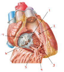
Interventricular septum
What is labeled 2?

Moderator band
What is labeled 3?

Posterior papillary muscle
What is labeled 4?

Anterior papillary muscle
What is labeled 5?
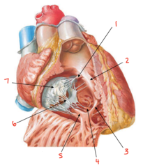
Chordae tendinae
What is labeled 6?

Tricuspid valve
What is labeled 7?

Trabeculae carneae
What term describes the muscular ridges found on the walls of the right and left ventricles?
Conus arteriosus
What structure in the right ventricular space narrows to funnel blood towards the pulmonary valve?
Pulmonic valve
What is labeled 1?

Conus arteriosus
What is labeled 2?

Right and left pulmonary arteries
What does the pulmonary trunk divide into?
4 pulmonary veins
(2 right/2 left)
How many veins feed into the left atrium?
Left auricle
What is labeled here?

Opening between the left atrium and left ventricle
Where is the left atrioventricular orifice located?
Bicuspic valve
What valve is located at the left atrioventricular orifice?
Anterior and Posterior
Where are the cusps of the bicuspid valve located?
Aortic trunk
What structure connects the left ventricle and ascending aorta?
Cardiac skeleton
What is the fibrous framework of dense connective tissue that anchors the heart valves and electrically insulates the atria from the ventricles?
Anterior pulmonic cusp and sinus
What is labeled 1?

Right pulmonic cusp and sinus
What is labeled 2?
