Respiratory Function Testing
 Call Kai
 Learn
 Practice Test
 Spaced Repetition
 Match
 Flashcards
 Knowt Play
1/60

There's no tags or description
Looks like no tags are added yet.
61 Terms
alveolar ventilation
Va
diffusing capacity of lung for carbon monoxide
DLCO
forced expiratory volume in 1 second
FEV1
forced vital capacity
FVC
functional reserve capacity
FRC
peak expiratory flow rate
PERF
pulmonary function tests
PFTs
total lung capacity
TLC
ventilation/perfusion scan
V/Q scan
inspiration
diaphragm muscle contracts and pulls downward while muscles pull upward; overcome elastic properties of lung and resistance to airflow
expiration
passive process in which the diaphragm relaxes; elastic recoil of lungs pulls lungs back to original size and shape
-evaluate physiologic processes
-diagnosis of symptomatic diseases
-screen for early, asymptomatic disease
-determine prognosis of disease
-monitor response to treatment
purpose of PFTs
obstruction to airflow, changes in lung size, decrease in gas transfer
types of abnormalities detected in PFTs
demographics (age, height, race, sex)
what normal PFT results are based off of
peak flow, spirometry, pulse ox, DLCO
common types of PFTs
arterial blood gas, exercise oximetry, six-minute walk test, maximum inspiratory/expiratory pressure
specialty types of PFTs
TV (tidal volume)
air exhaled during normal breathing
IRV (inspiratory reserve volume)
maximal air inhaled above tidal volume
ERV (expiratory reserve volume)
maximal air exhaled above tidal volume
RV (residual volume)
air remaining in the lung after maximal exhalation
TLC (total lung capacity)
sum of TV + IRV + ERV + RV
VC (vital capacity)
sum of TV + IRV + ERV
FRC (functional reserve capacity)
sum of RV + ERV
FEV1 (forced expiratory volume in 1 second)
volume of air exhaled during the first second of the FVC maneuver
FVC (forced vital capacity)
total amount of air that can be exhaled
FEV1/FVC
FEV1 as a ratio of FVC - more sensitive to measure obstruction as independent of patient's size or TLC
FEF (forced expiratory flow)
flow (speed) of air during a forced expiration
FEF 25%-75%
FEF during 25% to 75% of FVC - represents the mean flow during the middle half of the FVC
PEF (peak expiratory flow)
maximum flow obtained during forced expiration
obstructive lung disease
reduced capacity to get air out of the lungs
COPD, asthma, bronchiectasis, cystic fibrosis
types of obstructive lung diseases
FEV1/FVC < 0.7 (70%)
diagnosis of obstructive lung disease
restrictive lung disease
reduced capacity to get air into the lungs
interstitial lung disease (fibrosis, sarcoidosis), chest wall pathology (scoliosis), obesity, neuromuscular (ALS)
types of restrictive lung diseases
reduction in all expiratory volumes; FEV1/FVC is normal
diagnosis of restrictive lung diseases
pulmonary vascular disease
broad term including any condition that affects the blood vessels within the lungs
pulmonary hypertension, chronic thromboembolic disease
pulmonary vascular disease
spirometry
PFT in which a clip is placed on the nose and patient exhales hard and fast into a mouthpiece until no more air will come out
most lung volumes and capacities except residual volume (RV)
what spirometry measures
TLC, FRC
what CANNOT be calculated with spirometry?
reduced
FEV1 in obstructive lung disease
normal or reduced
FVC in obstructive lung disease
reduced
FEV1/FVC ratio in obstructive lung disease
normal or reduced
FEV1 in restrictive lung disease
reduced
FVC in restrictive lung disease
normal or reduced
FEV1/FVC ratio in restrictive lung disease
obstructive
obstructive or restrictive lung disease?

restrictive
obstructive or restrictive lung disease?
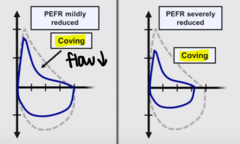
coving
hallmark of obstruction in a flow-volume loop
normal, just smaller
shape of flow-volume loop in restrictive lung disease
post-bronchodilator FEV1/FVC < 0.7
diagnosis of COPD with spirometry
>12% change in FEV1 pre vs post bronchodilator
diagnosis of asthma with spirometry
peak flow meter
PFT in which a patient blows into a meter as hard and fast as they can and determines how far the marker was moved
PEF (peak expiratory flow)
what peak flow meters measure
personal best
to find a patient's __________________ with peak flow meter, take their peak flow between noon and 2 PM each day, each time after they take their quick-relief medicine to relieve symptoms, or any other time their provider tells them to
DLCO (diffusing capacity of lung for carbon monoxide)
PFT that tests diffusing capacity of lungs for carbon monoxide, measuring ability of lungs to transfer gas from air to red blood cells across alveolar-capillary membrane
lung restriction, emphysema, interstitial lung disease
disease states where reduced diffusing capacity (DLCO) is seen
pulse oximetry
measures oxygen saturation via arterial saturation with a device that emits and reads the reflected light from capillary blood to estimate saturation
>95%
normal SpO2
V/Q scan (Ventilation Perfusion Scan)
method that uses radioactive gas and albumin to measure ventilation and perfusion with a series of two scans performed together: ventilation in which radioactive material is inhaled to measure where air flows in the lungs and perfusion in which radioactive material is injected into vein to measure where blood is flowing in lungs
PE and pulmonary hypertension
what are V/Q perfusion scans often used for?