Shoulder Pathology: Part 1 Motor Control and Mobility Deficits
 Call Kai
 Learn
 Practice Test
 Spaced Repetition
 Match
 Flashcards
 Knowt Play
1/53

There's no tags or description
Looks like no tags are added yet.
54 Terms
what are the 6 principles of shoulder abduction?
shoulder abducts due to scapulothoracic rhythm (120 GH abduction, 60 degrees scapular upward rotation)
60 degrees of scapular upward rotation is a result of SC joint elevation and AC joint upward rotation
clavicle retracts at the SC joint
upwardly rotating scapular posteriorly tilts and ER slightly
clavicle posteriorly rotates around the own axis
GH joint externally rotates
which conditions fall under shoulder pain with mobility deficits?
adhesive capsulitis
fractures
what is adhesive capsulitis?
aka frozen shoulder. Is classified as shoulder pain with mobility deficits. GH joint hypomobility due to thickened capsule and loss of axillary fold and decrease synovial fluid. prevalence for idiopathic is 2-5.3%, secondary is 4.3-38%
what are risk factors for primary adhesive capsulitis?
DM I and II
Hx (dupuytren’s contracture)
age (40-65)
thyroid
gender (more common in females)
Hx of adhesive capsulitis on 1 side
what are the causes and risk factors of adhesive capsulitis?
immobility, trauma, 2ndary to other NM issues causing capsuloligamentous fibrosis
what happens to the capsule in adhesive capsulitis?
capsule loses its pleating, the axillary and inferior pouch stiffens limiting the ability to elevate at the GHJ. the synovial fluid also gets thicker
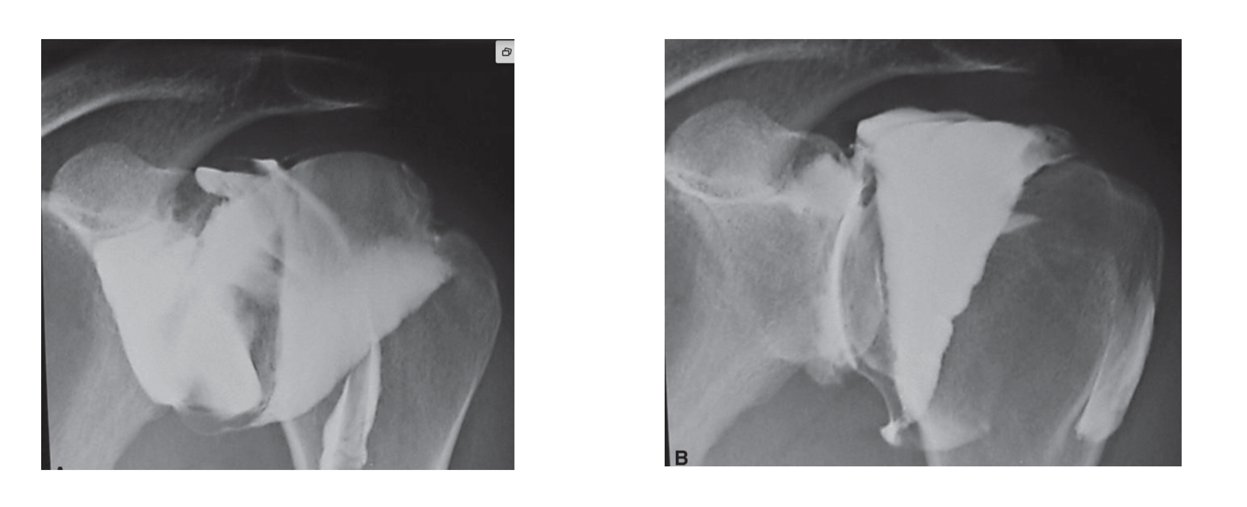
which image demonstrates adhesive capsulitis?
right image
how long does the freezing stage of adhesive capsulitis last for?
10-36 weeks
what is stage 1/freezing of adhesive capsulitis characterized by?
see the start of synovial inflammation
increased pain, esp at end range
primary complaint of ache then constant pain
ROM limitations gradually progress (initially dx is usually impingement)
minimal to no MS or postural changes beside pain/discomfort
aggressive angiogenesis/joint changes
loss of ROM in capsular patterns (ER > Abd > IR > Flex)
how long does stage 2 of adhesive capsulitis last?
4-12 weeks
what are the characteristics of stage 2/frozen stage?
physiological process of inflammation is ending
postural changes- looking for positions of comfort
MS performance/motor control changes
pain subsides to only at end of ROM
stiffness persists w significant loss of AROM/PROM
how long does stage 3 of adhesive capsulitis occur?
12-42 weeks
describe the characteristics of stage 3/thawing of adhesive capsulitis?
resolution begins
ROM begins to spontaneously improve
mean average of 30 weeks before symptoms commence resolving (from onset of symptoms)
comorbidities may delay timeframes
what is treatment like for adhesive capsulitis?
most effective in late frozen to thawing phase
joint mobilitizations are warranted
ROM activity repetitively though the day based on SINS
periscap strengthening as needed, mobility of the GHJ as needed, motor control for scapulo-thoracic and as GHJ resolves
what is the most common fracture in the shoulder region?
clavicle fracture because it is super stable medially via the coracoclavicular ligaments and since the force is designed to be transferred to the axial skeleton via the clavicle. the weakest portion is the middle and lateral 1/3 junction
what is the MOI of clavicle fractures?
fall on lateral shoulder
direct trauma
which demographic is clavicle fractures most common in?
children/adolescents
what are some associated injuries of clavicular fractures?
pneumothorax
rib fracture
what is the clinical presentation of a clavicle fracture?
pain, bruising, bone tenderness, visible step deformity maybe
what is the management of clavicle fractures like?
typically non surgical with figure 8 support for 6-8 weeks. begin AAROM and advance to AROM (clavicualr movements with association shoulder motion), MS performance interventions address musculature that attaches to the clavicle
describe proximal humeral fractures that are youth related
can occur due to trauma or excessive throwing. see epiphyseal stress fracture of shaft due to increase IR or ADD force, proximal fragments migrate upward due to pull of musculature
what is management like for proximal humeral fractures that are associated with youth?
rest initially, address the ROM, strength, motor control (coordination and proprioception/kinesthesia). progression will be guided by radiologic findings
describe proximal humeral fractures in the elderly
due to osteoporotic bone. see fractures most commonly in the surgical neck and more often in females. see increased risk with increased age. MOI often due to FOOSH or direct lateral trauma.
what nerve might a proximal humeral fracture at the surgical neck put into jeopardy?
The axillary nerve, which innervates the deltoid and teres minor muscles and is responsible for sensation over the lateral shoulder.
what is the clinical presentation of a proximal humeral fracture?
acute injury with pain, swelling, tenderness over the greater tubercle, crepitus, bruising possible to the forearm and hand. need to do an integrity assessment of the brachial artery, the median and musculocutaneous nerve, and assess the rotator cuff, deltoid, and pectoralis since they all attach on the proximal humerus
what is the management for non-displaced/minimally displaces proximal humeral fractures?
treated non-surgically
immobilization (slight distraction force) for 4 weeks bc need callous formation.
decrease pain/apprehension then able to begin PT
goal is to avoid capsular contraction/should stiffness which can happen quickly but pts generally have a lot of pain
describe the surgical management for proximal humeral fractures
used for fractures with significant displacement (closed recution with/wo percutaneous pinning, ORIF). PT management following will be rigorous and prolonged, many pts can not tolerate
what are the outcomes like for surgical management of proximal humeral fractures?
outcomes vary, poor outcomes may be due to hardware being closer to the joint space and limiting motion. also might see poorer outcomes systemic disease like rheumatological conditions, also less than 10 degrees of LR leads to poorer outcomes
describe the typical PT mangement for surgically repaired proximal humeral fractures
0-1 weeks pain management and early mobility with PROM
1-6 weeks AAROM with joint mobilizations and strength
6-12 weeks advance all activities and improve proprioception
12-24 weeks return to activity, functional positions, plyometrics
describe scapular fractures
uncommon injuries, due to direct HIGH trauma, may present with rib injury, pulmonary trauma (pneumothorax), is classified according to location (body, neck, glenoid fossa, acromion, spine or coracoid
what is management for scapular fractures like?
typically non surgical, progressive pendulum'/PROM, + healing (callus formation), AAROM to AROM, progressive ms strengthening, especially scapular ms. average healing time is 6 weeks, remodeling up to 1 year
when is surgical repair needed for scapular fractures?
if the fracture occurs in the acromion/spine, coracoid, glenoid rim, intraglenoid
why might GHJ OA develop?
repetitive microtrauma
macrotrauma
repetitive high load labor tasks
BMI > 30 increases risk due to increased axial load and systemic inflammation
strong correlation between rotator cuff health and decrease GHJ OA
which conditions fall under shoulder pain with movement coordination deficits?
dislocations and subluxations
labral tears
instabilities that affect the capsuloligamentous structures
where can dislocations occur in the shoulder?
SC joint tho is rare (3%)
AC joint which is shoulder separation
GH which is true dislocation
what is a 1st degree dislocation?
partial ligament tearing
what is a 2nd degree dislocation?
ligament is torn but the joint is intact
what is a 3rd degree dislocation?
ligament is torn, joint is detached
what direction does teh SC joint typically dislocate in?
anterior, MOI typically due to trauma (MVA or sports related)
what complications might a posterior SC joint dislocation lead to?
compression of vital organs (major blood vessles, esophagus, trachea, SOB, venous congestion, pneumothorax, dysphagai)
what is the clinical presentation of a SC joint dislocation?
deformity, localized swelling, ecchymosis, pain
what is management like for SC joint dislocations?
1st degree protection for 3-4 days
2-3rd reduction, protection due to instability for 2-3 weeks, may require surgical repair
what is the MOI for AC dislocation?
FOOSh or elbow of shoulder trauma, direct trauma. (most frequently injured)
which grades of dislocation of the AC joint typically require surgery?
possible grade 3, 4-6 require surgery (see image for reference)

describe an anterior dislocation of the shoulder
most common type of dislocation, called a bankart lesion is the labrum is torn. often recurrent dislocation
describe the complications that are associated with glenohumeral dislocations
nerve injury (axillary with inf or ant/inf, median with ant but uncommon, musculocutaneous with ant)
RC, bicep, or labrum may be affected
vascular changes (red flag)
what is the clinical presentation of GHJ dislocations?
palpation shows step off from acromion, humeral head is anterior (with subcoracoid dislocation), impaired ROM, possible muscular deficits
why might posterior GHJ dislocations occur (which are rare about 2% of cases)?
usually traumatic in nature, “cross body”, large force leads to a reverse hill sachs lesion (on medial side of the bone where get compressive fracture of the humeral head).
describe the clinical presentation of the posterior dislocation of the GHJ
decreased Extension/ER, horizontal adduction position
what is a SLAP lesion?
superior labrum anterior to posterior lesion- a tear of the superior labrum w or w/out proximal insertion of the long head of the biceps tendon
what are the gold standard diagnostic tests for SLAP tears?
MRA for imaging (has enhanced visualization due to radioisotope)
true gold standard is arthroscopic viewing
describe the 4 types of SLAP lesions
Type I: fraying of the superior labrum without biceps tendon involvement
Type II: tear of the superior labrum with biceps tendon detached
Type III: bucket handle tear of the superior labrum with intact biceps tendon
Type IV: bucket handle tear extending into the biceps tendon. part of the biceps anchor still intact
what is the MOI for labral tears?
multifactorial (young may be due to bicipital groove anomonlies, repetitive overhead or trauma, older may be degenerative changes)
FOOSH
deceleration injuries (increase tensile forces as the biceps attmepts to decelerate elbow extension)
acceleration injuries (late “cocking” phase of throwing, increased ER/ABD causing the tendon/labrum to be pulled off (peeled back) from the bone)

what does the image show?
labral tear- see the “oreo” sign inside the circled area- glenoid is dark, contrast is light, labrum is dark