Anatomy 2300 Unit 4: Spinal Cord
 Call Kai
 Learn
 Practice Test
 Spaced Repetition
 Match
 Flashcards
 Knowt Play
1/33

There's no tags or description
Looks like no tags are added yet.
34 Terms
CNS (Cord runs from where to about where? Function? )
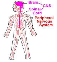
(central nervous system). The brain and spinal cord.
The cord sits in the vertebral canal, running from the foramen magnum to about L1-L2. The CNS integrates incoming information and sends out commands.

PNS (How many pairs? Function?)
(peripheral nervous system). All nervous tissue outside the brain and spinal cord: the 31 pairs of spinal nerves, 12 pairs of cranial nerves, and ganglia. It connects the CNS to the rest of the body.
Sensory Division (Other name? Function?)
(Afferent division). Carries information from receptors in the skin, muscles, joints, and organs toward the CNS. It includes somatic sensory fibers (touch, pain, position) and visceral sensory fibers (organ stretch).

Motor Division (Two Divisions? Other name? Function?)
(Efferent division). Carries commands from the CNS out to muscles and glands.
Its somatic part controls skeletal muscle voluntarily, and its autonomic part controls smooth muscle, cardiac muscle, and glands involuntarily.

Motor Neuron (Function? Cell bodies? Polar?)
An efferent neuron that carries signals away from the CNS to muscles or glands.
It is multipolar, with its cell body inside the CNS in the ventral horn, and its axon leaves through the ventral root.
Unlike sensory neurons, it is efferent and its cell body is in the CNS, not in the dorsal root ganglion.

Sensory Neuron (Function? Cell bodies? Polar?)
An afferent neuron that carries signals from receptors toward the CNS.
It is pseudounipolar, with one short process that splits into a peripheral branch and a central branch.
Its cell body is in the dorsal root ganglion (outside the cord), and the central branch enters the cord through the dorsal root. Unlike motor neurons, it runs toward the CNS and its cell body is in the PNS.

Interneurons (Polar? Sit where? Cell bodies? Function?)
Multipolar neurons that sit entirely within the CNS between sensory and motor neurons.
They are the most numerous type, and in the spinal cord their cell bodies are in the gray matter, especially the dorsal horn.
They integrate and relay information and form reflex circuits.

Spinal Cord (Runs from? Carries what information?
A cylinder of CNS tissue in the vertebral canal, running from the foramen magnum to about L1-L2 in adults (roughly 42-45 cm). It has 31 segments: 8 cervical, 12 thoracic, 5 lumbar, 5 sacral, and 1 coccygeal.
It carries sensory information up, motor commands down, and integrates reflexes. It is known as a REFLEX center. Because the cord ends higher than the vertebral column, segments do not line up with the vertebra of the same number, especially in the lower back.

Conus medullaris (Ends where?)
The tapered, cone-shaped bottom end of the spinal cord, at about L1-L2 in adults. It contains the lowest sacral and coccygeal segments.

Cauda equina (Below where? Sits?)
Latin for "horse's tail." It is a bundle of lumbar, sacral, and coccygeal nerve roots hanging below the conus medullaris, floating in CSF in the lumbar cistern in the subarachnoid space.
It is nerve roots, not cord, so it is relatively safe from a needle during a lumbar puncture.

Filum terminale (Sits? Function? Ends?)
A thin strand of pia mater extending down from the tip of the conus medullaris. It floats in CSF in the lumbar cistern in the subarachnoid space.
It anchors the cord, ending at the bottom of the dural sac (about S2) and then continuing to the coccyx.

Cervical enlargement (Spinal Cord levels? Function/Supple?)
A widening of the cord at about C5-T1.
It holds the extra neurons that supply the upper limbs through the brachial plexus.
This enlargement exists because the limbs need more motor neurons and take in more sensory input.

Lumbosacral enlargement (Spinal Cord levels? Function/Supply?)
A widening of the cord at about L1-S3.
It holds the extra neurons that supply the lower limbs through the lumbar and sacral plexuses.
This enlargement exists because the limbs need more motor neurons and take in more sensory input.

Meninges (Function? superficial to deep?)
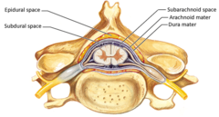
Three connective tissue layers around the spinal cord (and brain) that protect, support, and contain CSF.
From superficial to deep they are the dura, arachnoid, and pia.

Dura mater
The tough outermost meningeal layer. In the vertebral canal it is a single layer, separated from the bone by the epidural space.

Arachnoid mater
The thin, delicate, web-like middle layer lies against the inside of the dura. Thin strands cross the subarachnoid space from it to the pia.

Pia mater (carries? forms?)
The very thin innermost layer that clings tightly to the cord's surface and carries blood vessels. It forms the denticulate ligaments and the filum terminale.

Denticulate ligament (Function? Attaches)
A sheet of pia mater on each side of the cord, between the dorsal and ventral roots.
It has about 21 tooth-like extensions of the pia mater that attach to the dura mater and hold the spinal cord in place, preventing it from moving side to side.

Epidural space (Kind of space? Contains?)
(actual). The space between the vertebral bone and the dura, outside the meninges.
It contains fat and a venous plexus (a network of veins that drains blood.), and it is where epidural anesthesia is given.

Subdural space (Kind of space? )
(potential). The space between the dura and arachnoid. It is normally closed and only becomes a real space if fluid or blood collects there, as in a subdural hematoma.
Subarachnoid space (Kind of space? Contains? Holds?)
(actual). The space between the arachnoid and pia, containing CSF and blood vessels.
Below the conus medullaris, it widens to hold the cauda equina and filum terminale.

Spinal Tap (Inserted into? Spinal Cord levels? Uses?)
(lumbar puncture). A needle is inserted into the subarachnoid space in the lumbar cistern, usually between L3-L4 or L4-L5, below the end of the cord so the cord is not injured. It passes through skin, ligaments, the epidural space, dura, and arachnoid.
It is useful for collecting CSF to diagnose conditions like meningitis, subarachnoid hemorrhage, and MS, measuring CSF pressure, and injecting medication, anesthetic, or contrast dye. It is avoided when pressure inside the skull is raised, because of the risk of brain herniation.

Gray matter vs white matter (Composition? Functions?)
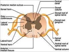
Gray matter is the central, butterfly-shaped region containing neuron cell bodies, dendrites, unmyelinated axons, and synapses. It handles integration and reflexes, and includes the dorsal and ventral horns.
White matter is the outer region of myelinated axons organized into ascending sensory and descending motor tracts, which conduct signals up and down the cord.

Dorsal Horn (Receives information from? Contain cell bodies of?)
The posterior projection of the gray matter. It receives incoming sensory information from the dorsal root, and its cell bodies belong to multipolar interneurons that process that input.
Sensory neuron cell bodies are not here, since they are in the dorsal root ganglion.

Dorsal Root (fiber type? Carries info. into?)
The posterior root attaching to the cord.
It contains sensory (afferent) fibers only, the axons of pseudounipolar sensory neurons carry information into the spinal cord, entering the dorsal horn.
Dorsal Root Ganglion (Contains cell bodies of?)
A swelling on the dorsal root, in the intervertebral foramen just before the roots join.
It contains the cell bodies of pseudounipolar sensory (afferent) neurons. No synapses occur here.

Ventral Horn (Contains cell bodies of? Send signals to?)
The anterior projection of the spinal cord's gray matter. It contains the cell bodies of multipolar somatic motor (efferent) neurons that send signals out of the spinal cord through the ventral roots to control skeletal muscles.
It is larger in the spinal cord enlargements, where nerves supplying the limbs originate.

Ventral Root (Send signals from? Fiber type?)
The root that exits the spinal cord and contains only motor (efferent) fibers.
These are the axons of ventral horn motor neurons that carry commands from the spinal cord (ventral horn) to skeletal muscles, causing them to contract.

Spinal Nerve (Carries? How many pairs?)
Formed where the dorsal and ventral roots join in the intervertebral foramen.
It is a mixed nerve, carrying both sensory and motor fibers. There are 31 pairs, and each splits almost immediately into a dorsal ramus and a ventral ramus.

Dorsal Ramus (Carries? Supplies?)
(A) The smaller, posterior branch of the spinal nerve.
It is mixed (sensory and motor) and supplies the skin and deep muscles of the back.

Ventral Ramus (Carries? Supplies? Form what?)
(B) The larger, anterior branch of the spinal nerve. It is mixed (sensory and motor) and supplies the front and sides of the trunk and the limbs.
Ventral rami form the nerve plexuses (cervical, brachial, lumbar, sacral), except in the thoracic region where they stay separate as intercostal nerves.

Central canal of spinal cord (Continuous with?)
A tiny CSF-filled channel running through the center of the spinal cord's gray matter, in the gray commissure.
It is continuous with the fourth ventricle at the top of the cord.

Spinal Cord Fissure
: A deep groove on the front of the spinal cord that partially separates the right and left halves, with the anterior white commissure at its base where some axons cross sides.

Spinal Cord Sulcus
A shallow groove along the back midline of the spinal cord that separates the right and left halves and lies between the two dorsal white matter columns.
