ANS 206 Final Practical
 Call Kai
 Learn
 Practice Test
 Spaced Repetition
 Match
 Flashcards
 Knowt Play
1/65

There's no tags or description
Looks like no tags are added yet.
66 Terms
blood
connective tissue composed of a liquid portion and a cellular portion
Fluid that circulates through the heart, arteries, capillaries, veins, and constitutes the chief means of transport within the body
blood vessels
where is the majority of the blood?
functions of blood
transport of gases, nutrients, hormones, heat, and wastes
regulation of pH, body temperature, and water content of cells
anti-clotting mechanisma nd body defenses
plasma
whole, intact, watery component of blood (gases, nutreints, waste, ions, salt, hormones)
formed elements
cells and formed elements,
99% RBCs and 1% WBCs and platelets
serum
plasma minus clotting factors, collected without anti-coagulant
enriched sample, not as many components
platelets
not nucleated, they have vesicles
vesicles contain heparin, histamine, and other proteins and enzymes so they can stick together and form a plug when clotting occurs
RBCs
do not have nucleus when they are mature
erythrocytes
live around 120 days
macrophages
monocytes that leave ciruclation and enter a tissue
T lymphocytes
immune cell found in the thymus
B lymphocyte
immune cell found in the bursa of chicken
leukocytes
nucleated cells, combat infection, start inflammation, 2 types
Granulocytes
type of leukocyte
cytoplasmic granule containing cell
neutrophils, eosinophils, basophils
Agranulocyte
type of leukocyte
monocytes and lymphocytes
absence of granules
Eosin
pink acidic dye
methylene blue
blue basic dye
megakaryote
parent cell of thrombocyte
platelets
disc-shaped, no nucleus, release substance to stimulate clotting and constriction of blood vessels
hemoglobin
protein that carries o2 and co2 in the blood
heme binds to o2 and globin binds to co2
heparin
naturally present anti-coagulant on endothelial cells, one of the main anti-clotting mechanisms inside the body of the animal
Warfarin
rat poisoning, chemicals that prevent coagulation
internally bleeding out of their blood vessels and their heart explodes
EDTA
irreversibly binds to calcium and topples clotting cascade
best anticoagulant known to man
chelator
chemical substance that binds irreversibly to calcium
citrate
mild anticoagulant
oxalate
in the gray tubesm we use this for sugar assessment, also find NaF in it, NaF prevents glucose breaking enzymes.
silica
red, gel, no AC, serum, for serum chemistry and serology assays
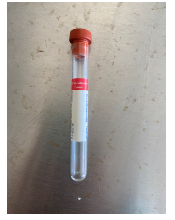
SST
tiger top, gel plug, no AC, serum, for serum separation, ensures complete clotting in <5 minutes

lithium/sodium heparin
green top, yes AC, plasma, for plasma assays in chem

EDTA
purple top, yes AC, plasma, for whole blood analysis, prevents clotting

citrate
blue top, liquid, yes AC, plasma, for coagulation spcimens assays

sodium fluoride/potassium oxalate
gray top, powder, yes AC, plasma, for glucose determination, has EDTA as well

acid citrate dextrose
yellow top

veins
superficially located
anatomically large diameter
walls are thin
low blood pressure - due to thin walls
arteries
deeper seated
narrow diameter
thicker walls
higher blood pressure, much harder to stop blood flow
myocardium
muscle heart is made out of
pericardium
a double walled sac around the heart composed of
a parietal layer lines the internal surface of the fibrous pericardium
the visceral layer or epicardium lines the surface of the heart
pericardial cavity
fluid filled layer that seperates the parietal layer and visceral layer
serosal layer
flush on the outside, folds over itself
viscera
girth of the body
atria
top two chambers that receive the blood
1/3 of the heart
ventricles
bottom two chambers
2/3 volume of the heart
heart
hollow muscular pump
arteries
take the blood away
pulmonary artery
on the right hand side
takes blood away from the right ventricle to the lungs
aorta
largest artery in the body that is taking blood away from the heart from the left ventricle
vena cavae
two large veins that are bringing deoxygenated blood to the heart. Top is superior and bottom is inferior
superior vena cava
drains the head and the neck region
inferior vena cava
draining the rest of the body below the heart
pulmonary veins
bring oxygenated blood to the left atrium
apex
located on the left side of theheart and is formed by the left ventricle entirely, sits on the diaphragm
base
located in the cranial region and includes all the blood vessels
auricle
small ear-shaped muscular pouch that projects outwards from each of the two atria of the heart. Purpose is expansion and blood reservoir. Acts as extra storage space, expanding blood-holding capacity and contractile surface of the atria.
chordae tendineae
tendinous cords which open and close the valves
papillary muscles
these mark the ventricular walls, walls of myocardium are thrown into pillars called these.
abdominal aorta
largest part of the aorta
thoracic aorta
thinner, because it doesn’t have to go too far
atrioventricular valves
valves between atria and ventricles
tricuspid and bicuspid
prevents backflow into the atria when ventricles contract
semilunar valves
guards the entry of the blood in the arteries
prevents backflow of blood into the ventricles
3 half-moon cup-shaped flaps
aortic semilunar valve and pulmonary semilunar valve
pulmonary circuit
sends blood into lungs, does not need much force
systemic circuit
needs to have a lot more force as it needs to send blood to the head, tail, limbs, etc.
ejection fraction
60% of the total blood that had collected in the ventricles has now left the building and ventricles have done their job
upper respiratory
nose, nasal cavity/paranasal sinuses,
nasopharynx
larynx
lower respiratory
trachea
lungs
bronchi/bronchioles/alveoli
functions of the respiratory system
deliver oxygen to the cells of the body and remove carbon dioxide from the cells
regulate pH of body fluids and blood
assist in temperature control
phonation
panting
the mechanism used to cool off, dissipating the heat that was created by the skeletal muscles
nose
immense variability in the external organ of the respiratory