Neuro exam 2
 Call Kai
 Learn
 Practice Test
 Spaced Repetition
 Match
 Flashcards
 Knowt Play
1/113

There's no tags or description
Looks like no tags are added yet.
114 Terms
Ion channels are composed of
transmembrane proteins with a channel that allows facilitated diffusion for a select type of ion or ions
What are the five primary types of ion channel selectivity
potassium chanel, soidum channel, calcium channel, cation channel, chloride channel
Difference between macroscopic currents and microscopic currents
macroscopic currents - current flowing through a large number of channels distrubuted over a more extensive region of surface membrane
microscopic currents- currents flowing through a single channel
What are the three states that ion channels exist in
open, closed, inactive
what is the open state for an ion channel
open pore that allows passage of ions
What is the closed state of an ion channel
closed pore that can be open upon a new stimulus
What is the inactivated state of an ion channel
a closed pore that cannot be reopened until a new physical state is achieved
not all ion channels have an inactivated state
the inactivated state of the Na+ channel is responsible for the refractory period of the action potential .
The inactivated state of what channel is responsible for the refractory period of the action potential
the Na+ channel
gating is
the movement between the different states that ion channel exist in
What do states represent
specific,semi-stable, conformations of the channel protein
what is kinetics?
the rate at which a channel moves between states
In the context of ion channel conformations and state transitions, why are only certain states accessible from a given state?
Because the channel must change its physical shape to transition between functional states, and only certain conformational shapes can be reached directly from a given shape.
what are the two methods to study ion channel behavior
V-clamp and patch clamp
patch clamp purpose
to measure currents flowing through single ion channels and test theoretical deductions about channel characteristics
microscopic currents
currents flowing through single, individual ion channels
macroscopic currents
currents flowing through a large number of channels across an extensive region of the cell membrane
patch clamp technique
a laboratory technique used to study the electrical properties of individual ion channels by applying mild suction to form a tight contact and then isolating a small patch of membrane and measuring ionic currents. (inside out recording)
4 reasons why the microcurrents are due to opening of single, voltage-activated sodium channels
the currents are carried by Na+; thus, they are directed inward when the membrane potential is more negative than ENa, reverse their polarity at ENa, are outward at more positive potentials, and are reduced in size when the Na+ concentration of the external medium is decrease
the channels have a time course of opening, closing, and inactivating that matches the kinetics of macroscopic Na+ currents (4.1c)
both the opening and closing of the channels are voltage-dependent; thus, the channels are closed at –80 mV but open when the membrane potential is depolarized. In fact, the probability that any given channel will be open varies with membrane potential (4.1E)
tetrodotoxin, which blocks the macroscopic Na+ current (see Figure 3.5), also blocks microscopic Na+ currents.

How did patch clamp experiments reveal why the microcurrents are due to opening of single, voltage-activated sodium channels
Testing K+ Channels: Patch clamp experiments on membranes (with blocked sodium channels) show that microscopic outward potassium currents share the exact same properties as macroscopic potassium currents.
Key Shared Features: Both types of potassium currents fail to inactivate during brief depolarizations, respond to the same drugs and ionic changes, and are voltage-dependent.
Conclusion: Macroscopic potassium currents responsible for action potential repolarization are simply the combined result of many individual voltage-sensitive potassium channels opening at the same time.


In the patch-clamp experiment shown in Figure 4.1, why do the individual microscopic traces (INa) look sporadic and different from trial to trial, even though the same voltage step is applied?
Because individual ion channels open and close probabilistically and stochastically. Summing up the behavior of many of these individual stochastic openings yields the smooth macroscopic current.
What does the sigmoidal curve on the graph represent regarding voltage-gated sodium channels, and how does channel gating behave fundamentally?
The graph shows that the open probability (PoP_oPo) of sodium channel opening increases as the membrane becomes more depolarized. Fundamentally, channel gating is a stochastic (probabilistic) process.
What happens to the open probability (Po) curve of voltage-gated channels when affected by most chemicals or drugs, and what does this shift indicate?
Leftward Shift: The curve moves toward more negative membrane potentials. This means the channels open more easily at lower (more negative) voltages than normal.
Rightward Shift: The curve moves toward more positive membrane potentials. This means the membrane has to be depolarized more than usual for the channels to open.
How can you visually tell the difference between a non-inactivating ion channel (KV2.1) and an inactivating ion channel (KV4.1) on a current-versus-time graph during a sustained membrane depolarization?
A non-inactivating channel's current stays elevated and flat for the entire duration of the voltage step, whereas an inactivating channel's current spikes to a peak and then automatically decays back down to baseline while the voltage is still held.

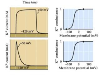
is the potassium channels open or not
no because the conductance is 0 on the chart
How can you tell if an ion channel is open or closed on a conductance graph?
If the conductance line is at zero (baseline), the channel is closed. If it spikes upward, the channel is open.
How do you distinguish between a channel closing due to repolarization versus inactivating during sustained depolarization?
If the current drops to zero because the voltage returns to rest, the channel closed. If the current drops to zero while the voltage step is still held positive, the channel inactivated.
How do you distinguish between a channel closing due to repolarization versus inactivating during sustained depolarization?
If the current drops to zero because the voltage returns to rest, the channel closed. If the current drops to zero while the voltage step is still held positive, the channel inactivated.
How do diverse ion channel protein structures impact neuronal behavior, and what are a few examples of the physical or chemical stimuli that activate them?
specific protein structures give rise to diverse channel behaviors that govern neuronal response properties. Examples of activating stimuli include voltage, neurotransmitters, light/cyclic nucleotides, temperature, and mechanical stretch.
in general where would you find neurotransmitter receptor channels
Neurotransmitter receptor channels can generally be found on the postsynaptic membrane of neurons, where they respond to specific neurotransmitters released by presynaptic neurons.

in general where would you find v-gated na and k channels?
along the axon

in general where woudl you find v gated calcium channels
located in the presynaptic terminals and dendrites

in general where would you find leak channels
in the postsynaptic membrane near the soma
what is the purpose of leak channels
keep cell membrane at resting potential
what is the purpose of calcium channels
to translate electric signals to chemical signals
what are the three main types of stimuli or mechanisms by which most ion channels are "gated"?
Voltage, ligand, and physical (or mechanical) stimuli.
How is a functional ion channel composed regarding its subunits, and what feature is present in all cases?
It is composed of one or more subunits forming the complete channel, and in all cases, a central pore is formed.
What is the function of an auxiliary (beta) subunit when it associates with a primary channel subunit?
It affects channel function by aiding in processes such as intracellular trafficking, cellular localization, and protein complexing.
What are the two primary structural architectures used by ion channels, using KcsA and sodium channels as examples?
Multimeric/Homotetrameric channels built from 4 identical separate subunits (e.g., KcsA K+ channel), and 2) Single-polypeptide channels built from one continuous chain containing multiple domains plus auxiliary subunits (e.g., Na+ channel).S

Why can’t you just have ions enter through channels based on size?
if you were to allow a big ion through any ion smaller than that, should we also be able to get through. If we are being selective to only one ion, we ONLY want that ion to be able to get through, not anything else
Walk through the step-by-step mechanism of how a potassium channel achieves ion selectivity, from the initial hydration shell to the final movement through the filter.
The Hydration Shell Problem: In water, ions don't travel completely bare; they are surrounded by a tight cage of water molecules (their "hydration shell"). Because sodium is smaller than potassium, its water shell is actually held tighter.
Entering the Pore: A hydrated K+ ion enters the wide, water-filled cavity of the channel. But to pass through the narrow selectivity filter, it has to shed that water shell (it gets "dehydrated").
The Carbonyl Backbone Trick: The narrow filter is lined with carbonyl groups from the protein's amino acid backbone. The exact spacing and chemical properties of these carbonyl oxygens are custom-built to perfectly mimic the water molecules that potassium lost. This makes the energetic cost of stripping potassium's water shell "worth it" because the carbonyl groups take over and stabilize the ion.
Why Sodium Gets Blocked: Sodium (Na+) is too small. Because its radius is smaller, it cannot make proper contact with all the carbonyl groups at the same time. It can't shed its water shell energetically, so it stays trapped in its hydration shell and bounces back out.
Pushing Through: Once the potassium ions line up inside the selectivity filter, electrostatic repulsion (positive charges repelling each other) helps push them right through to the other side.
alternate mechanism for sodium selectivity
Attraction: Negative charges near the external pore attract the sodium ion (Na+).
Partial Dehydration: Instead of getting completely stripped bare the way potassium does, sodium gets partially dehydrated and moves into an internal hydrated pore.
Favorable Interactions: The specific chemical interactions inside the channel favor sodium over all other ions.
Electrostatic Push: Just like with potassium, electrostatic interactions help push the ions along to increase the overall ion flux.

common elements between both types of selectivity filteres

what are the four types of gating mechanisms
voltage, ligand, heat, mechanical displacement
voltage gated mechanism
The Voltage Sensors (Top Right / Bottom Right): The protein has specific segments known as voltage sensors that contain positively charged amino acids (like Arginine and Lysine).
The Electrical Pull (Bottom Right): At rest, the inside of the cell is negatively charged. Because opposites attract, that internal negativity physically pulls down on those positively charged amino acids, keeping the channel gate shut.
The Depolarization Trigger (Left Box): When a depolarization event happens (meaning the inside of the cell becomes less negative), that electrical "pull" on the voltage sensor is relieved.
The Conformational Change (Middle Box): As that tension is released, the movement of the voltage sensor shifts the protein's structure via a linker domain, physically pulling the gate open so ions can flow through.
ligand gated mechanism
conformational Change: The binding of a ligand molecule to the receptor induces a structural change that swings the pore open.
Binding Sites: Ligands can bind to either side of the membrane:
Extracellular Side: Typically binds neurotransmitters (such as the nicotinic acetylcholine receptor shown in the slide's diagram).
Intracellular Side: Can bind molecules originating from intracellular signaling cascades.
how does glutamate alllow its channel to open its pore to allow cations to transit the membrane?
Glutamate binds to extracellular ligand binding domains, causing the Y-shaped channel to change shape via an "induced fit" mechanism. This conformational change opens the pore.
According to the current slide on Heat & Mechanical Displacement, how are TRP channels and Piezo channels activated by physical forces?
TRP channels open in response to heat via the proposed mechanism of a lipid molecule dissociating when temperatures rise. Mechanosensitive Piezo channels use blade-like structures that act as levers to open the channel when curved membranes are physically flattened.
How do auditory hair cells open mechanosensitive ion channels in response to sound waves
When the hair bundle is displaced in the direction of the tallest stereocilium, more transduction channels open, which causes depolarization as K+ and Ca2+enter the cell (Figure 13.10). Depolarization in turn opens voltage-gated calcium channels in the hair-cell membrane, and the resultant Ca2+ influx causes transmitter release from the basal end of the cell onto the auditory nerve endings (see Figure 13.9), similar to chemical neurotransmission elsewhere in the central and peripheral nervous systems. This process involves mechanical stimulation of hair cells, leading to the opening of ion channels and ultimately resulting in signal transduction into the auditory nerve.
Because some transduction channels are open at rest, the receptor potential is ____. where movement toward tallest stereocilia ___ cell, while movement away from tallest stereocilia __ cell
biphastic, depolarize, hyperpolarize
Why are perturbations of specific ion channel functions often robustly observable in physiology and behavior?
Because ion channels are essential to physiological function. These perturbations can be caused by natural defenses/offense (venoms), genetic mutations, or pharmacological interventions.
What is the primary action of alpha-toxin from the scorpion Leiurus quinquestriatus?
It slows down the inactivation of voltage-gated sodium channels, prolonging the inward Na+ current and action potential duration.

How do scorpion toxins (alpha- and beta-toxin) cause paralysis despite initially causing hyperexcitability?
Continuous Na+ influx traps the membrane in a permanently depolarized state (depolarization block). This forces voltage-gated Na+ channel inactivation gates to freeze shut and prevents them from resetting, halting all further action potentials and causing flaccid paralysis.
How are the effects of ion channel mutations classified in terms of channel function?
They are classified as gain-of-function or loss-of-function mutations, and their exact physiological effects depend on which specific channel is altered and where in the body it is located.
What are the 5 ways disease related mutations change on or more of the properties of channel behavior
activation
shifts in voltage-dependent properties
blocking
open time
inactivation
Draw an example of a Na current graph and a membrane potential graph for a gain of function mutation?
A shift to more negative potentials should be seen

Draw an example of a Na current graph and a membrane potential graph for a loss of function mutation?

what is the purpose of active transporters?
to establish concentration gradients by transporting ions against their concentration gradient, thus requiring an energy source
What are the two classes of active transporters
ATPase pumps (ATP energy)
ion exchangers (gradient energy, down/up)
ATpase pump (Na+/k+ pump) stucture
two subunis (alpha and beta). the alpha subunit is responsible for ion translocation and spans the membrane ten times, with most of the molecule found on the cytoplasmic side. The Beta side spans the membrane only once and is predominantly extracellular
How do ATP binding and phosphorylation control ion transport in the sodium-potassium pump?
ATP binding drives intracellular sodium binding and potassium release, whereas phosphorylation triggers extracellular sodium release and potassium binding, allowing the pump to move ions against their electrochemical gradients.
How does phosphorylation mediate ion translocation in the sodium-potassium pump?
Transfer of a phosphate group from ATP onto the pump's phosphorylation domain induces a structural change (via the actuator domain) that forces extracellular release of bound Na+ and enables the binding of K+.
What are the full sequential steps of the sodium-potassium pump's ion transport cycle?
ATP Binding: ATP docks onto the pump, which drives the binding of intracellular Na+ and the release of K+.
Phosphorylation: The pump transfers a phosphate group from ATP onto itself (via its phosphorylation domain).
Ion Translocation & Release: Phosphorylation triggers a structural rearrangement (via the actuator domain) into an occluded state, forcing the extracellular release of Na+ and the binding of K+.
Dephosphorylation & Reset: Hydrolysis/dissociation resets the pump back to its initial state to repeat the cycle, moving ions uphill against their electrochemical gradients.
What are the key properties of the Na+/K+ pump regarding ion movement, charge, and energy requirements
Ion Movement: Pumps 333 Na+\text{Na}^+Na+ ions out of the cell and 222 K+\text{K}^+K+ ions into the cell per cycle, moving them against their concentration gradients to maintain resting gradients against leakage.
Electrogenic Nature: It is electrogenic because it creates a net outward movement of positive charge.
Energy Requirement: Requires ATP hydrolysis (active transport).
Na/K= atpase cycle
Step 1: High-affinity internal sodium (Na+\text{Na}^+Na+) binding sites are exposed to the inside of the cell.
Step 2: Three Na+\text{Na}^+Na+ ions and one ATP molecule bind to the pump.
Step 3: The pump undergoes phosphorylation and changes shape (conformation).
Step 4: The Na+\text{Na}^+Na+ binding sites face the exterior, their affinity drops, and the three Na+\text{Na}^+Na+ ions are released outside the cell.
Step 5: High-affinity potassium (K+\text{K}^+K+) binding sites become exposed to the outside, and two K+\text{K}^+K+ ions bind.
Step 6: Inorganic phosphate (PiP_iPi) is released, causing the pump to change back to its original conformation.
Step 7: The K+\text{K}^+K+ binding sites now face the interior with low affinity.
Step 8: The two K+\text{K}^+K+ ions are released inside the cell, ADP is released, and the cycle is ready to repeat.


What does the experimental graph demonstrate regarding the efflux of sodium Na+ when external potassium K+is removed or restored, and when an inhibitor is added? (label everything happening in the graph)

Where are the Na+ and K+ binding sites located on the Na+/K+ pump?
Na+ binding sites: Located on the inside (intracellular) side.
K+ binding sites: Located on the outside (extracellular) side.
Hodgkin and keynes key findings
Steady-State Efflux: There is a steady-state efflux of Na+.
Potassium Dependency: Na+ efflux is dependent on external potassium.
Energy Dependency: ATP synthesis blockers inhibit Na+ efflux.
Core Conclusion: Sodium and potassium are coupled in a linked transport process which requires ATP energy.
Why is the sodium-potassium pump dependent on external potassium?
Answer:
Binding Requirement: The pump cannot complete its functional cycle without binding potassium from the outside.
Conformational Reset: Once the pump releases sodium to the extracellular side, it must bind external potassium ions in order to trigger the release of the attached phosphate group and reset its shape back toward the interior of the cell.
Where can synapses occur?
throughout neuronal structure not just dendrite and axon but also at the soma and other sites.
what are the two types of synapses
electrical (direct electrical coupling between cells, typically involves touching)
chemical (chemical transmitters delivered to target cells)
how do electrical synapses work
work via a gap junction that joins the pre and postynaptic membrane. this creates a LARGE PORE that allows passive current and bi-directional electrical coupling between neurons
where are electrical synapses found
motor areas and other areas where coordinated signaling is utilized
structure of gap junctions at electrical synapses
gap junctions form when connexons from two cells attach forming a complete channel.
Connexons are formed from connexin subunits

why are electrical synapses important?
Fast transmission of signal: Allows electrical current to pass directly between neurons with virtually no delay, enabling rapid reflex circuits.
Coordinated action of multiple cells: Synchronizes the activity of groups of neurons or muscle cells so they fire together as a unified network.
Coordinated intracellular signaling: Facilitates the sharing of metabolic and second-messenger signals across connected cellular interiors.This allows connected cells to coordinate not just their electrical firing, but also their internal metabolic states and biochemical signaling pathways as a unified group.
chemical synapse [5]
What was the significance of Otto Loewi's experiment in relation to chemical neurotransmission?
It provided the first definitive proof of chemical neurotransmission by demonstrating that stimulating the vagus nerve of one frog heart released a chemical substance (later identified as acetylcholine) into a fluid bath, which was then able to slow down the heart rate of a second, isolated frog heart.
What are the classical criteria that define a neurotransmitter?
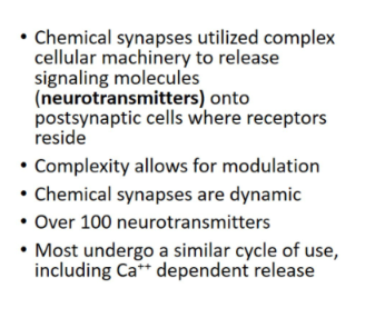
Synthesis & Storage: Must be synthesized and stored within the presynaptic neuron.
Release: Must be released by the presynaptic neuron in response to depolarization (calcium-dependent).
Receptors: Specific postsynaptic receptors must be present to receive it (and application of the transmitter, agonist, or antagonist should mimic or block its physiological effects).
classically,neurotransmitters are either ___ or ____
small molecules or peptides
steps to a synapse
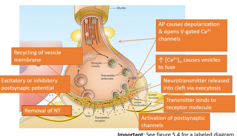
1. Transmitter is synthesized and then stored in vesicles. (Note: Referenced from earlier text context)
2. An action potential invades the presynaptic terminal.
3. Depolarization of presynaptic terminal causes opening of voltage-gated Ca2+Ca^{2+}Ca2+ channels.
4. Influx of Ca2+Ca^{2+}Ca2+ through channels.
5. Ca2+Ca^{2+}Ca2+ causes vesicles to fuse with presynaptic membrane.
6. Transmitter is released into synaptic cleft via exocytosis.
7. Transmitter binds to receptor molecules in postsynaptic membrane.
8. Opening or closing of postsynaptic channels causes a change in membrane conductance.
9. Postsynaptic current causes excitatory or inhibitory postsynaptic potential that changes the excitability of the postsynaptic cell.
10. Removal of neurotransmitter by glial uptake or enzymatic degradation.
11. Retrieval of vesicular membrane from the plasma membrane.
What are the main structural features and differences regarding synapse ultrastructure at chemical synapses?
Active Zone: The region in the presynaptic terminal where vesicles fuse to release their neurotransmitters.
Vesicle States: Some vesicles are primed and ready for release (often highlighted in purple), while others are linked to cytoskeletal elements until needed.
Postsynaptic Density: A dense network of proteins typically observed at excitatory synapses.
Asymmetric vs. Symmetric Synapses:
Asymmetric synapses are generally excitatory and retain plastic potential.
Symmetric synapses are generally inhibitory.

How was the role of calcium in vesicle release found ?
probing a squid giant synapse
What discovery by Katz and Ricardo Miledi provided the first indication of presynaptic calcium channels?
first they probed and voltage clamped a squid’s giant synapse
and then they used tetrodotoxin to block na and K+ channels to observe the effects of calcium only
when doing that they realized that there was a remaining inward current (inward because graph dips below 0mv) while the membrane was being depolarized
they also realized that the postsynaptic cell induced an excitatory response suggesting that inward calcium current triggers neurotransmitter release.
to confirm that it was calcium they added a calcium channel blocker and found that when the calcium channel blocker was introduced, there was no inward current and no postsynaptic response
conclusion: this proved that the voltage gated calcium channels are involved in synaptic transmission

how does this graph prove that presynaptic calcium triggers transmitter release
As shown in the graph on your slide, when researchers experimentally inject calcium chelators or inhibitors into the presynaptic terminal, it blocks the calcium signal and inhibits postsynaptic depolarization, confirming that transmitter release fails without free Ca2+
What happens at certain synapses during high stimulation regarding neurotransmitter release?
Co-release of additional or secondary transmitters becomes possible under conditions of high-frequency or intense stimulation.
To study how neurtransmitters were released what was used
frog NMJ by Bernard katz colleagues
What experimental setup is used to record end plate potentials (EPPs) during synaptic transmission at the neuromuscular junction?
An axon is stimulated to trigger an action potential, while a recording electrode is placed in the muscle cell to measure the resulting postsynaptic membrane potential at the end plate.This setup allows for the observation of the electrical signals generated in response to neurotransmitter release at the neuromuscular junction.
What did scientists discover about how neurotransmitters are released (contrasting the idea of a "continuous flood"), and how do MEPPs and low-calcium experiments prove it?
The Myth: Neurotransmitters aren't released in a continuous, smooth flood.
The Reality (Quantal Release): They are released in tiny, discrete packets called quanta.
The Proof: When scientists lowered extracellular calcium to shrink the signals, they found that the smallest possible evoked EPP is the exact same size as a single MEPP (Miniature End Plate Potential)—meaning a single MEPP is just one individual packet of neurotransmitter being released all by itself.
What does the 0.4 mV peak represent in the quantized distribution of EPP amplitudes?
It represents the size of a single quantum (one vesicle opening), proving that neurotransmitters are released in discrete packets rather than a continuous stream.
How does the voltage change of a single quantum relate to vesicle content, and how did Heuser and Reese prove it?
Vesicle Content: A single quantum corresponds to the release of about 10,000 ACh molecules (which fit into one vesicle) and causes a ~0.4 mV potential change.
Heuser and Reese Experiment: They used rapid freeze-fixation techniques combined with stimulation to directly correlate vesicle fusion events with the quantal load released (The physical number of little sacs (vesicles) popping open matches the exact mathematical "packets" (quanta) measured by the electrical voltage.)
conclusion: found that the number of vesicles fusing correlated perfectly with the number of quanta measured
what are MEPPs (mini endplate potentials)?
spontaneous changes in muscle Vm
what is quanta
signals that are released in defined packets
How does the number of acetylcholine molecules in a single core vesicle relate to the size of a MEPP?
The number of ACh molecules packed inside a single core vesicle matches the exact amount needed to produce a single miniature end plate potential (MEPP).
how do vesicles fuse with membrane
they have specific proteins that target the membrane and fusion, and other fucntions
Give the steps for how vesicle attaches to plasma membrane
overall : hydrophobic effect plays out the closer the vesicle membrane and the plasma membrane get together, causing polar molecules to be directed outward and nonpolar to be directed inward
in this case the hydrophobic heads of the lipids will face the solution so then you get fusion of the membranes
not a spontaneous process (have to force them together, ie takes energy
mechanism: two sets of proteins
vesicle : V-snares ; synpatobrevin and synaptotagmin
target: t-snares ; snap-25, syntaxin
v-snares and t-snares associate bringing vesicle to presynaptic membrane (AKA the docking phase)
synpatotagmin binds to the snare complex, priming the vesicle for function (AKA docked to membrane)
need something to twist the two. this is done by having in influx of calcium that binds to synpatotagmin
membranes in close apposition fuse leading to exocytosis
steps to vesicle membrane endocytosis after transmitter release
clathrin coated pits form
clathrin cage constricts membrane into a stalk
dynamin forms a ring that pinches stalk off
actin filaments guide vesicle into terminal
Hsc70 and auxilin remove clathrin s

what is the fast of vesicular membrane after fusion
membrane is returned to recycling endosomes via clathrin mediated endocytosis
What are the key structural and functional differences between ionotropic and metabotropic receptors?
Feature | Ionotropic Receptors | Metabotropic Receptors |
Mechanism | Ligand-gated ion channels; neurotransmitter binding directly opens or closes the pore. | G-protein-coupled receptors (GPCRs); neurotransmitter binding activates a signaling cascade via second messengers. |
Speed | Very fast onset (milliseconds). | Slower onset (hundreds of milliseconds to days). |
Duration | Short-lasting effects. | Longer-lasting, modulatory effects. |
How does release of neurotransmitters elicit postsynaptic potentials?
there are EPSP and IPSP on dendrites
ionic selectivity of receptor determines which ions flow through it
direction of flow and charge of ion = effect of postsynaptic membrane potential
What does an outside-out patch clamp experiment reveal about single-channel activity?
The setup: Isolating a tiny patch of membrane with an ACh receptor and exposing it to a low concentration of ACh.
The result: Individual channels open and close in brief bursts, creating tiny, discrete currents measured in picoamperes (pA).

explain the graph
1) If a single channel is examined during the release of ACh from the presynaptic terminal, the channel can be seen to open transiently. (2) If several channels are examined together, ACh release opens the channels almost synchronously. (3) The opening of a very large number of postsynaptic channels produces a macroscopic EPC.