Lateral Elbow Errors voicethread
 Call Kai
 Learn
 Practice Test
 Spaced Repetition
 Match
 Flashcards
 Knowt Play
1/18

There's no tags or description
Looks like no tags are added yet.
19 Terms
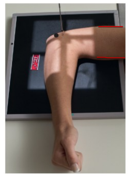
Lateral elbow ( marker should be anterior)
Shoulder, elbow and wrist on same plane
Elbow flexed 90 degrees
Wrist in lateral position
CR to mid elbow joint
1.5 inches medial to the posterior surface of the olecranon process
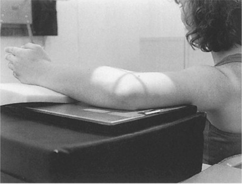
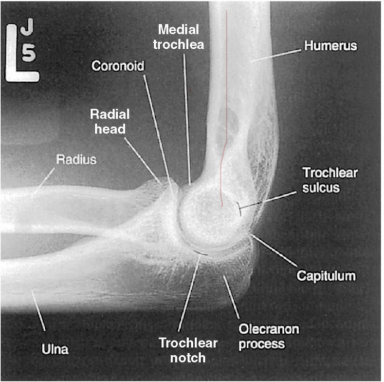
Good lateral elbow example
Lateral elbow image analysis criteria: Position
elbow flexed 90 degrees
about one half of the radial head should be superimposed by the coronoid process
the olecranon process should be visualized in profile
Radial tuberosity is superimposed
True lateral view is indicated by three concentric rings of trochlear sulcus , double ridge of Capitulum and trochlea , and the trochlear notch of the ulna
Superimposition of the humeral epicondyles
Lateral elbow image analysis criteria: Collimation and CR
collimation visible on four sides
elbow joint,1/4 of proximal forearm , distal humerus and lateral soft tissue included within collimation field
CR to the midpoint of the elbow joint
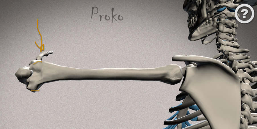
Posterior aspect of what the lateral elbow would look like and how humeral epicondyles are superimposed
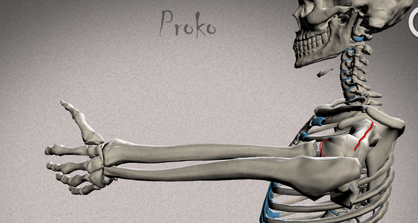
Perspective of lateral elbow and the relationship of the Ulna and Radius
Look at radial head and capitulum
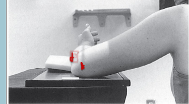
Mispositioning of the Proximal Humerus ( TILT ERROR)
relationships b/w radial & coronoid process
Distal surface of the capitulum to the distal surface of the trochlea
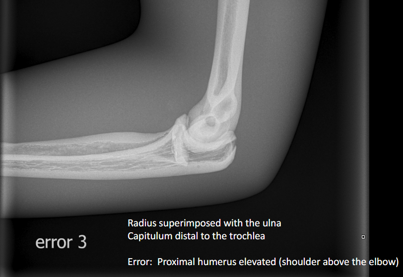
Proximal humerus elevated
Radial head appears superimposed w/ the ulna
Capitulum is more distal than the trochlea ( Trochlea appears superior and capitulum appears inferior)
Elbow is depressed
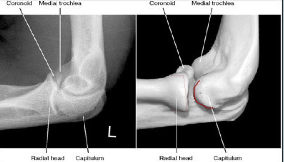
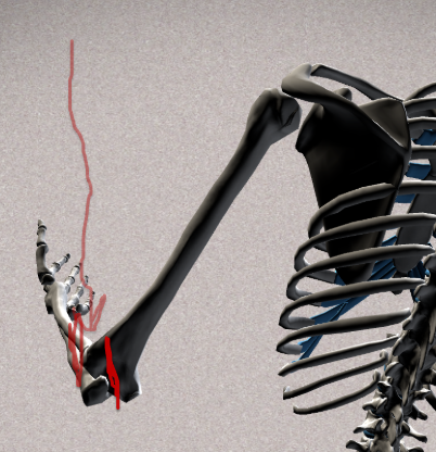
Posterior aspect of Proximal humerus elevated : How capitulum appears inferior to Trochlea
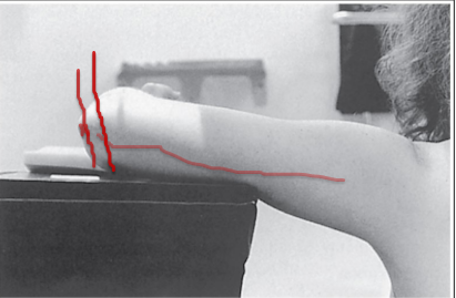
Mispositioning of the Proximal Humerus ( TILT ERROR)
relationships b/w radial & coronoid process
Distal surface of the capitulum to the distal surface of the trochlea
Proximal humerus depressed
Radial head appears barely superimposed with ulna ( they separated)
Distal capitulum appears proximal to the trochlea ( Capitulum appears superior to Trochlea and the trochlea appears inferior to capitulum)
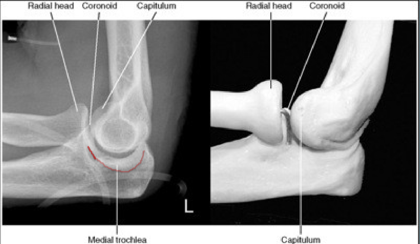
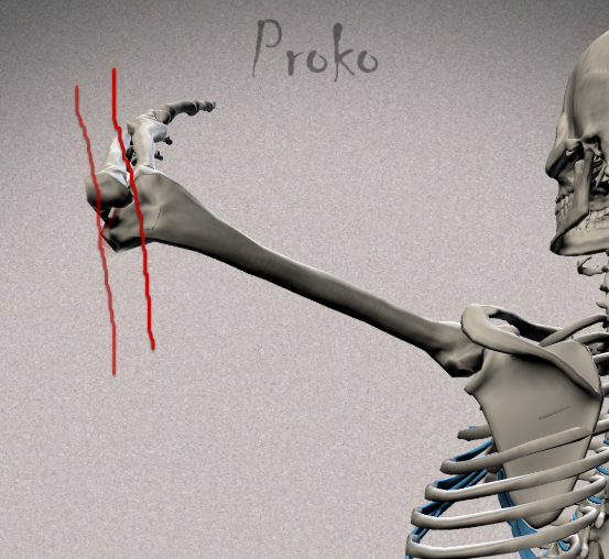
Posterior aspect of Proximal humerus depressed : How capitulum appears superior to Trochlea
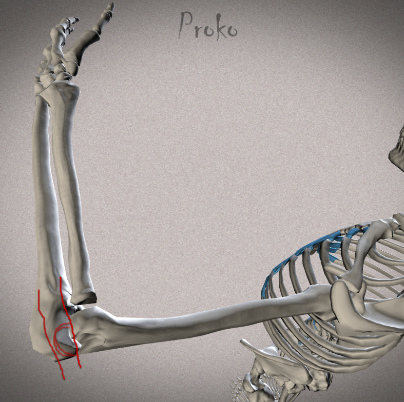
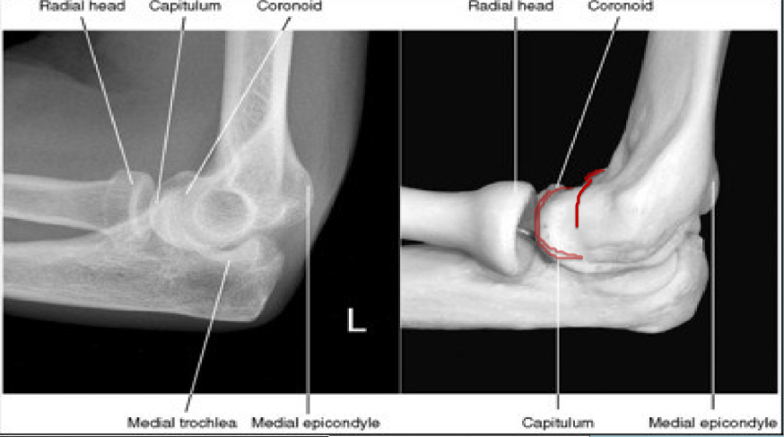
Mispositioning of Distal forearm ( Rotational error )
relationships b/w radial head & coronoid process
Capitulum to trochlea
Forearm depressed
Radial head is distal to coronoid process ( top of radial head can be seen & coronoid process can be outlined w/ no superimposition of the radial head)
Capitulum is anterior ( superimposed with coronoid process)
trochlea moving posteriorly
Medial epicondyle can be seen
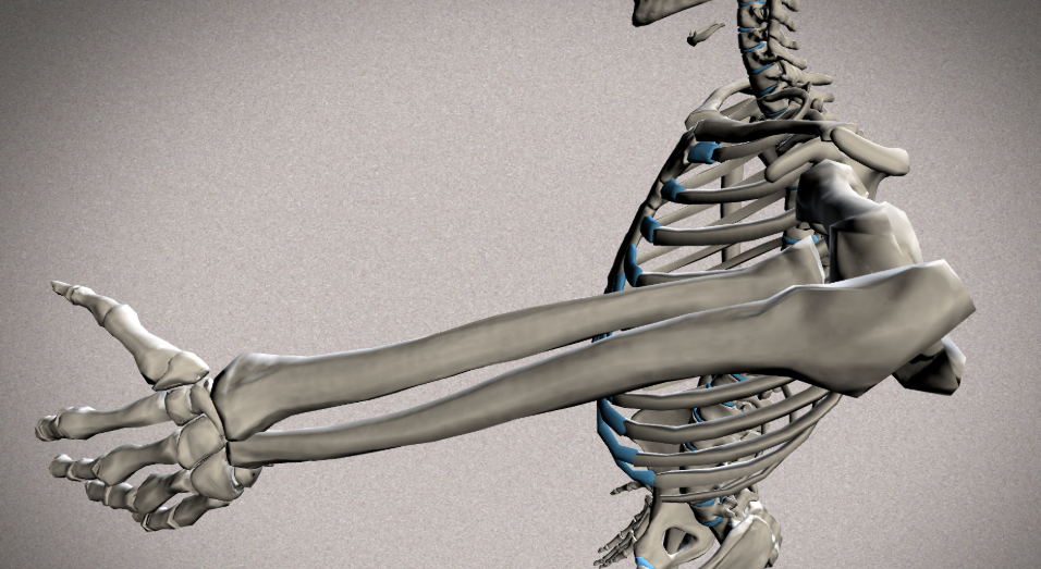
Forearm is depressed and how it looks
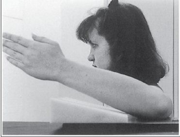
Mispositioning of Distal forearm ( Rotational error )
relationships b/w radial head & coronoid process
Capitulum to trochlea
Forearm elevated
Radial head is proximal to the coronoid process ( coronoid process can be outlined closer to radial neck & completely superimposed by the radius)
Trochlea is anterior ( superimposed w/ the radial head )
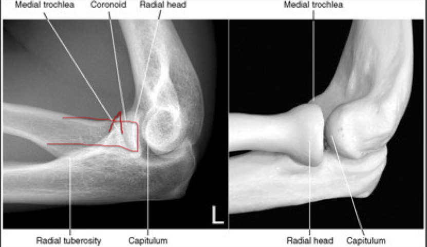
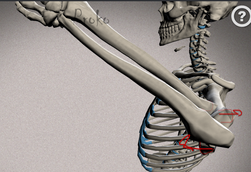
Forearm is elevated and how it looks
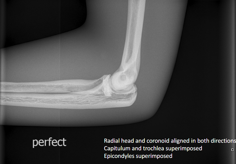
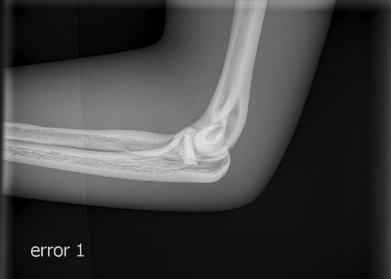
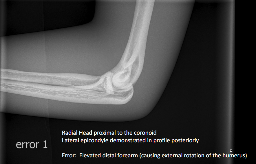
Error : Elevated Distal forearm ( Slanting error)
Radial head proximal to the coronoid
Lateral epicondyle demonstrated in profile posteriorly
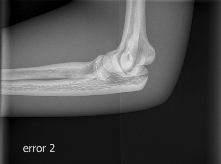
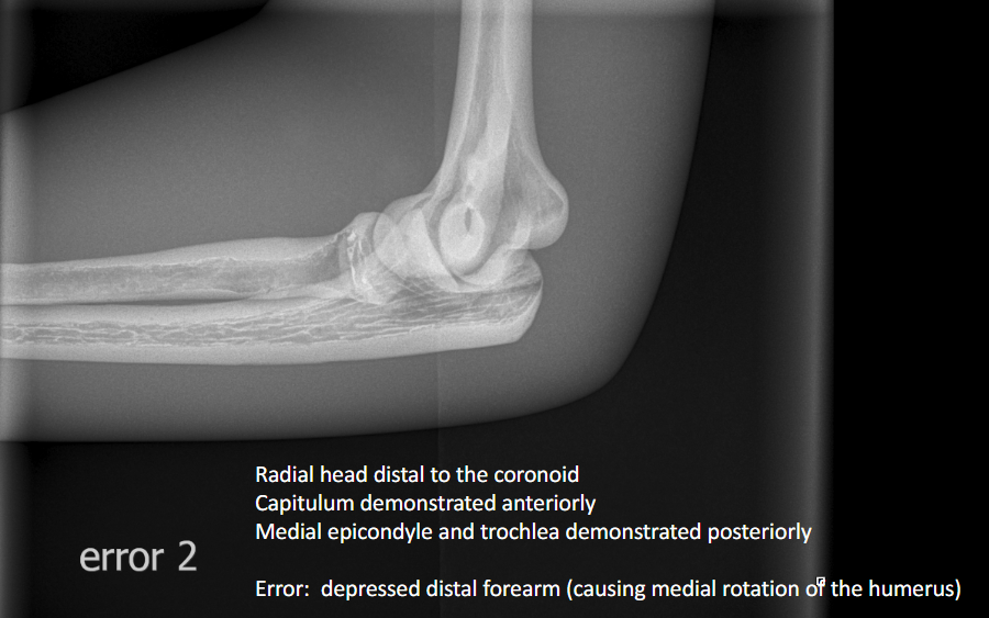
Error: Depressed Distal Forearm ( Slanting error)
Radial head distal to coronoid
Capitulum demonstrated anteriorly
Medial epicondyle and trochlea demonstrated posteriorly
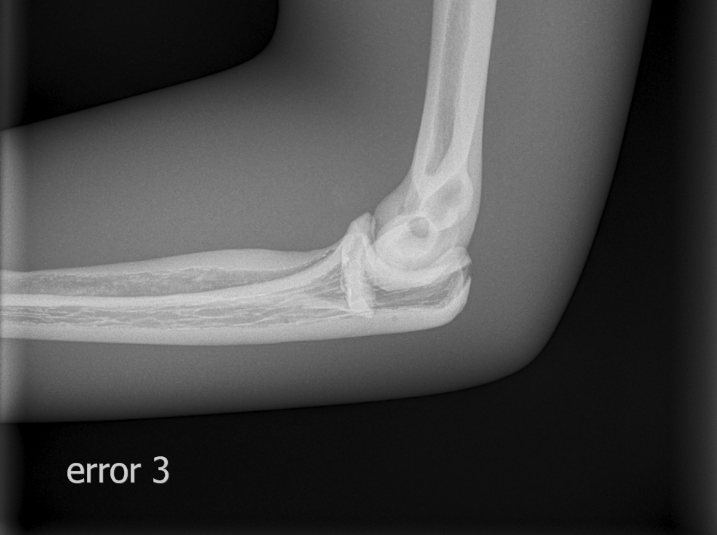
Error: Proximal Humerus elevated ( shoulder above elbow)
Radius superimposed with Ulna
Capitulum distal to the trochlea
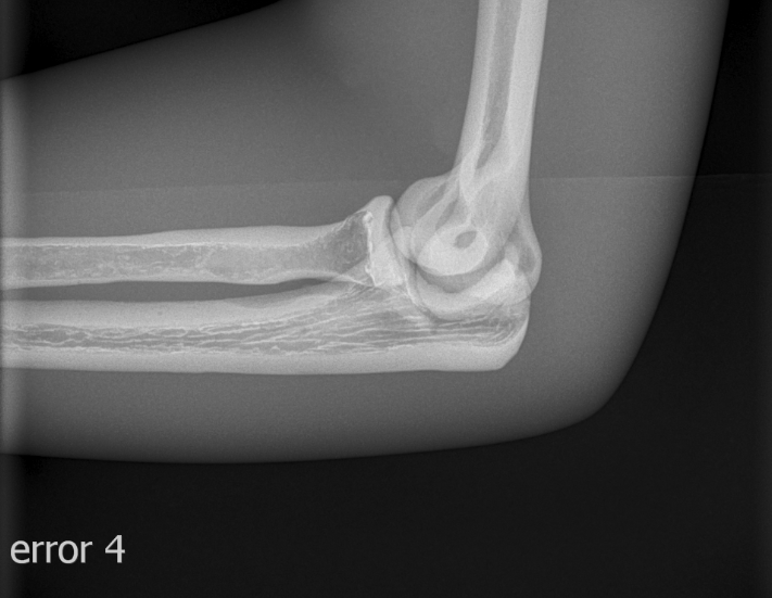
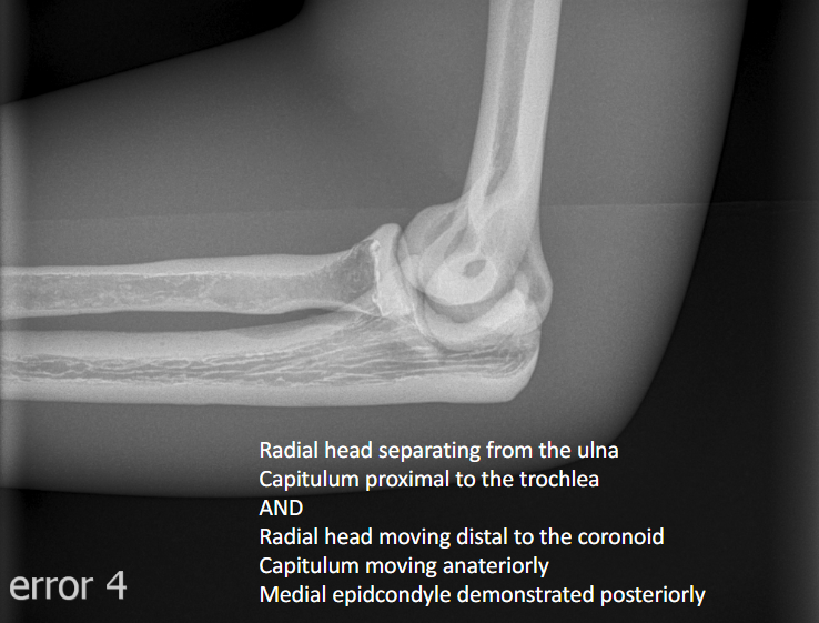
Error: Proximal Humerus depressed and forearm slightly depressed
Radial head separating from ulna
Capitulum proximal to the trochlea
Radial head moving distal to the coronoid
Capitulum moving anteriorly
Medial epicondyle demonstrated posteriorly