Biopsy and medical management of oral pathoses
 Call Kai
 Learn
 Practice Test
 Spaced Repetition
 Match
 Flashcards
 Knowt Play
1/53

Earn XP
Description and Tags
lectures given 10/27/2026
54 Terms
biopsy
removal of tissue from a living body for microscopic diagnostic examination
primary purpose- determine diagnosis to facilitate treatment
rule out malignancy / pre malignant lesions
most oral lesions are benign
what are the indications for biopsy?
any pathological condition that cannot be diagnosed clinically
lesions with no identifiable cause that persists for more than 10-14 days despite local therapy
intra bony lesions that appear to be enlarging
visible / palpable submucosal swelling
lesions with pre-malignant / malignant characteristics
lesions that have grown rapidly
red, white lesions or pigmented lesions where cause is unknown
lesion that is firmly attached or fixed to surrounding structures
ulceration
induration (lesion and surrounding tissue are firm to touch)
associated with lymph node enlargement (infections, TB)
clinical exam and work up to establish differential diagnosis
health history- lesion under investigation may be manifestation of systemic disease (leukemia, crohn’s disease)
medications- any medications that may cause oral manifestations
history of smoking- pack years
history of lesion
duration of lesion- lesion present for several years might be congenital and is more likely to be benign
change in size- clinical/radiographic, rapidly developing lesion is more ominous
change in character- lump becoming an ulcer? / vesicle that turned into ulcer
symptoms associated with lesion
pain- associated with inflammatory / infectious lesions
sensory nerve changes- parasthesia is seen with malignant / inflammatory lesions
swelling- could suggest a cyst / tumor, bony expansion could suggest a chronic process or a fibro osseous lesion
anatomic location involved
keratinized vs non keratinized
unilateral vs bilateral
salivary gland tumor will occur in an area with salivary gland
some lesions have predilection for certain areas or oral cavity
associated systemic signs / symptoms
weight loss- commonly associated with malignant lesions
neck nodes often get enlarged in pts with oral malignancies
fever and malaise are associated with infectious process
viral lesions like HIV may have oral manifestations
some immune mediated diseases manifest in oral cavity in addition to skin lesions
any historical event associated with the onset of lesions
history of trauma / cheek biting in case of ulcers
application of medications not recommended for intra oral use
history of trauma in past- leading to pulp necrosis and manifesting as radicular cyst
clinically examining and documenting the findings
examination includes inspection and palpation
anatomic locations and what type of tissues make up the area? ie lower lip and abundance of minor salivary glands
physical characteristics- ulcer / nodule / soft (lipoma) / firm (neoplasm) / fluctuant (cystic due to fluid) / hard (bony lesions and osseous growth)
single vs multiple lesions, unilateral vs bilateral
raised vs flat, exophytic vs endophytic, sessile (broad based) vs pedunculate
colored lesions that blanch on pressure and fill back up when pressure is released- likely to be vascular
vascular lesions may be pulsatile- invasive procedures such as biopsy are to be avoided in these lesions
lymph node examination
in adults, normal lymph nodes are not palpable
lymph nodes can become enlarged due to inflammation and malignant lesions
surgical procedures do induce inflammation thus causing lymphadenitis
important to establish baseline before doing biopsy- location, size, tenderness, mobile vs fixed, soft vs firm
imaging
radiographs tell us if lesion is arising from bone, eroding bone or causing bony expansion / periosteal reaction
osteomyelitis can be hard to distinguish from malignancy- what can help?
osteomyelitis is more acute and have an inciting event
malignancy is more chronic- but not too long
lab investigations
oral lesions as manifestation of systemic disease- multiple myeloma, hyperparathyroidism, lymphoma, HIV
for hyperparathyroidism- calcium , phosphorus, and alkaline phosphatase
if you decide to refer…
referral appt arranged before pt leaves office
follow up with specialist and make sure pt keeps the appointment with their office
copy of all correspondence kept on records
also the communication from specialist office, biopsy results and recommendations should all be kept
failure to diagnose and refer a pt with pathological condition is a leading cause of litigation
to biopsy or to refer…
depends on pts systemic condition, dentist’s skill and comfort level
surgical difficulty, access, risk of bleeding (vascular lesion?)
iin case of lesions thought to be malignant- perform a biopsy then refer to specialist OR refer to specialist whol will be providing the definitive treatment so that he can evaluate and does the biopsy (chance for the surgeon to see the lesion before any surgical intervention has been compromised)
if pt and dentist choose to not do a biopsy and follow up…
shared responsibility- plan for surveillance must be documented and adhered to
baseline clinical picture and imaging kept on record
making the pt aware of any alarming signs- change in size, shape, color, development of pain/discharge
what are the types of biopsies?
excisional, incisional, aspiration biopsy (radiolucencies, salivary gland swellings, rule out vascular lesions)
excisional biopsy
lesions < 1cm
definitive treatment
excision will not compromise function, risk injury to vessels or nerves
incisional biopsy
small part of lesion is removed
adequate depth- deepn and narrow specimen should be taken
can take multiple samples of characteristics of lesion differ from one site to another
armamentarium
blade
biopsy
sutures to lift, stabilize the tissue
biopsy for ulcerative lesions that are suspected to be carcinoma?
incised tissue should be included edge to ulcer base without extending into normal tissue
extending to include the normal tissue, will risk spreading the tumor
biopsy for immune based lesions
pemphigus
include perilesional tissue (normal appearing mucosa)
how do you send tissue to the lab?
send the tissue in 10% buffered formalin (pH 7), preserves cellular morphology and architecture by forming gel between aldehydes and proteins
SEND PICTURES
how do you send vesiculobullous tissue to the lab? (like pemphigus)
michel’s soultion aka michel’s transport medium
salt solution used to preserve and transport biopsy specimens for immunofluorescence studies
post biopsy
follow up in 10-14 days
next steps will depend on results- referral vs close follow up with imaging as needed
radicular cyst
most common cyst
non-vital tooth / previous trama / endodontically treated tooth

dentigerous cyst
associated with impacted tooth
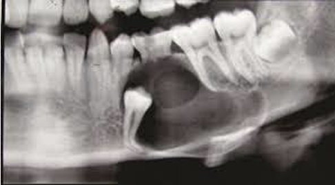
odontogenic keratocyst
high risk for recurrence
aspiration will produce thick yellow cheesy material (keratin)

marsupialization of cyst
useful for very large cyst- where removal will risk jaw fracture
useful in young pts- allows teeth eruption
ideal to decompress cyst so that bone can fill in
can enucleate once cyst is smaller
only part of lining goes for biopsy- disadvantage
cyst enucleation
removal of entire cyst lining- thus the initial step is curvative, also the entire lining is given for pathological exam
ameloblastoma
true neoplasm of odontogenic epithelium
persistent, locally invasive and aggressive
treatment involves resection of the lesion and 1cm margin around the radiographic margins of the lesion

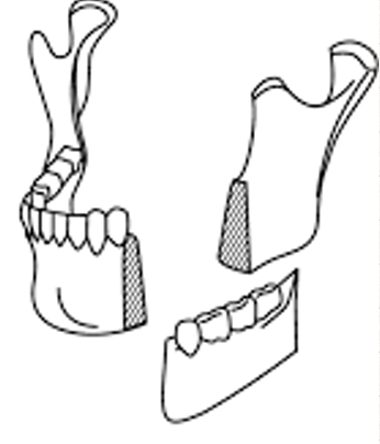
marginal mandibular resection
preserves the lower border

segmental mandibular resection
does not preserve the lower border
mandibular tori

palatal tori

cemento-osseous dysplasia

lots of options / differentials

stafne’s defect

lateral periodontal cyst

cemento-osseous dysplasia
check viability of teeth

pyogenic granuloma
dd- PCGC, POF

epulis fissuratum

apthous ulcers
herpetic ulcers?

dentigerous cysts

squamous cell carcinoma

what are reasons to consider medical therapy over surgery?
bleeding risk
aesthetic zone
high risk for post-traumatic trigeminal neuropathy
patient fear
peds pt
surgery already failed
pts unable to tolerate extensive healing process- extensive scarring, limitation in alimentation, immunosuppression
reduced procedure time
overlap conditions
pyogenic granuloma
leukoplakia/dysplasia
post-radiation fibrosis/trismus
hemangioma
pyogenic granuloma
medical therapies- topical agents (timolol, corticosteroids, imiquimod), sclerotherapy, silver nitrate
dental therapy- scaling and root planning + chlorohexidine glyconate soaks
recurrence after excision ~6% / giant (>2cm) lesions ~22%
risk factors- age (20-30s), female sex, mandibular location
leukoplakia / dysplasia
goal- prevent scc
5 therapeutic options- periodic surveillance and biopsy, excision, lesion destruction (laser, cryotherapy), topical chemotherapy (5-fluorouracil + calcipotriene), topical immunotherapy (imiquimod)
radiation therapy induced trismus
fibrosis of mucosa, skin, salivary gland, bone- hyposalivation, dysphagia, obstructive sleep apnea, edema, mucositis, pain
stretching- manual, pt, appliances, therabite, restorabite (10-14mm over 10 weeks to 12 mo)
low level laser therapy- 400-1500nm and <25 j/cm2 (5-10mm)
pentoxifylline and vitamin E- 400mg PO twice daily and vitamin E 1000 IU PO once daily for 6-24 months (10-15mm over 6 mo)
pirifenidone- 800 mg TID for 3-6 mo
hemangioma
positive on diascopy
require doppler US prior to excision- high flow v low flow
congenital hemangioma- RICH, PICH, NICH
infantile hemangioma- rapidly grows, plateaus, then involutes by school age
therapeutic options for infantile hemangioma
first-line treatement- oral propanolol 2-3mg/kg/day for at least 6 mo (95% clearance)
topical timolol- highly effective for small, superficial lesions, esp in infants younger than 8mo
intralesional pingyangmycin, corticosteroids, and vincristine are reserved for refractory pts
laser and surgery- mainly used for residual changes after involution or where medical therapy is contraindicated or ineffective
study with 853 infants treating with topical timolol, oral propanolol, intralesional pingyangmycin, or IV vincristine - there was an excellent response to therapy, esp in pts under 8 mo (89.1%)
therapeutic options for congenital hemangioma
RICH may involute spontaneously and often require no intervention
NICH and PICH may persist and, due to high flow vascularity and risk of bleeding, are more often managed surgically
sclerotherapy may be considered for select pts, but evidence is limited
propanolol and topical timolol are not effective
surgical excision is often curative for NICH and PICH, with success dependent on lesion size and location