Action Potentials (L6-
 Call Kai
 Learn
Learn Practice Test
 Spaced Repetition
 Match
Match Flashcards
 Knowt Play
1/65

There's no tags or description
Looks like no tags are added yet.
Name | Mastery | Learn | Test | Matching | Spaced | Call with Kai | Chat |
|---|
No analytics yet
Send a link to your students to track their progress
66 Terms
primary ions carry electrical signals in neruons
Na+ , K+ , Cl- , and Ca2+
Mechanism of Na+ and K+ channel opeing during action potential
channels detect changes in membrane potential and reversibly change shape, allowing transient current slow within a narrow time window
direction of current and ion movement in voltage-gated Na+ channels
inward current; Na+ ions enter the cell
response speed and gating characteristics of voltage-gated Na+ channels
fast response time; controlled by voltage-regulated and time-dependent gates
direction of current and ion movement in voltage-gated K+ channels
putward current; K+ ions exit the cell
response speed and regulation of voltage-gated K_ chanels
slow response time; both opening and closing depend on membrane potential
number of conformation states: voltage-gated Na+ channels vs voltage-gated K+ channel
Na+ channels have 3 states (Closed/resting, open, inactivated); K+ channels have 2 states (closed open)
ball and chain model of Na+ channel inactivation
when the channel is closed, the “ball” tethered by a peptide chain floats freely. When the pore opens, the ball is attached to the open pore and physically blocks it
experimental evidence 1 for the ball and chain model
enzymatic removal or deletion via site-directed mutagenesis of the ‘ball’ structure eliminates channel inactivation
experimental evidence 2 for the ball and chain model
adding a synthetic peptide matching the ‘ball’ amino acid sequence restoes inactivation in channels where the ball was previously removed
effect of increasing axonal membrane depolarization on channel conductance
greater membrane depolarization increases the open probability, recruitment, and conductance of both voltage-gated Na+ and K+ channels
comparison of Na+ vs K+ conductance changes over time during depolarization
Na+ conductance increases rapidly immediately after depolarization and inactivates in under 1 ms. K+ conductance increases after a delay and does not rapidly inactivate
conductance changes during the Threshold phase
Initial depolarizing stimulus opens enough voltage-gated Na+ channels so that inward Na+ current exceeds outward background currents, triggering a positive feedback loop
conductance changes during the depolarization phase
Na+ conductance increases rapidly due to fast-opening voltage-gated Na+ channels, causing massive inward Na+ influx K+ channels begin slowly transitioning toward open
Conductance changes at the peak of the action potential
Na+ conductance reaches its max and begins rapidly declining due to ball-and-chain inactivation; delayed voltage-gated K+ channels fully open, raising K+ conductance
Conductance changes during the repolarization
Na+ conductance is minimal due to channel inactivation; high K+ conductance causes outward K+ exit, driving membrane potential back toward equilibrium
conductance changes during After-hyperpolarization (undershoot)
slowing-closing voltage-gated K+ channels remain open, keeping K+ conductance temporarily higher than at resting potential and driving VM toward EK
Stages of the action potential:
Resting membrane potential is near E(K), P(K) > P(Na) due to leaky K+ channels. K+ and Na+ voltage-gates are closed although Na+ time-dependent gate is open
Membrane is brought to threshold voltage by depolarizing stimulus causing the Na+ voltage-dependent gate to open
Na+ flows through open time-dependent and voltage dependent gates. Flow of Na+ into the cell rapidly depolarizes the membrane, causing more Na+ channels to open
Closure of time-dependent Na+ gate and delayed opening of voltage-gated K+ channels halts membrane depolarization
Open voltage-gated K+ channels repolarize the membrane back to a negative membrane potential
Slowly closing voltage-gated K+ channels hyperpolarize membrane towards E(K); Na+ channels return to closed resting value
Negative feedback because of hyperpolarizing causes gate closure for K+
1st stage of the action potential
Resting membrane potential is near E(K) , P(K) >P(Na) due to leaky K+ channels. K+ and Na+ voltage-gates are closed although Na+ time-dependent gate is open
2nd stage of action potential
Membrane is brought to threshold voltage by depolarizing stimulus causing the Na+ voltage-dependent gate to open
3rd stage of action potential
Na+ flows through open time-dependent and voltage dependent gates. Flow of Na+ into the cell rapidly depolarizes the membrane, causing more Na+ channels to open
4th stage if action potential
Closure of time-dependent Na+ gate and delayed opening of voltage-gated K+ channels halts membrane depolarization
5th stage of action potentiak
Open voltage-gated K+ channels repolarize the membrane back to a negative membrane potential
6th stage of action potential
Slowly closing voltage-gated K+ channels hyperpolarize membrane towards E(K); Na+ channels return to closed resting value
Negative feedback because of hyperpolarizing causes gate closure for K+
What will lowering external Na+ concentrations do to the driving force on sodium ions?
Lower driving force
If Na+ in fact the ion responsible for the rising phase of the action potential, how will this affect the action potential?
The peak of action potential becomes smaller
Lower peaks b/c lower concentrations
Activation of Na+ and K+ voltage gated channels and the action potential:
positive feedback: Na+ entry causes depolarization, which opens more voltage gated Na+ channels, which causes more depolarization
negative feedback: voltage gated K+ channels bring the action potential to an end and induce their own closing
Stimulus intensity signaled by:
rate and population coding
rate coding
Strong stimulus allows threshold to be reached earlier in the refractory period
Stronger stimulus elicits larger receptor potential and higher firing rate
population coding
Individual receptors differ in their sensory thresholds
Stronger stimulus excites more receptors‹
encoding mechanism for stimulus strength given the All-or-None Principle
because individual action potentials have fixed amplitude and duration, stimulus intensity, and duration are encoded by the frequency (rate_ and temporal pattern of action potentials, not by potential size
Labelled Line Coding
a neural coding strategy where individual neurons are highly specific/tuned to a single distinct stimulus or feature (eg, a neuron that fires exclusively in response to a bee)
determing stimulus identity in a Labelled line system
Identity is determined by identifying which specific dedicated neuron (or group of identical line-specific neurons) displays the highest firing rate.
Distributed/ Population coding
A coding mechanism where information is represented across the joint pattern of firing rates in an ensemble (population) of broadly tuned neurons, rather than by single highly specific cells.
Advantage of population coding over specificity/labelled-line coding
It allows a small population of broadly-tuned feature neurons to represent a vast number of unique stimuli combinatorially, without requiring a unique dedicated neuron for every object.
combinatorial decoding example using feature-tuned neurons
A stimulus like a 'fly' can be represented when a danger-tuned neuron fires low, a 'big'-tuned neuron fires high, and a 'small'- tuned neuron fires low, identifying the object without a fly- specific cell.
Refractory Period
During an action potential a second stimulus will not produce an action potential
Limits the rate potentials are generated and key in determining the direction of propagation ensure only one direction
Have to wait to have an amplitude like first one --> can’t make action potential
Action potentials can vary across cell types → neurons generate action potentials but differ in way action potentials are generated
Absolute Refractory Period:
It begins with the activation of voltage-gated Na+ channels. Na+ channels either open or inactivate from first action potential
Ends when their inactivation is removed
Neuron incapable of generating another action potential no matter how strong the stimulus
Ensures each action potential is separate and distinct
Relative Refractory Period:
A brief period after the absolute refractory period when repolarization is occurring
It begins when Na+ channel inactivation is removed and ends when the voltage-gated K+ channels deactivate
Threshold for action potential significantly elevated, a strong stimulus can reopen the Na+ gates and allow another action potential
absolute and relative refractory period

Refractory Period during falling phase
Inactivation of the Na+ channels is maximal- few if any channels are available to increase Na+ conductance
K+ conductance is very large
During relative refractory periods, a very large increase in the Na+ conductance is required to override the K+ conductance and initiate a regenerative depolarization

Action potentials can vary across cell types:
Duration of action potentials can range from as little as 200 microseconds to many milliseconds
Rate can vary from a few action potentials/seconds to frequencies near 1000/second
Differences are related to variations in the channel types underlying depolarization and repolarization
How would rapid repolarization influence the action potential frequency
by shortening the overall duration of each individual spike.
When a cell repolarizes quickly, it accelerates the cycle and allows for a higher rate of firing due to the following mechanisms:
Shortened Refractory Period
Faster Membrane Resetting
Shortened refractory period
Fast repolarization enables voltage-gated sodium channels to recover from inactivation much sooner, directly reducing the cell's absolute and relative refractory periods.
Faster Membrane Resetting
The membrane potential returns to its negative resting state rapidly, allowing the cell to be re-stimulated and fire a subsequent action potential almost immediately.
action potentials
code stimulus intensity through change in frequency

graded potential
code stimulus intensity through changes in amplitude
Graded Potential Leading to an Action Potential
If a graded potential is strong, it will cause the cell to fire an action potential(s)
Graded vs action potential
Graded Potential | Action Potential | |
Type of Signal | Input signals | Regenerating conduction signal |
Takes Place | Dendrites, cell body | Axon trigger zone |
Ion Channels Involved | mechanical , chemical, voltage-gated | Voltage-gated channels |
Ions Involved | Na+ , Cl- , Ca2+ | Na+ and K+ |
Type of Signal | Depolarizing or hyperpolarizing | depolarizing |
Strength of Signal | Stimulus dependent, can be summed | All or none, cannot be summed |
Initiator of Signal | Entry of ions through channel | Above threshold stimulus |
Unique Characteristics | Summation, Stimulus Strength | Threshold, Refractory Period |
magnitude | Graded (different amplitudes) | Fixed amplitude |
Other names | PSP (postsynaptic potentials), APP (end-plate potentials, receptor potentials) | Spike or impulse |
Action Potential Propagation
The action potential flows along the nerve axon
Some current is passively lost through the membrane. Two strategies to improve conduction properties:
Increasing diameter of the axon-decreasing the internal resistance
Myelination-increases electrical insulation around the axon
Concentric wrappings of the membrane of glial cells
Act as high resistance, low capacitance electrical insulators
Allows rapid nerve impulse conduction- faster propagation of than non-myelinated fibers of the same axon diameter (10-120 m/sec compared to 1 m/sec)
2 strategies to improve conduction properties
Increasing diameter of the axon-decreasing the internal resistance
Myelination-increases electrical insulation around the axon
Glial Cells
Non-neuronal cells in the brain: neuroglia (or nerve glue)
outnumber neurons
Support cells that provide nutrients to neurons (astrocytes) and the removal of damaged neurons and infection agents (microglia)
Myelin sheaths are made from glia: oligodendrocytes in the CNS and Schwann cells in the PNS
Also participate in the uptake and metabolism of the neurotransmitters and intracellular signaling
Myelination
the process of wrapping nerve fibers in a protective, fatty layer called a myelin sheath, which helps electrical signals travel much faster and more efficiently through the brain and body
The Wire (Axon): Think of a nerve cell's long fiber (the axon) as a copper wire carrying an electrical message.
The Insulation (Myelin): Myelination is like wrapping that wire in rubber electrical tape.
The Result: Just as tape keeps electricity from leaking out of a wire, myelin keeps neural signals strong and lets them "jump" down the fiber, making messages move up to 100 times faster.
Oligodendrocytes:
myelin forming glial cells in the central nervous system. A single oligodendrocyte uses multiple branch like extensions to myelinate multiple separate axons (up to 60) simultaneously
Schwan cells
myelin-forming glial cells in the peripheral nervous system. A single Schwann cell forms myelin around just one axon segment
how does myelination act as an insulator
A multilayered sheath of plasma membrane that wraps around axons and acts as an insulator. Thus, even though the axial path is high resistance due to the small axon diameter, current flows down the length of axon, not through the membrane

Explain how refractory periods limit firing frequency and help produce unidirectional action-potential propagation
Refractory periods set a strict upper limit on how fast a neuron can fire and ensure that action potentials travel in only one direction down the axon.
Here is how they accomplish both:
1. Limiting Firing Frequency
During an action potential, voltage-gated sodium Na+ channels open and then rapidly enter a closed, inactive state.
Absolute Refractory Period: While these Na channels are inactivated, they cannot be reopened by another stimulus, no matter how strong. This creates a hard physical limit on the maximum number of action potentials a neuron can send per second (firing frequency).
Relative Refractory Period: Immediately following, voltage-gated potassium K+ channels remain open, causing hyperpolarization. A new action potential can only be triggered by a much stronger stimulus than usual.
2. Ensuring Unidirectional Propagation
An action potential moves down an axon because the depolarization of one membrane segment generates local currents that depolarize the adjacent patch of membrane.
As the action potential moves forward to Segment B, the previous Segment A enters its absolute refractory period.
When current passively spreads backward from Segment B to Segment A, Segment A's Na+ channels are inactivated and cannot depolarize again.
Therefore, the action potential can only successfully excite the resting, excitable membrane ahead of it (Segment C), forcing the electrical impulse to travel exclusively in a single, forward direction toward the axon terminal.
Limiting Firing Frequency
During an action potential, voltage-gated sodium Na+ channels open and then rapidly enter a closed, inactive state.
Absolute Refractory Period: While these Na channels are inactivated, they cannot be reopened by another stimulus, no matter how strong. This creates a hard physical limit on the maximum number of action potentials a neuron can send per second (firing frequency).
Relative Refractory Period: Immediately following, voltage-gated potassium K+ channels remain open, causing hyperpolarization. A new action potential can only be triggered by a much stronger stimulus than usual.
Ensuring Unidirectional Propagation
An action potential moves down an axon because the depolarization of one membrane segment generates local currents that depolarize the adjacent patch of membrane.
As the action potential moves forward to Segment B, the previous Segment A enters its absolute refractory period.
When current passively spreads backward from Segment B to Segment A, Segment A's Na+ channels are inactivated and cannot depolarize again.
Therefore, the action potential can only successfully excite the resting, excitable membrane ahead of it (Segment C), forcing the electrical impulse to travel exclusively in a single, forward direction toward the axon terminal.
2 types of signal propagation
Continuous (Direct) conduction for unmyelinated axons
Saltatory conduction for myelinated axons
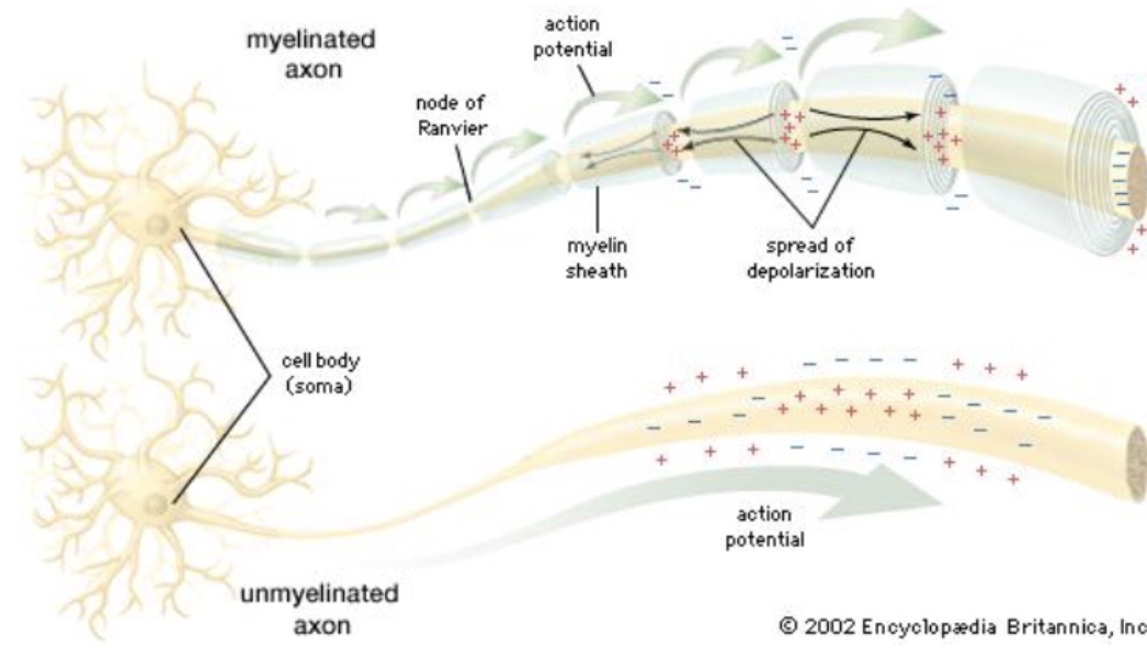
Continuous Conduction
Unmyelinated Axons
Voltage-gated channels must open sequentially all the way down the entire axon membrane.
The action potential travels like a slow wave because every single patch of membrane must regenerate the signal.
speed of propagations are slow, typically ranging from 0.5 to 10 m/s.
Salvatory Conduction
Myelinated Axons
Voltage gated K+ and Na+ channels are located at the nodes
The myelin sheath acts as an electrical insulator preventing ion leakage through the membrane.
Myelin prevents current flow through the membrane
Current moves rapidly and passively beneath the insulated sections.
The signal is regenerated only at exposed gaps, creating a rapid "jumping" effect from node to node.
Speeds reach up to 150 m/s. [1, 2, 3, 4]
Nodes of Ranvier
The myelin sheath is interrupted at regular intervals, forming short uncovered regions
How Myelin and Nodes of Ranvier Increase Speed and Efficiency
Myelin Insulation: Myelin wraps around the axon to increase membrane resistance and decrease capacitance, allowing passive electrical signals to travel fast and far without fading.
Nodes of Ranvier: These short, uncovered gaps interrupt the myelin sheath at regular intervals and contain dense clusters of voltage-gated Na+ and K+ channels.
Regenerative Leaps: When the passive current reaches a node, it triggers these channels to fire, regenerating a full-strength action potential that swiftly moves to the next gap.
Refractory Period: Backward propagation is prevented because the Na+ channels in the recently fired section behind the signal are inactivated in a refractory state.
In what case would it not be helpful to have myelinated neurons?
When the distance the signal has to travel and the available space is very small (eg, gray matter of the brain and spinal cord)
What prevents the action potential from spreading backwards in an unmyelinated axon?
Depolarization of action potential spreads in both directions, but doesn’t cause a backward propagation because previous section just completed an action potential - Na+ channels are inactivated (refractory)