A & P (CH 8 Joints) need chapter 7)
 Call Kai
 Learn
Learn Practice Test
 Spaced Repetition
 Match
Match Flashcards
 Knowt Play
1/127

There's no tags or description
Looks like no tags are added yet.
Name | Mastery | Learn | Test | Matching | Spaced | Call with Kai | Chat |
|---|
No analytics yet
Send a link to your students to track their progress
128 Terms
What are joints? What are they also called?
•also called articulations: sites where two or more bones meet
What is the function of joints?
give skeleton mobility and hold skeleton together
What are the 2 classifications of joints and their subtopics?
1.Structural: three types based on what material binds the joints and whether a cavity is present
▪Fibrous
▪Cartilaginous
▪Synovial
2.Functional classifications: three types based on movement joint allows
▪Synarthroses: immovable joints
▪Amphiarthroses: slightly movable joints
▪Diarthroses: freely movable joints
•Structural classifications are more clear cut, so these will be used here
What are bones joined by in fibrous joints? Do they have a cavity? Are they moveable? What are the different types of fibrous joints?
•Bones joined by dense fibrous connective tissue
•No joint cavity
•Most are immovable
–Depends on length of connective tissue fibers
•Three types of fibrous joints
–Sutures
–Syndesmoses
–Gomphoses
What are sutures? What fibers do they contain? What happens to sutures in middle age and what do they become? What movement?
•Sutures= Rigid, interlocking joints of skull
•Allow for growth DURING YOUTH
–Contain short connective tissue fibers that allow for expansion
•In MIDDLE AGE, sutures ossify and fuse, become SYNOSTOSES (immovable sutures)
–Immovable joints make skull into one unit that protects brain
What are syndesmoses (how are bones connected)? What type of fibers and what are some examples? What movement?
•Bones connected by ligaments, bands of fibrous tissue
•Fiber length varies, so movement varies
–Short fibers offer little to no movement
▪Example: inferior tibiofibular joint
–Longer fibers offer a larger amount of movement
▪Example: interosseous membrane connecting radius and ulna
What are gomphoses? What is an example? What is the fibrous connection?
•Peg-in-socket joints
•Only examples are the teeth in alveolar sockets
•Fibrous connection is the periodontal ligament
–Holds tooth in socket
What are bones joined by in cartilaginous joints? Do they have a cavity? Are they moveable? What are the different types of cartilaginous joints?
•Bones united by cartilage
•Like fibrous joints, have no joint cavity
•Not highly movable
•Two types
Synchondroses
Symphyses
What are bones united by in synchondroses? What type of movement do they have? What are some examples?
a bar or plate of hyaline cartilage
Examples:
Temporary epiphyseal plate joints
▪Become synostoses after plate closure
Cartilage of 1st rib with manubrium of sternum
What are bones united by in symphyses? What type of movement do they have, are they weak or strong? what other cartilage is present? What are some examples?
fibrocartilage in symphysis joint
Symphyses are strong, amphiarthrotic (SLIGHTLY MOVABLE) joints
Hyaline cartilage also present as articular cartilage on bony surfaces (end plates, superior and inferior - for the intervertebral discs)
Examples:
–Intervertebral joints
–Pubic symphysis
What are bones separated by in synovial joints? What type of movement do they have?
•Bones separated by fluid-filled joint cavity
•All are diarthrotic (FREELY movable)
what type of joint contain almost all limb joints?
synovial
What are some characteristics of synovial joints?
general features
–Have bursae and tendon sheaths associated with them
Stability is influenced by three factors
▪shape of joint,
▪ligaments,
▪muscles
–Classified into SIX different types, depending on movements allowed
What are the 6 general features of synovial joints?
Articular cartilage
Joint (synovial) cavity
Articular (joint) capsule
Synovial Fluid
Ligaments
Nerves and blood vessels
Describe each of the general features.
1.Articular cartilage: hyaline cartilage covering ends of bones
▪Prevents crushing of bone ends
2. Joint (synovial) cavity: small, fluid-filled potential space unique to synovial joints
3. Articular (joint) capsule: two layers thick
▪External fibrous layer: dense irregular connective tissue
▪Inner synovial membrane: loose connective tissue that makes synovial fluid
4.Synovial fluid: viscous filtrate of plasma and hyaluronic acid
▪Lubricates and nourishes articular cartilage
▪Contains phagocytic cells to remove microbes and debris
5. Different types of reinforcing LIGAMENTS
▪Capsular: thickened part of the capsule
▪Extracapsular: outside the capsule
▪Intracapsular: deep to capsule; covered by synovial membrane
6.Nerves and blood vessels
▪Nerves detect pain, joint position and stretch
▪Capillaries supply filtrate for synovial fluid
What are 2 other features of synovial joints?
fatty pads
articular discs
these are for cushioning between fibrous layer of capsule and synovial membrane or bone
fatty pads
these are fibrocartilage that separates articular surfaces to improve “fit” of bone ends, stabilize joint, and reduce wear and tear
Articular discs (menisci)
These are:
Not strictly part of synovial joints, but closely associated
Bags of synovial fluid that act as lubricating “ball bearing”
Bursae and tendon sheath
reduce friction where ligaments, muscles, skin, tendons, or bones rub together
Bursae
elongated bursae wrapped completely around tendons subjected to friction
tendon sheath
What are the 3 factors that determine the stability of joints to prevent dislocations?
1.Shape of articular surface (minor role)
▪Shallow surfaces less stable than ball-and-socket
2.Ligament number and location (limited role)
▪The more ligaments, the stronger the joint
3.Muscle TONE keeps tendons taut as they cross joints (most important)
▪Extremely important in reinforcing
–shoulder
–knee joints
arches of the foot

•All muscles attach to bone or connective tissue at no fewer than _____ points
2
this is the attachment to immovable bone
origin
this is the attachment to movable bone
insertion
What does muscle contraction cause?
insertion to move toward origin
What are the 3 planes of motion?
transverse
frontal
sagittal
What are the 4 different kinds of range of motion (ROM) allowed by synovial joints?
–Nonaxial: slipping movements only
–Uniaxial: movement in one plane
–Biaxial: movement in two planes
–Multiaxial: movement in or around all three planes
What are the 3 general types of movements?
–Gliding
–Angular movements
–Rotation
One flat bone surface glides or slips over another similar surface
gliding movement
What are examples of gliding movements?
▪Intercarpal joints
▪Intertarsal joints
▪Between articular processes of vertebrae
changes angle between 2 bones.
angular movement
What do angular movements include in the sagittal plane?
▪Flexion: decreases angle of the joint
▪Extension: increases angle of the joint
–Hyperextension: movement beyond the anatomical position
What do angular movements include in the frontal plane?
–Abduction: movement along frontal plane, away from the midline
–Adduction: movement along frontal plane, toward the midline
What does circumduction involve?
▪Involves flexion, abduction, extension, and adduction of limb
▪Limb describes cone in space
•: turning of bone around its own long axis, toward midline or away from it
rotation
rotation toward the midline.
rotation away from the midline.
medial
lateral
What are some examples of rotation in the body?
▪Rotation between C1 and C2 vertebrae
▪Rotation of humerus and femur
Describe supination and pronation
–Supination and pronation: rotation of radius and ulna
▪Supination: palms face anteriorly
–Radius and ulna are parallel
▪Pronation: palms face posteriorly
–Radius rotates over ulna
(transverse plane)
What is flexion and extension called in the foot?
–Dorsiflexion and plantar flexion of foot
▪Dorsiflexion: bending foot toward shin
▪Plantar flexion: pointing toes
Describe inversion and eversion
▪Inversion: sole of foot faces medially
▪Eversion: sole of foot faces laterally
mandible juts out
protraction
mandible is pulled towards neck
retraction
what plane do protraction and retraction occur in?
movement in lateral plane (scapula too!)
lifting body part superiorly
elevation
lowering body part
depression
movement of the thumb
opposition
▪Example: touching thumb to tips of other fingers on same hand or any grasping movement

What are the six different types of synovial joints?
–Categories are based on shape of articular surface, as well as movement joint is capable of
▪Plane
▪Hinge
▪Pivot
▪Condylar
▪Saddle
Ball-and-socket
What are examples of plane joints? What is their movement?

Movement: nonaxial- gliding
What are examples of hinge joints? What is their movement?

What are examples of pivot joints?

What are examples of condylar joints? What is their movement?

What are examples of saddle joints?What is their movement?

What are examples of ball and socket joints? What is their movement?
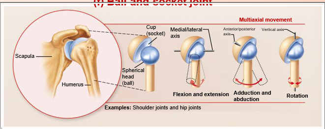
What are the 5 main synovial joints?
Jaw (Temporomandibular Joint)
Shoulder
Elbow
Hip
Knee
•Synovial joints are diverse
•All have general features, but some also have unique structural features, abilities, and weaknesses
What is the most easily dislocated joint in the body?
The temporomandibular joint
What is type of joint is the temporomandibular joint (TMJ)? What articulates with what in it? Does it have a strong or weak ligament?
•Modified hinge joint
•Mandibular condyle articulates with temporal bone
•Strong lateral ligament
In TMJ, What does the posterior temporal bone form and what does the anterior portion of the temporal bone form? What happens when your mouth opens and when you bite something big?
–Posterior temporal bone forms mandibular fossa, while anterior portion forms articular tubercle.
When the mouth OPENS, the condyle moves FORWARD, on the articulartubercle – when you BITE something big, the condyle presses against the tubercle, NOT the weak spot in the middle of the mandibular fossa!
This separates a superior and inferior compartments of the synovial cavity in the TMJ.
articular disc
What type of movements occur in the temporomandibular joint?
–Hinge: depression / elevation of mandible
–Gliding: side-to-side (lateral excursion) grinding of teeth
–Sliding: protraction and retraction
Why is dislocation most common in the TMJ? How does it typically dislocate? What are some symptoms of a dislocated TMJ?
•Dislocation of TMJ is most common because of shallow socket of joint
•Almost always dislocates anteriorly, causing mouth to remain open
–To realign, physician must push mandible back into place
•Symptoms: ear and face pain, tender muscles, popping sounds when opening mouth, joint stiffness
What often causes TMJ? What are some possible treatments?
•Usually caused by grinding teeth, but can also be due to jaw trauma or poor occlusion
–Treatment for grinding teeth includes bite plate
–Relaxing jaw muscles helps
What is the most freely moving joint in the body?
the shoulder (Glenohumeral) joint
What type of joint is the shoulder (glenohumeral) joint? What articulates with what?
•BALL-AND-SOCKET joint
–Large, hemispherical head of humerus fits in small, shallow glenoid cavity of scapula
▪Like a golf ball on a tee
What is sacrificed for what in the glenohumeral joint?
•Stability is sacrificed for freedom of movement
What is the glenoid labrum? What is its role in the glenohumeral joint?
•fibrocartilaginous rim around glenoid cavity
–Adds depth to shallow cavity
–Cavity still ONLY HOLDS ONE-THIRD OF HEAD of humerus
_______ ________ enclosing cavity is also thin and loose
–Contributes to freedom of movement (in the shoulder joint)
articular capsule
Where are reinforcing ligaments primarily in the shoulder joint? What are the two kinds of reinforcing ligaments in the shoulder? What are each of their roles?
–Primarily on anterior aspect
–CORACOHUMERAL ligament
▪Helps support weight of upper limb
–Three GLENOHUMERAL ligaments
▪Strengthen anterior capsule,
but are weak support
•Reinforcing _____ _______ contribute most to joint stability
–__________ muscle is “superstabilizer” (in the shoulder joint)
•Reinforcing muscle tendons contribute most to joint stability
–Tendon of long head of biceps brachii muscle is “superstabilizer”
–Tendon of long head of biceps brachii muscle is “superstabilizer”
▪Travels through _________
▪Secures _______ to ___________
–Tendon of long head of biceps brachii muscle is “superstabilizer”
▪Travels through intertubercular sulcus
▪Secures humerus to glenoid cavity
What are the 4 rotator cuff tendons that encircle the shoulder joint?
▪Subscapularis
▪Supraspinatus
▪Infraspinatus
▪Teres minor
What articulates with what in the elbow joint? What type of joint is the elbow joint?
•Humerus articulates with radius and ulna
Hinge joint
What is the hinge joint in the elbow primarly formed from? What type of movement does a hinge joint allow for in the elbow?
•Hinge joint formed primarily from trochlear notch of ulna articulating with trochlea of humerus
–Allows for flexion and extension only
________ surrounds head of radius- _______ joint for head of radius rotation around its axis
Anular ligament surrounds head of radius- PIVOT joint for head of radius rotation around its axis
What are the two capsular ligaments in the elbow? What do they restrict?
•Two capsular ligaments restrict side-to-side movement
–Ulnar collateral ligament
–Radial collateral ligament
In the elbow joint BOTH ________ (ulnar and radial) actually join _____ and _______, on lateral and medial side (the radial collateral is on the lateral side, where radius is located, hence the name)
BOTH collaterals (ulnar and radial) actually join ulna and humerus, on lateral and medial side (the radial collateral is on the lateral side, where radius is located, hence the name)
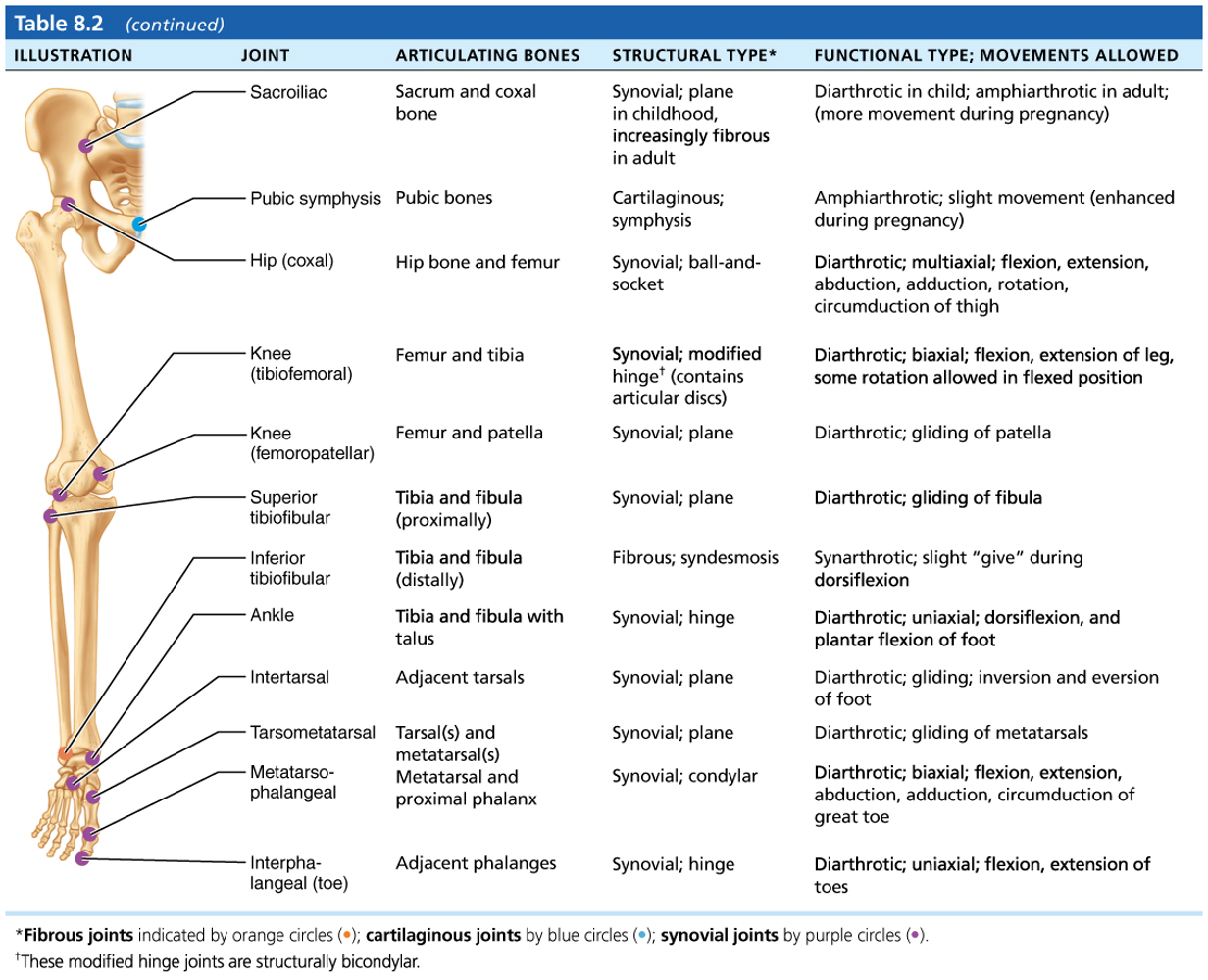
What type of joint is the hip joint? What articulates with what in the hip joint? What is the range of motion like?
•Ball-and-socket joint
•Large, spherical head of the femur articulates with deep cup-shaped acetabulum on hip bone
•Range of motion limited by the deep socket
rim of fibrocartilage that enhances depth of socket (hip dislocations are rare)
acetabular labrum
What are the 4 reinforcing ligaments in the hip and coxal bones? What does the last one contain?
–Iliofemoral ligament - inverted Y-shaped
–Pubofemoral ligament
–Ischiofemoral ligament
–Ligament of head of femur (ligamentum teres)
▪Slack during most hip movements, so not important in stabilizing
▪Does contain artery that supplies head of femur
What does the greatest stability come from in the hip and coxal bones?
the deep ball and socket joint
What is the largest and most complex joint of the body?
Knee joint
What are the 3 joints surrounded by a single cavity in the knee?
1. Femoropatellar joint
2. Lateral joint and
3. Medial joint
What type of joint are each of the three? What movement do they allow for?
1. Femoropatellar joint
▪Plane joint
▪Allows gliding motion during knee flexion
2. Lateral joint and
3. Medial joint
▪Lateral and medial joints together are called tibiofemoral joint
▪Joint between femoral condyles, tibial condyles, and lateral and medial menisci of tibia
▪Hinge joint that allows flexion, extension, and some rotation when knee partly flexed!
Knee joint:
•Capsule is thin and ____ _______
•Anteriorly, _____ ________ gives rise to three broad ligaments that run from patella to tibia
•Capsule is thin and absent anteriorly
•Anteriorly, quadriceps tendon gives rise to three broad ligaments that run from patella to tibia
What are the three broad ligaments that run from patella to tibia. Whick does a doctor tap to test knee jerk reaction?
–Medial and lateral patellar retinacula that flank the patellar ligament
▪Doctors tap patellar ligament to test knee-jerk reflex
•At least ___ bursae associated with knee joint
•At least 12 bursae associated with knee joint
What ligaments stabilize the knee joint?
•Capsular, extracapsular, or intracapsular ligaments stabilize the knee joint
What do capsular and extra capsular ligaments help prevent?
hyperextension
What are the 3 examples of capsular and extracapsular ligaments in the knee?
–Fibular and tibial collateral ligaments: prevent rotation when knee is extended
–Oblique popliteal ligament: stabilizes posterior knee joint
–Arcuate popliteal ligament: reinforces joint capsule posteriorly
•INTRACAPSULAR ligaments reside within capsule, but ____ ______ cavity
•INTRACAPSULAR ligaments reside within capsule, but outside synovial cavity
What do intracapsular ligaments help prevent?
anterior - posterior displacement
What are the 2 main intracapsular ligaments in the knee? Where do each of them attach and what do they prevent?
–Anterior cruciate ligament (ACL)
▪Attaches to anterior tibia
▪Prevents forward sliding and internal rotation of tibia, stops hyperextension of knee
▪
–Posterior cruciate ligament (PCL)
▪Attaches to posterior tibia
▪Prevents backward sliding of tibia
What are common joint injuries?
Cartilage tears
- Sprains
- Dislocations
–Due to compression and shear stress
–Fragments may cause joint to lock or bind
cartilage tears
–______ rarely repairs itself
–Repaired with _____ _____
–PARTIAL menisci removal renders joint less ___ but mobile; COMPLETE REMOVAL leads to __________
–Meniscal _______ possible in younger patients
Cartilage rarely repairs itself
–Repaired with arthroscopic surgery
–PARTIAL menisci removal renders joint less stable but mobile; COMPLETE REMOVAL leads to osteoarthritis
–Meniscal transplant possible in younger patients
ligaments are stretched or torn
sprains
what are common sites of sprains?
–Common sites are ankle, knee, and lumbar region of back