Rad Bio - Dose (Unit 1-4)
 Call Kai
 Learn
 Practice Test
 Spaced Repetition
 Match
 Flashcards
 Knowt Play
1/173

There's no tags or description
Looks like no tags are added yet.
174 Terms
Patient exposure can be substantially reduced by (FILSERP):
Adequate filtration of the x-ray beam
Use of proper body or body part immobilization and other motion reduction techniques
Proper x-ray beam limitation devices
Use of specific area shielding
Selection of suitable technical exposure factors
Elimination of repeat radiographic exposures
Use of appropriate digital image processing
_________ communication:
Encourages reduction in __________________.
Increases the chance for successful completion of x-ray reducing the potential for ______________.
Good
Anxiety and emotional stress
Repeat exposures
AIDET
Acknowledge, Introduce, Duration, Explanation, Thank You
If the procedure will cause pain, discomfort, or any strange sensations, ______________ is essential.
Constant communication
Inadequate instructions may prevent the patient from ______________________.
Being able to cooperate as needed.
Filtration
Hardening the beam because it removes low-energy (soft) photons.
Low-energy photons cause _________ radiation dose to the patient because _______________.
Increased
They have insufficient energy to exit the patient.
Aluminum filters are used to reduce ___________ by removing low-energy photons, thereby decreasing ___________.
Patient dose
Beam intensity
Beam filtration results in an x-ray beam of ______ average energy.
Higher
The purpose of built-in filtration is to _______________________.
Reduce patient skin dose
Filtration does not affect _____________ because techniques are calibrated higher to compensate.
Receptor exposure
Motion obliterates ____________.
Recorded detail
Good communication is the most effective method of reducing _________________.
Voluntary motion
Most important method to reduce involuntary motion is to use ________________________.
Shortest possible exposure time.
___________________ is probably the single most important factor in keeping patient dose to a minimum.
Beam restriction/limitation of field size
Beam restriction does what?
Reduces patient dose
Reduces production of scattered radiation
Improves image quality
Types of beam limitation:
Aperture diaphragm
Cone/cylinder
Collimator
______ do little or nothing to benefit the patient and should not be used routinely.
Shields
There is _______ evidence that radiation from medical imaging damages reproductive cells.
No direct
Fetal and gonadal shielding should/should not be routinely used, regardless of patient's age, sex, or pregnancy status?
Should not
In very limited circumstances, shielding may be carefully used if in the best interest of ______________.
An extremely anxious patient
FOR ARRT EXAM:
Shield the patient ____, ____, ____, and ____.
Gonads
Lens of the eye
Breasts
Thyroid gland
In an effort to keep radiation dose to a minimum, use the ______ mAs and the ________ kV that will maintain image quality.
Lowest
Highest
When an air-gap technique is used, much of the scattered radiation generated within the patient __________ the IR.
Never reaches
An air-gap technique may be occasionally used in place of a ______.
Grid
ABC
Automatic Brightness Control. Type of fluoroscopic AEC.
In stationary fluoro, positioning the image intensifier closer to the patient ________ the SID and ________ the beam intensity.
Decreases
Increases
When ABC is activated, the mA is _______ to compensate for the increase in intensity.
Decreased
To maintain an optimal level of brightness, the x-ray tube mA is decreased, which decreases the ___________.
Patient ESE dose
Moving the II closer to the patient results in a shorter OID, and therefore decreased ____________ and improved _________.
Magnification
Image quality
ESE for a single radiograph exposure = ________x____
Output intensity
mAs
For live fluoro, we rely on the accepted standard that the average ESE is _______.
40 mGya/min
A fluoro procedure requires 2.5 min at 90 kVp and 2 mA. What is the approximate ESE?
100 mGya

Exams with the highest repeat rates include: (4)
Lumbar spine
Thoracic spine
Chest
Abdomen
Repeat rates should not exceed _____.
5%
Unnecessary Patient Radiation Dose
Any radiation dose that is not required for the patient’s well-being or for proper management and care.
Routine x-ray exam should not be performed when there is no precise ______________.
Medical indication
A clinician may be sued if lack of an exam results in ____________________________.
Improper management of the patient’s condition.
Chest x-ray screening may be appropriate in which high-risk groups?
Medical and paramedical personnel
Service personnel posing a potential community hazard (ex. food handlers, teachers)
At-risk occupational groups
Artifacts
Unwanted densities in the image that are not part of the patient’s anatomy and may negatively affect the ability of a radiologist to interpret the image correctly.
A quality control program includes regular monitoring and maintenance of all __________ and ____________ equipment.
Processing
Image display
A quality control program promotes quality assurance by decreasing the likelihood of producing ___________ or _________.
Suboptimal-quality images
Repeat doses
Which of the following are methods of reducing patient dose? (select all that apply)
Reducing filtration of the x-ray beam
Immobilization of patient
Increasing beam restriction
Eliminating repeats
High mAs and low kVp
Use of appropriate digital image processing
Immobilization of patient
Increasing beam restriction
Eliminating repeats
Use of appropriate digital image processing
Amount of radiation recieved by a patient form diagnositc imaging procedures may be presented in three ways:
Entrance skin exposure (ESE)
Gonadal dose
Bone marrow dose
___ is the most frequently reported dose to patient because it is the simplest to determine.
ESE
_____ refers to the dose to the epidermis.
ESE
_____________ are the sensing devices most often used to determine skin dose.
Thermoluminescent Dosimeters (TLD)
________ is a family of curves from which one can estimate the output intensity of a radiographic unit with a particular technique.
Nomogram
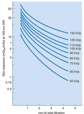
Referring to the nomogram, what is the approximate patient ESE from an AP projection of the abdomen made at 105 cm using 80 kVp, 300 mA, 50 ms, and 2.5 mm Al filtration?
90 mGy (15 mAsx 6 mGy/mAs)

During radiography and fluoro, radiation scatters from the patient ______________.
In all directions
The approximate intensity/quantity of scattered radiation at 1 m from the patient is ___ of ESE dose.
0.1%
Whatever the ESE value, the exit skin dose will be approximately ___ of the ESE.
1%
Currently, the AAPM no longer subscribes to the long-held theory that genetic effects may result from exposure to routine x-ray exams, requiring _________________.
Protection of reproductive organs
Bone marrow contains large numbers of stem blood cells that could be either ________ or ___________ by substantial exposure to ionizing radiation.
Depleted
Eliminated
Bone marrow dose may also be described in terms of ____________, which is defined as the average radiation dose to the entire active bone marrow.
Mean marrow dose
The most radiosensitive of bone marrow cells are _________.
Lymphocytes
The threshold for lymphocytes to demonstrate depletion is ____, although all cells will decrease in number following a large dose of ionizing radiation.
0.25 Gy/25 rad
___% of bone marrow in adults is in the thoracic vertebrae, ___% is in lumbar vertebrae, ___% is in the sacrum, ___% is in the lower limb girdle.
13
11
11
29
The exam with the highest ESE dose is ____, with a dose of ___.
CT Head
40.0 mGy
The exam with the highest bone marrow dose is ________ with a dose of ___.
Lumbar spine
0.60 mGy
The exam with the highest gonadal dose is ________ with a dose of ___.
CT Pelvis
20.0 mGy
Which of the following exams produce the highest patient ESE?
CT Head

Referring to the nomogram, what is the approximate patient ESE from a particular projection made at 105cm using 60 Kvp, 300 mA, 5 ms, and 2.5 mm Al total filtration?
4.5 mGy

ALARA
As low as reasonably achievable
Most occupational exposure comes from ____________.
Patient scatter
3 Cardinal Principles of Radiation Protection
Time
Distance
Shielding
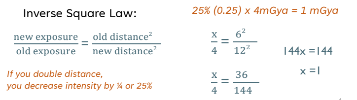
Inverse Square Law Equation

If a radiographer stands 6 m away from an x-ray tube and receives an exposure rate dose of 4.0 mGya/hr, what will the exposure rate dose be if the same radiographer moves to stand at a position located 12 m from the x-ray tube?
1 mGya/hr
Occupational Effective Dose
50 mSv
Occupation Dose for Lens of Eye
150 mSv
Occupational Dose for Skin, Hands, Feet
500 mSv
Cumulative Occupation Dose
10 mSv x age
Public Effective Dose
1 mSv
Public Dose Lens of Eye
15 mSv
Public Dose for Skin, Hands, and Feet
50 mSv
Total Equivalent Dose for Embryo
5 mSv
Equivalent Dose 1 Month for Embryo
0.5 mSv
Negligible Individual Dose Limit
0.01 mSv
Effective Dose (EfD) annual limits are used for ______________________.
Whole body and whole body cumulative
Equivalent Dose (EqD) annual limits for __________________.
Tissues and organs (more localized)
_______________ is potential for death or overall poorer quality of life.
Occupational risk
Methods of reducing scatter radiation also reduce the occupational hazard for the radiographer:
Beam limitation devices
Reduction of repeats
Higher kVp and lower mA techniques
Appropriate beam filtration
Proper utilization of protective apparel
At least _____, aprons and gloves should be put under fluoro to check for cracking.
Once a year
Lead aprons are what type of radiation barrier?
Secondary
Federal regulation requires at least ____________ equivalent for lead aprons.
0.25 mm Pb
Per NCRP recommendations, most departments use ______________ lead aprons.
0.5 mm Pb
Pregnant rad techs should wear a fetal dosimeter at ____ level, ___ lead shielding to ensure monthly EqD does not exceed ____.
Waist
Under
0.5 mSv
True or False: The pregnant radiographer needs to be reassigned to a lower radiation exposure position.
False. This would put non-pregnant technologists at higher occupational risk.
Primary Barriers
Any surface that can be struck by the primary beam.
What is the height and lead required for primary barriers?
1.6 mm (1/16”) of lead up to 7 ft high
Secondary Barriers
Any surface that can be struck with scatter/leakage radiation.
What is the height and lead required for secondary barriers?
Includes that portion of the walls above 7ft high and the control booth. 0.8 mm (1/32”) Pb
Use Factor
Describes the % of time that the primary beam is directed toward a particular wall.
Workload
Determined by the # of x-ray exposures made per week.
mAs/wk
Controlled Area
Occupied by trained radiation workers wearing radiation montiors.
Uncontrolled Area
Occupied by the general population
Leakage radiation must be less than _________________ at a distance of 1 m from the protective housing.
1 mGya/hr
The intensity of scatter radiation 1m from the patient is approximately ________ of the intensity of the incident beam at the patient.
The Code of Federal Regulations (CFR) states that monitoring should be provided for occupationally exposed individuals who are likely to receive ______ the dose-equivalent limit.
>1/10
What are the types of personal radiation monitors: (5)
Optically Stimulated Luminescence (OSL)
Thermoluminescent Dosimeter (TLD)
Film Badge
Pocket Dosimeter
Direct Ion Storage Device (DIS)