Ch. 7 - Skeletal System Essentials of Anatomy and Physiology - Holes
 Call Kai
 Learn
 Practice Test
 Spaced Repetition
 Match
 Flashcards
 Knowt Play
1/70

There's no tags or description
Looks like no tags are added yet.
71 Terms
epiphysis
expanded end; distal and proximal

diaphysis
bone shaft

Articular cartilage
cartilage at the end of epiphysis

articular cartilage
covers epiphysis

Periosteum
strong membrane around bone (dense irregular connective tissue)

Endosteum
thin membrane that lines the medullary cavity (reticular connective tissue)

compact bone
consists of osteons; weight bearing, resist compression (wall of diaphysis)
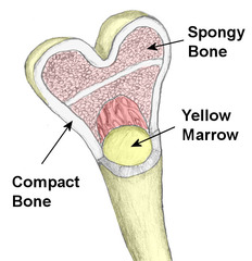
spongy bone
consists of trabeculae; somewhat flexible, nutrients diffuse through canaliculi (makes up epiphysis)

trabeculae
branching bony plates, make up spongy bone

medullary cavity
hollow chamber in diaphysis; contains yellow marrow

bone marrow
red or yellow marrow; spongy bone and medullary cavity
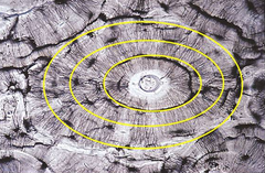
lamellae
Concentric rings of bone matrix around the central canals
osteocytes
mature bone cell that is surrounded by matrix

osteoblasts
bone forming cell; deposits bone matrix around themselves

osteoclasts
break down calcified matrix

lacunae
chambers that contain osteocytes

osteon
cylinder shaped unit

canaliculi
tiny passageways that osteocytes use to exchange nutrients and wastes

central canal
tubes running through compact bone that contains blood vessels and nerves; centrally located within an osteon

collagen
structural protein that is part of extracellular matrix that gives bone its strength and resilience
calcium phosphate
salt that is most abundant in extracellular matrix; makes bone hard and resistant to crushing
perforating canals
join adjacent central canals

epiphyseal plate
growth zone of hyaline cartilage

Intramembranous bone
bone that forms from membrane like layers of primitive connective tissue; become the flat bones of the skull.
endochondral bone
bone that begins as hyaline cartilage that is subsequently replaced by bone tissue

endochondral ossification
process of replacing hyaline cartilage to form an endochondral bone

Bone Resorption
removal of bone, action of osteoclasts
Bone Deposition
formation of bone, action of osteoblasts
Vitamin D
calcium absorption; deficiency causes rickets or osteomalacia
Physical Stress
stimulates bone growth
major bone functions
Provide shape to body, support body structures, protect body structures, aid body movement, contain tissue that produces blood cells, store inorganic salts
Hematopoiesis
Blood cell formation
Red bone marrow
formation of platelets, red blood cells, and white blood cells
Yellow bone marrow
stores fat
calcitonin
hormone which lowers blood calcium levels; activates osteoblasts for bone deposition.
parathyroid hormone
hormone which increases blood calcium levels; activates osteoclasts for bone resorption.
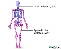
Axial Skeleton
skull, middle ear bones, hyoid bone, vertebral column, and thoracic cage

Appendicular Skeleton
Pectoral girdle, upper limbs, pelvic girdle, lower limbs
costal cartilage
hyaline cartilage; connects rib to sternum

Sternum (3 parts)
manubrium, body, xiphoid process

True Ribs
7 pairs; directly connected to sternum

False Ribs
5 pairs; connected to sternum via cartilage (see Floating ribs)

Floating Ribs
2 pairs; not connected to sternum (part of false ribs)

Cervical vertebra
7; transverse foramina, spinous processes (mouse)

Thoracic vertebra
12; transverse process that project posteriorly at sharp angles, pointed spinous processes that slope down, facets that articulate with ribs (giraffe)

Lumbar vertebra
5; large bodies, thinner transverse processes that project laterally, short/thick spinous process that project posteriorly (moose)

Sacrum
5 fused; posterior sacral foramina, sacral hiatus, anterior sacral foramina

Coccyx
4 fused; attached to the margins of the sacral hiatus

Atlas
C1; supports head, articulates with occipital condyle

Axis
C2; pivots around the dens

Joints
Articulations between adjoining bones
cartilaginous joints
allow only slight movement and consist of bones connected entirely by cartilage

synovial joints
freely movable joints; contain capsules filled with synovial fluid.

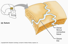
fibrous joints
generally immovable joints; held together by dense connective tissues
flexion
decreases the angle of the joint and reduces the distance between the two bones

extension
increasing the angle between two bones and the parts move farther apart.

dorsiflexion
bending of the foot or the toes upward

plantar flexion
bends the foot downward at the ankle

Hyperextension
extension beyond anatomical position

abduction
Movement away from the midline of the body

adduction
Movement toward the midline of the body

rotation
circular movement around an axis
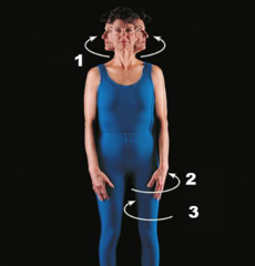
circumduction
circular movement of a limb at the far end

pronation
movement that turns the palm down

supination
turning the palm upward

eversion
turning the sole of the foot faces laterally

inversion
Turning the sole of the foot medially

retraction
moving a part backward

protraction
Moving a part forward

elevation
raising a body part

depression
lowering a body part
