Muscle Practical Lab
 Call Kai
 Learn
 Practice Test
 Spaced Repetition
 Match
 Flashcards
 Knowt Play
1/91

Earn XP
Description and Tags
Key guide: O- origin and I-Insertion
92 Terms
Origin
portion of the muscle attached to an immovable structure (usually bone)

Insertion
portion of muscle attached to the movable structure (usually bone)

Agonist (brachii)
(Prime mover) the main muscle responsible for a given movement

Synergist (biceps brachii)
muscles that assist and stabilize the prime mover

Antagonist (triceps brachii)
muscle that resists the prime mover; responsible for the opposite motion

Occipitofrontalis (epicranius)
O: occipital; I: muscles around eye; top of cranium
raises eyebrows, pulls scalp posteriorly
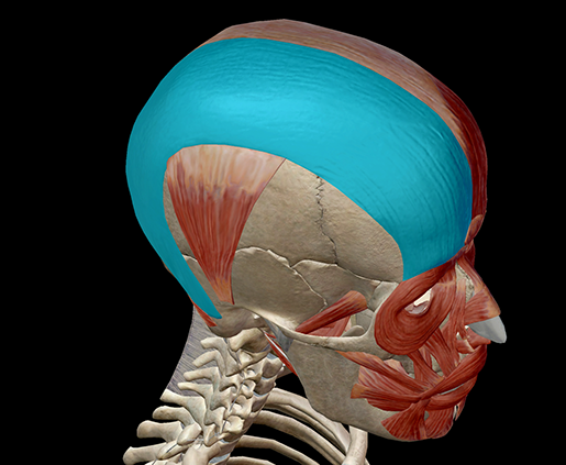
Frontalis

Occipitalis

Epicranial aponeurosis

Orbicularis oculi
O: frontal bone & maxilla, orbit ligament; I: eyelid
closes eye; the "squinting muscle"

Sphincter
(muscle which decreases the size of an opening) surrounding eye

Orbicularis oris
O: maxilla & mandible, orbit ligament; I: muscle & skin encircling lips and mouth
sphincter muscle surrounding mouth;
purses lips, the "kissing muscle"

Buccinator
O: alveolar processes of maxilla & mandible; I: orbicularis oris
parallels body of mandible and forms the wall of cheek;
compresses cheek (as in blowing or sucking), holds food between teeth

Zygomaticus
O: zygomatic; I: orbicularis oris
muscles between cheek & mouth; the "smiling muscle" - raises lateral corner of mouth

Risorius
o: fascia of masseter; I: lateral mouth
parallel to mouth beneath zygomaticus, The “laughter muscle”

Platysma
O: fascia of upper chest; I: lower mandible
large sheet like muscle; pulls lower lip downward, "pouting muscle"

Mastication
Chewing Muscles

Masseter
O: zygomatic arch & maxilla, I: ramus & body of mandible
thick and powerful muscle anterior to the ear
closes the jaw and elevates mandible

Temporalis
O: temporal; I: coronoid process & ramus of mandible
broad muscle in the temple region
closes the jaw, elevates mandible, aids in grinding movements

Pterygoids
O: sphenoid, I: mandible
wing-like muscle (pterygoid = "wings") deep to masseter, synergist of masseter & temporalis

Medial pterygoids

Lateral pterygoids

Sternocleidomastoid
O: sternum & clavicle, I: mastoid process
long oblique muscles of lateral neck
flexes neck when both contract, rotate head when acting

Capitis group
extend, bend & rotate head

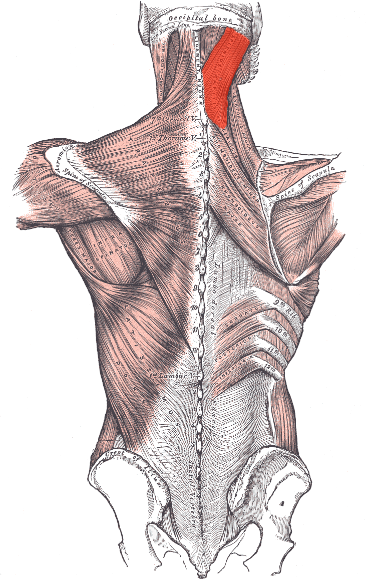
Splenius capitis
O: C7 - T3 spinous processes; I: mastoid process & occipital
oblique-band-like (= "splenium") muscle in back of head (=capit) most superficial of capitis group
Semispinalis capitis
deeper capitis muscle “along the spine” part of erector spinae group (below)

Longissimus capitis

Erector spinae group
O: lower spine, ribs & pelvis; I: ribs, C & T vertebrae
semispinalis, longissimus, & iliocostalis muscles
important group, muscle which lie along either side of the vertebral column
all extend and laterally bend the vertebral column, etc.

Semispinalis

Longissimus

liocostalis

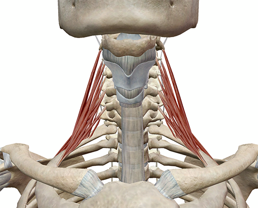
Scalene group
O: transverse processes of c vertebrae; I: ribs 1 and 2
no need to distinguish – just know as a group 3 very deep muscles of lateral neck flex and slightly rotate the neck
External oblique
O: ribs 5-12, I: iliac crest & linea alba (center "line" of abdominal aponeurosis)
largest and most superficial abdominal muscle

Internal oblique
O: iliac crest, I: cartilage of ribs 10-12, linea alba deep to external oblique

Transversus abdominis
O: iliac crest, cartilage of ribs 6-12, I: linea alba, pubis, xiphoid process
deepest of the abdominal muscles
the obliques & transversus abdominis all compress the abdomen

Obliques
also bend the vertebral column laterally when the R or L contracts independently

Rectus abdominis
O: pubis; I: xiphoid process, ribs 5-7
straight (= rectus) large, superficial muscle separated by horizontal bands of CT
compresses abdominal contents, flexes vertebral column

Diaphragm
Separates abdominal and thoracic cavity; not on models. involuntary skeletal muscle for inhalation

External Intercostals
O: superior border of rib below; I: inferior border of rib above
11 pairs of muscles on the external rib cage
pull the rib cage up and out and aid in inhalation

Internal Intercostals
O: inferior border of rib; I: superior border of rib below
11 pairs of muscles on the internal rig cage–pull the rib cage down and in and aid in exhalation

Trapezius
(Scapula spine)

Serratus anterior
O: ribs 1-8, I: ventral scapula
large saw-like ("serratus") muscle along upper lateral thoracic cage, pulls shoulder anteriorly and inferiorly ("pushing / boxing muscle"), stabilizes shoulder

Rhomboideus
O: spinous process of T2-T5; I, vertebral border of scapula
large oblique (rhomboid: parallelogram) muscles between vertebral column & scapula;
steadies, elevates and retracts scapula

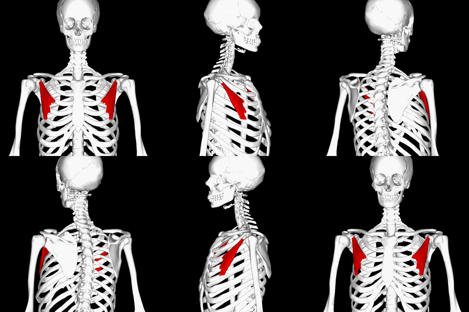
Pectoralis minor
O: anterior ends of rib 3-5; I: I: coracoid process
tripartite oblique muscle deep to the pectoralis major draws scapula anterior and inferior (with ribs fixed), draws ribs upward
Levator scapulae
O: transverse processes of C1 - C4, I: vertebral border of scapula
narrow muscle adjacent to capitis group elevates ("levator") scapula and bends neck laterally

Coracobrachialis (flexor)
O: coracoid process, I: medial surface of shaft of humerus
small muscle from scapula to medial upper arm toward the axillary region flexes and adducts

Pectoralis major (flexor)
O: clavicle, sternum, cartilage of ribs 2-6; I: greater tubercle
complex thick fan-shaped muscle of upper chest flexes, adducts (as in swimming) & rotates medially; pulls body up when arm is fixed (as in climbing)

Teres major (extensors)
O: inferior posterior scapula, I: lesser tubercle
thick round (= teres) muscle inferior to teres minor
extends, adducts and rotates medially

Latissimus dorsi (extensors)
O: spinous processes of T6-L5, sacrum, iliac crest, ribs 10-12,
I: intertubercular groove;
broad flat complex superficial muscle of lower back and sides
extends, adducts (climbing) and rotates arm medially

Supraspinatus (Abductors)
O: supraspinous fossa, I: greater tubercle
deep to deltoid & trapezius
abducts arm – very important stabilizer of the shoulder

Subscapularis (Rotators)
O: anterior scapula, I: lesser tubercle
large muscle covering the entire anterior surface of scapula
medial / internal rotation of arm

Infraspinatus (Rotators)
O: posterior scapula below spine, I: greater tubercle of humerus
superior to teres major & minor
lateral / external rotation of arm

Teres minor (Rotators)
O: axillary border of scapula, I: greater tubercle of humerus
inferior to infraspinatus and superior/deep to teres major
lateral / external rotation of arm

Rotator cuff
all 3 rotators and supraspinatus
“SITS” (supraspinatus, infraspinatus, teres minor, subscapularis)
it encircles most of the shoulder joint and holds the head of the humerus in the glenoid fossa
very common sight of muscle and tendon tearing in athletic injuries

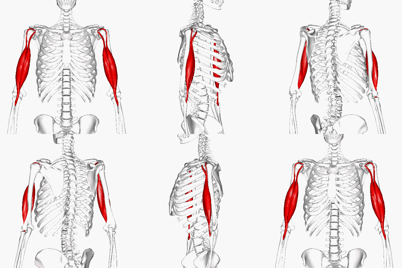
Biceps brachii (flexor)
O: coracoid process (short head); above glenoid cavity of scapula (long head)
I: radial tuberosity
long two-headed superficial muscle of anterior upper arm
flexes and supinates forearm; weakly flexes upper arm
Brachialis (flexor)
O: distal anterior humerus, I: coronoid process of ulna
muscle deep to biceps brachii
most powerful forearm flexor

Brachioradialis (flexor)
O: proximal to lateral epicondyle of humerus, I: distal radius
flexes forearm

Triceps brachii (extensor)
O: 3 separate – proximal humerus and glenoid area I: olecranon
the main powerful extensor of posterior humerus

Supinator (rotators)
O: lateral epicondyle & proximal ulna, I: lateral proximal radius
deep oblique muscle in lateral elbow/forearm region
important supinator!!!

Pronator teres (rotators)
O: medial epicondyle of humerus, coronoid process of ulna, I: lateral mid-radius
muscle that is medial to brachioradialis
pronates forearm

Pronator quadratus (rotators)
O: distal anterior ulna, I: distal anterior radius
deep and transverse/oblique muscle of anterior wrist
pronates forearm

Flexor carpi radialis (flexor)
O: medial epicondyle humerus; I: 2nd & 3rd metacarpal
Located on the anterior forearm
medial to pronator teres, crosses forearm / toward lateral side (along the radius)
flexes hand (also abducts)

Palmaris longus (flexor)
O: medial epicondyle humerus; I: fascia / aponeurosis of palm
medial to flexor carpi radialis
flexer wrist (& tenses the palmar fascia)

Flexor carpi ulnaris (flexor)
O: medial epicondyle humerus, olecranon of ulna;
I: carpals metacarpals
Located on the anterior forearm
most medial of the flexor group
flexes the hand (also adducts)

Extensor carpi radialis: Longus and Brevis (Extensors)
know as the pair, no need to distingish
O: distal humerus; I: 2nd & 3rd metacarpals
Located on the posterior forearm
next to brachioradialis along the lateral posterior forearm – along the radius
extends hand (also abducts)

Extensor digitorum (extensors)
O: lateral epicondyle of humerus; I: tendons to phalanges 2-5
Located on the posterior forearm
multipart muscle on center of posterior forearm
extends fingers (also flares fingers)

Extensor carpi ulnaris (extensors)
O: distal humerus; I: 3rd metacarpal
most medial of the posterior group – along the ulna
extends hand (also abducts)

liopsoas
both (psoas major and iliacus ) flex the thigh
("walking muscles")
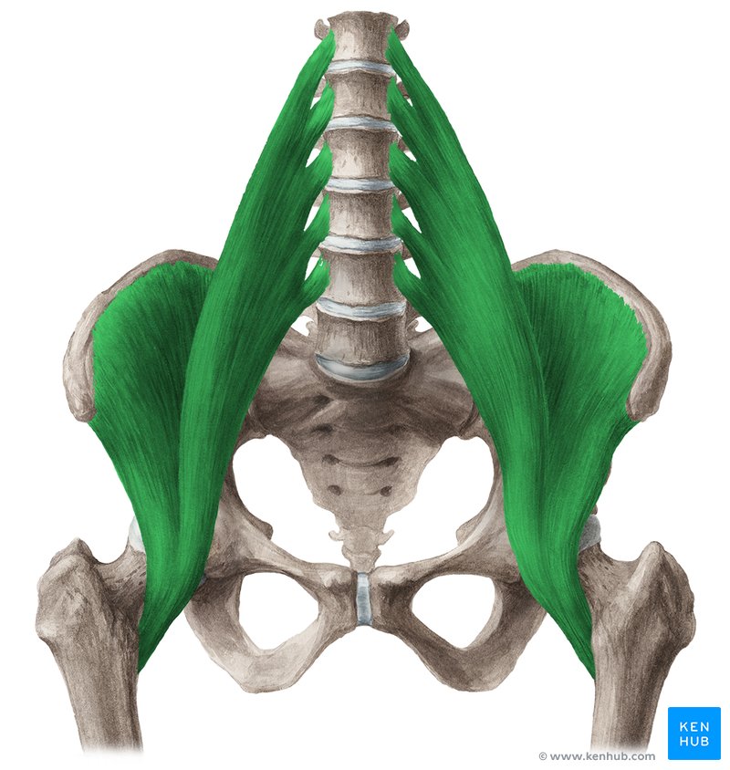
Psoas major (anterior)
O: L vertebrae, I: lesser trochanter
long thick muscle which passes through pelvis to the superior medial thigh

Iliacus (anterior)
O: ilium, I: lesser trochanter
large fan shaped muscle in the iliac fossa; lateral to the lower psoas major
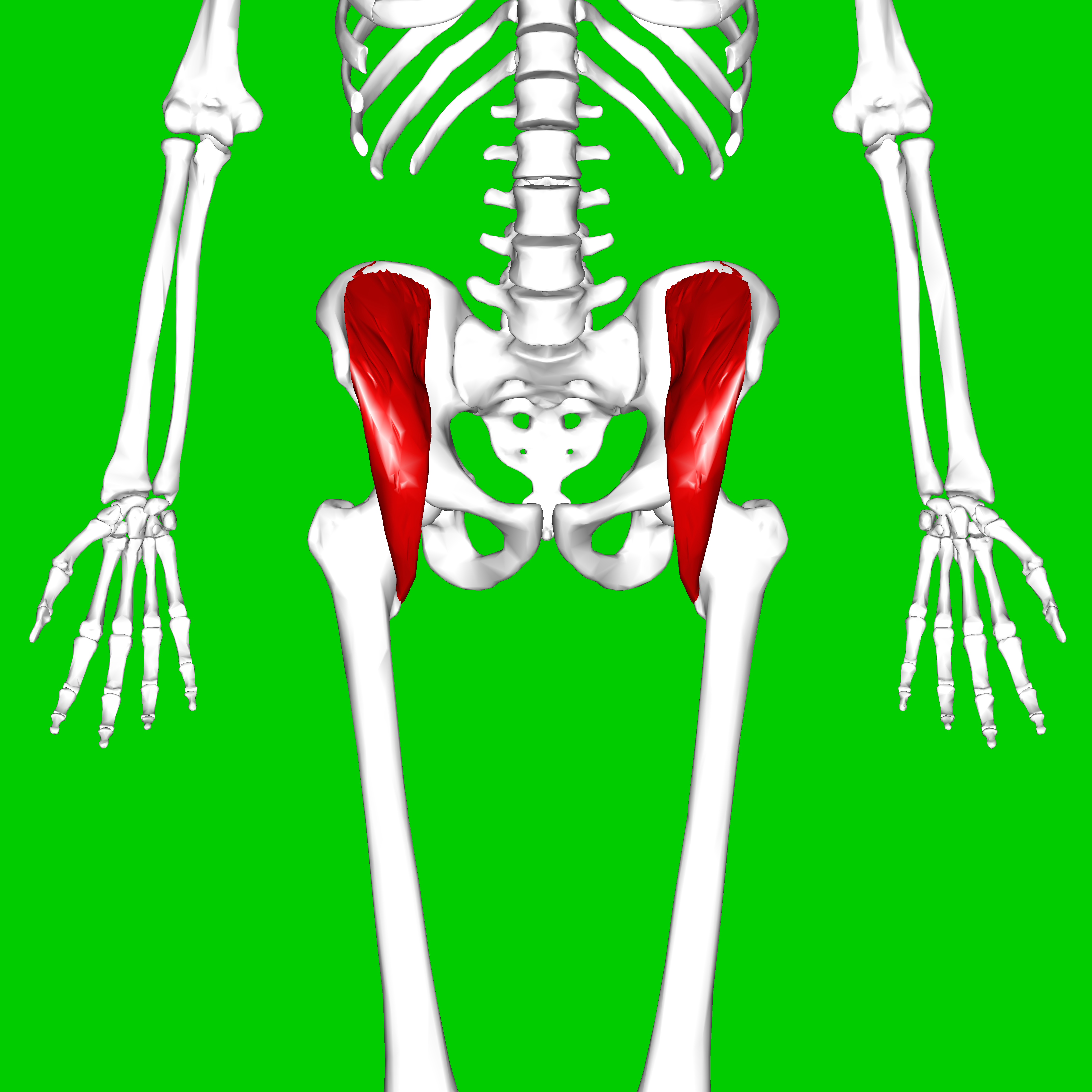
Tensor fasciae latae (lateral)
O: iliac crest, I: iliotibial tract to tibia
superficial lateral hip muscle at the top of the iliotibial tractlateral leg fascia) tenses iliotibial tract (hence name), abducts, medially rotates and flexes thigh

Gluteus maximus (posterior)
O: sacrum, coccyx, & iliac crest, I: mid-iliotibial tract
largest most superficial gluteal muscle, oblique, forms
buttock mass extends and rotates thigh laterally

Gluteus medius (posterior)
O: lateral ilium, I: greater trochanter
thick muscle on back of hip, smaller & partially covered by gluteus maximus abducts and medially rotates thigb

Adductor longus (adductors)
O: anterior pubis, I: linea aspera
long medial thigh muscle
adducts, flexes and medially rotates thigh

Adductor magnus (adductors)
O: pubis & ischial tuberosity, I:linea aspera & medial epicondyle
slightly deeper and has more inferior boundary than adductor longus adducts, also extends, medially rotates and stabilizes thigh

Gracilis (adductors)
O: pubis, I: medial proximal tibia
strap-like (gracilis = slender), superficial muscle of medial thigh; adducts leg, also flexes knee and medially rotates thigh a “pulled groin” muscle is usually an adductor muscle

Hamstrings
Contains: biceps femoris, semitendinosus, semimembransus (BSS)
fleshy muscles of posterior thigh; flex knee and extend thigh

Biceps femoris (Flexors)
O: ischial tuberosity (long head) & linea aspera of femur (short head)
I: head of fibula & lateral condyle of tibia
the most lateral hamstring, the anterior portion deep to
gluteus maximus

Semitendinosus (Flexors)
O: ischial tuberosity, I: medial proximal tibia
medial to biceps femoris, has a long tendon at its distal end ("half tendon")

Semimembranosus (flexors)
O: ischial tuberosity, I: medial proximal tibia
deepest and flattest hamstring; partially more medial to
semitendinosus

Sartorius (flexors)
O: anterior superior iliac spine; medial proximal tibia
strap-like, superficial and oblique muscle;
flexes thigh & knee, abducts & lateral rotation of thigh;
"tailors (= sartor) muscle"

Quadriceps femoris
large and fleshy muscle group that covers the front and sides of the thigh
all have a common insertion: a tendon which continues as the patellar ligament to the tibial tuberosity
all 4 (rectus femoris, vestus lateralis, vastius medialia, vastus intermedius) extend the leg at the knee
rectus also flexes the thigh

Rectus femoris (extensors)
O: anterior inferior iliac spine, I: tibial tuberosity
runs straight (= "rectus") down center of thigh

Vastus lateralis (extensors)
O: greater trochanter & linea aspera, I: tibial tuberosity
forms lateral thigh and is lateral to the rectus femoris
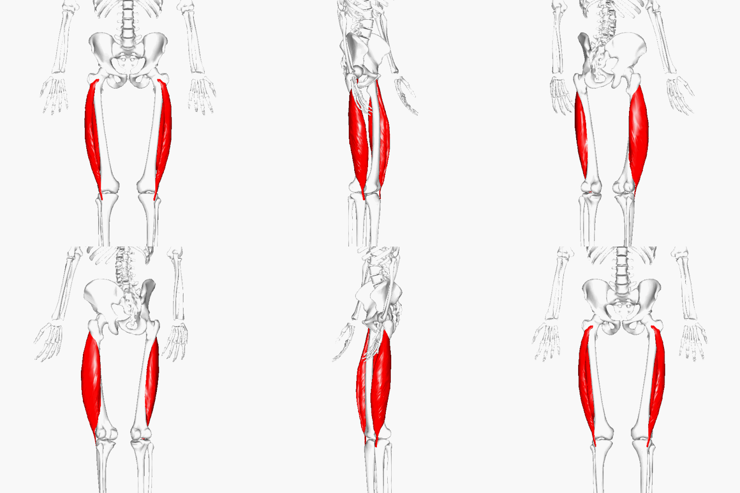
Vastus medialis (extensors)
O: linea aspera, I: tibial tuberosity
forms the medial thigh & is medial to the rectus femoris

Vastus intermedius (extensors)
O: anterior/superior femur, I: tibial tuberosity
deep to the rectus femoris; between (= "intermedius") vastus (lateralis & medialis)
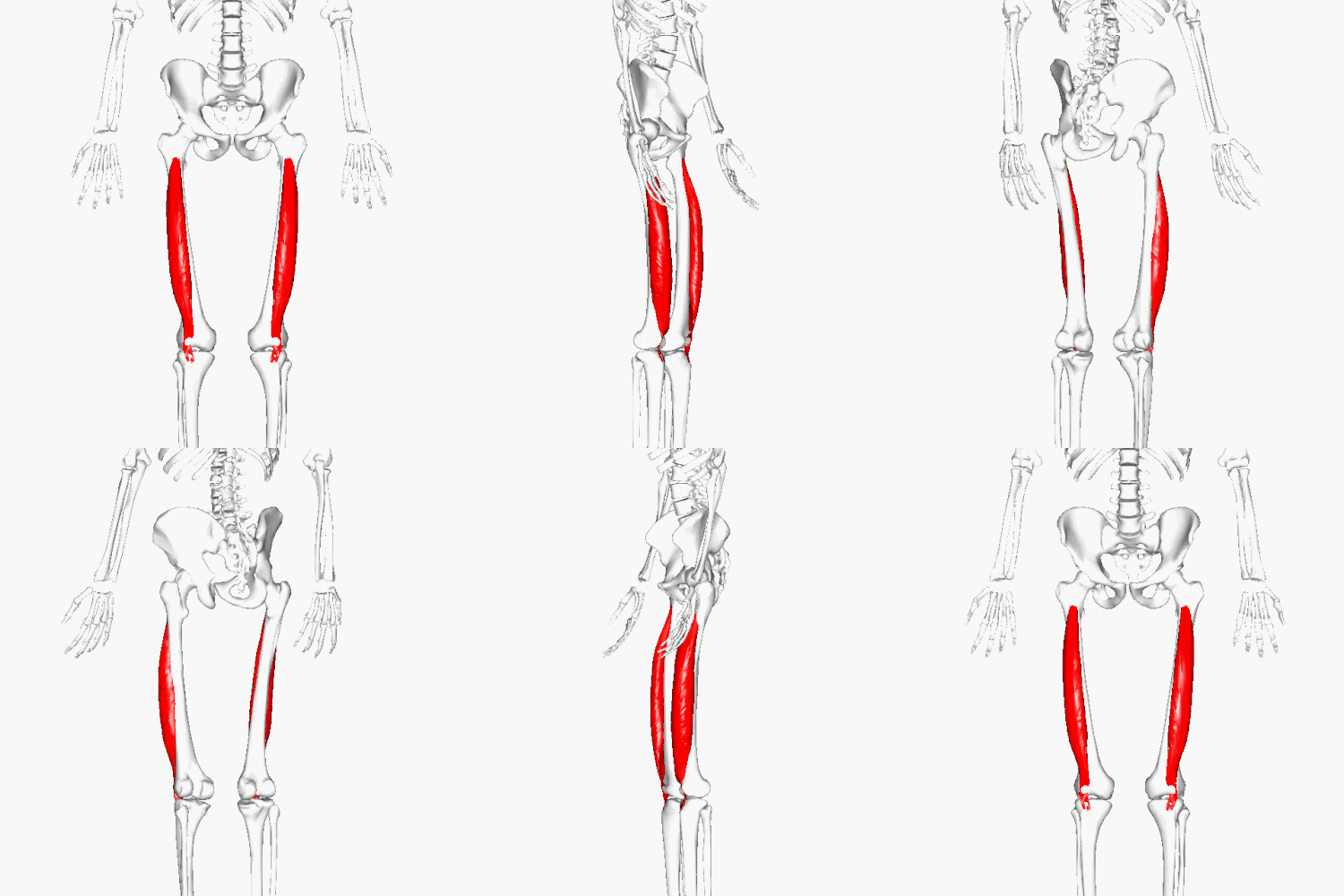
Tibialis anterior (dorsal flexor)
O: lateral condyle & lateral proximal tibia, I: tarsal (first cuneiform)
dorsiflexes and inverts foot

Extensor digitorum longus (dorsal flexor)
O: distal tibia; I: phalanges of toes 2-5
extends toes, dorsiflexes foot

Gastrocnemius (plantar flexor)
O: lateral & medial condyles of femur, O: calcaneus (Achilles / calcaneal tendon)
two headed superficial calf muscle, forms a prominent
bulge in the upper calf; plantar flexes foot and flexes knee

Soleus (plantar flexor)
O: superior fibula & medial tibia,
I: calcaneus via Achilles tendon;
broad calf muscle deep to gastrocnemius; plantar flexes foot

Tibialis posterior (inverter)
O: proximal fibula & tibia, I: tarsals & metatarsals
runs along the lateral tibia, deep to soleus
inversion & plantar flexion of foot

Peroneus / fibularis longus (everter)
O: superior fibula; #1 metatarsal; superficial lateral muscle eversion and plantar flexion of the foot
