mod 1.4a-c reading quiz (PSYCH)
 Call Kai
 Learn
Learn Practice Test
 Spaced Repetition
 Match
Match Flashcards
 Knowt Play
1/98

There's no tags or description
Looks like no tags are added yet.
Name | Mastery | Learn | Test | Matching | Spaced | Call with Kai | Chat |
|---|
No analytics yet
Send a link to your students to track their progress
99 Terms
ancient Greek physician ___ correctly located the mind in the brain
his contemporary, ___, thought it was sited in the heart
ancient Greek physician Hippocrates correctly located the mind in the brain
his contemporary, Aristotle, thought it was sited in the heart
phrenology
the studying of bumps on the skull — the belief it could reveal a person’s mental abilities + character traits
proposed by Frank Gall, German physician
disproven / primitive method today
succeeded in focusing attention on the localization of function — the idea that various brain regions have particular functions (though, not the functions Gall proposed)
biological psychology
the scientific study of the links between biological (genetic, neural, hormonal) + psychological processes
biological psychologists’ different names
behavioral neuroscientists
neuropsychologists
behavior geneticists
physiological psychologists
biopsychologists
biopsychosocial approach
an integrated approach that incorporates biological, psychological, and social-cultural levels of analysis
we are biopsychosocial systems
what 3 levels of analysis does the biopsychosocial approach integrate
biological
psychological
social-cultural
levels of analysis
the differing complementary views, from biological to psychosocial to social-cultural, for analyzing any given phenomenon
neuroplasticity
the brain’s ability to change, especially during childhood, by reorganizing after damage or by building new pathways based on experience
greatest in childhood, but persists throughout life
enables us, more than other species, to adapt to our changing world
London’s taxi driver trainees who spend years learning + remembering the city’s 25,000 street locations + connections are rewarded w/ better income + enlarged hippocampus
well-practiced pianists are rewarded with a larger-than-usual auditory cortex area, a sound processing region
T or F — the brain you were born in is not the brain you will die in
true
T or F — even limited practice times cannot produce neural benefits
false
__ said a brain simple enough to be fully understood is too simple to produce a mind able to understand it
John Barrow
how were early explorers mapping the brain?
by localizing brain functions through early case studies
ex
damage to one side of the brain = numbness / paralysis on opposite side — suggests the body’s right side is wired to the brain’s left side, vise versa
damage to the back of brain = disrupted vision
damage to left-front part of brain = speech difficulties
lesion
tissue destruction
brain lesions may occur naturally (from disease or trauma), during surgery, or experimentally (using electrodes to destroy braincells)
T or F — unlike early explorers, scientists now can selectively lesion tiny clusters of normal / defective braincells to observe any effect on brain function
true
studies revealed that damage to one area of a rat’s hypothalamus reduces eating to the point of starvation, while damage to another area produces overeating
3 ways today’s neuroscientists can stimulate brain parts (aside from lesions)
electrically
chemically
magnetically
in order to note effect
modern electrodes can __ (2)
detect the electrical pulse in a single neuron
ex: detects exactly where the info goes in a rat’s brain when someone tickles its belly
eavesdrop on chatter of billions of neurons
optogenics
a technique allowing neuroscientists to control the activity of individual neurons
ex: programming neurons to become receptive to light can help researchers determine / examine biological bases of
sensations, fear, depression, substance abuse disorders
electroencephalography (EEG)
electrodes placed on scalp
measures electrical activity in neurons
an amplified recording of the waves of electrical activity sweeping across brain’s surface
waves are measured by electrodes placed on scalp
brainwaves recorded through shower-cap-like hat filled w/ electrodes covered w/ constructive gel
studying EEG is like studying a blender’s motor by listening to its hum
researchers can present a stimulus repeatedly + filter out brain activity unrelated to stimulus

MEG (magnetoencephalography)
a head coil
measures magnetic fields from brain’s natural electrical activity during certain tasks
head coil like salon hair dryer that participants sit under
special rooms to isolate brain’s magnetic fields + cancel out other magnetic signals (ex: Earth’s magnetic field)
participants complete activities —> tens of thousands of neurons generate electrical pulses —> create magnetic fields —> speed + strength of magnetic fields enable research to understand how certain tasks influence brain activity

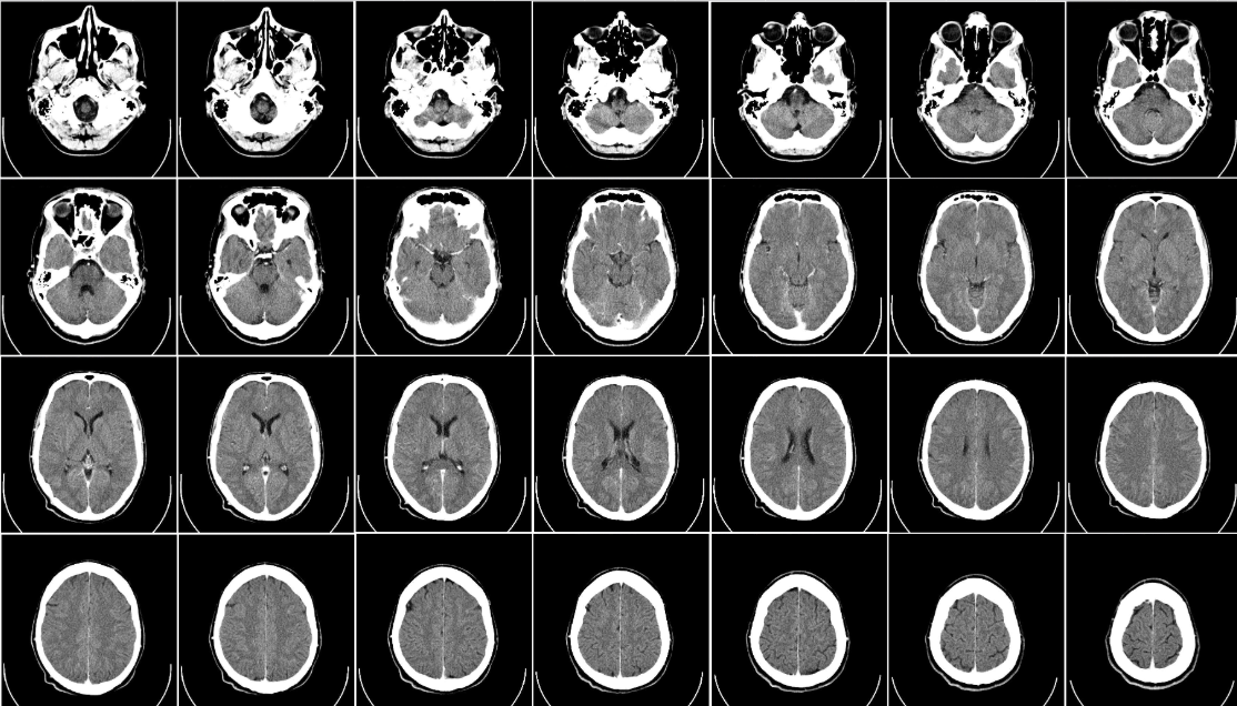
CT (computed tomography)
X-rays of the head generate images that may locate brain damage
a series of X-ray photos taken from different angles + combined by computer into a composite representation of a slice of the brain’s structure
PET (position emission tomography)
technique for detecting brain activity that displays where a temporarily radioactive form of glucose goes while the brain performs a given task
depicts brain activity by showing each brain area’s consumption of its chemical fuel — sugar glucose
monitors while participants do math, look @ face images, daydreams
brain = ~2% body weight, but consumes 20% of calorie intake

brain is ~ __% body weight, but consumes _% of calorie intake
brain = ~2% body weight, but consumes 20% of calorie intake
MRI (magnetic resonance imaging)
a technique where people sit / lie down in a chamber that uses magnetic fields + radio waves to provide a map of rain structure
computer generated images of soft tissue
head put into strong magnetic field, aligns spinning atoms in brain molecules
radioactive pulse momentarily disorients atoms —> when back to normal spins, signals omitted to provide detailed picture of soft tissues, including brain
ex: enlarged ventricles (fluid filled brain areas) in some people w/ schizophrenia; larger neural area in left hemisphere of musicians
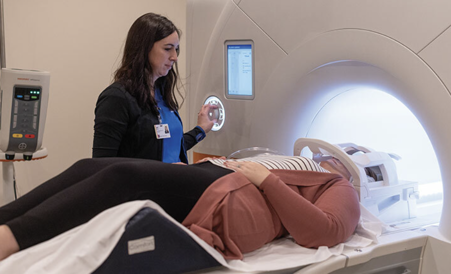
CT vs MRI
CT
X-ray photos that are composited into 3d image
generally better at distinguishing between the internal structures of the body
MRI
magnetic waves + radio waves used to produce images
generally better at distinguishing between different types of tissue in the body
fMRI (functional MRI)
special application of MRI
by comparing continuous MRI scans, it measures blood flow to brain regions
reveals blood flow —> brain function + structure
where brain is active, blood goes
fNIRS (functional near-infrared spectroscopy)
shines infrared light on blood molecules to identify brain activity
equipment can fit in large backpack, enabling researchers to study biology of mind in difficult-to-reach populations
__% accuracy of neuroscience team when identifying which activities the study participants were doing when 129 people’s brains were scanned while doing 8 different tasks
80% accuracy
T or F — today’s techniques for peering into brain are doing for psychology what microscopes did for biology + telescopes did for astronomy
true
T or F — learning about neurosciences now is like studying world geography when Magellan explored the seas
true
__ said, “individually, we contribute little or nothing to the truth. by the union of all a considerable amount is amassed”
Aristotle
vertebrate brain’s 3 divisions
hindbrain
midbrain
forebrain
hindbrain
contains brainstem structures that direct essential survival functions
breathing, sleeping, wakefulness, arousal, coordination, balance
consists of medulla, pons, cerebellum
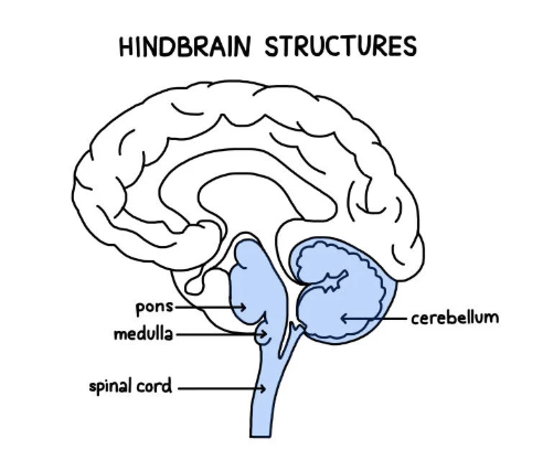
what 3 structures does the hindbrain contain
pons
medula
cerebellum
midbrain
connects hindbrain w/ forebrain
controls some motor movement
transmits auditory + visual information
atop the brainstem

forebrain
manages complex cognitive activities, sensory associative functions, and voluntary motor activities
consists of the cerebral cortex, thalamus, hypothalamus

T or F — organisms’ brains have evolved to suit their environment
true
humans have extremely developed forebrains — ability to make complex decisions + judgements
predator sharks have complex hindbrains — ability to hunt prey
brainstem
responsible for automatic survival functions
a crossover point where most nerves to and from each side of the brain connect w/ the body’s opposite side (contralateral hemispheric organization)
the brain’s innermost region
the central core of the brain, beginning where the spinal cord wells as it enters the skull
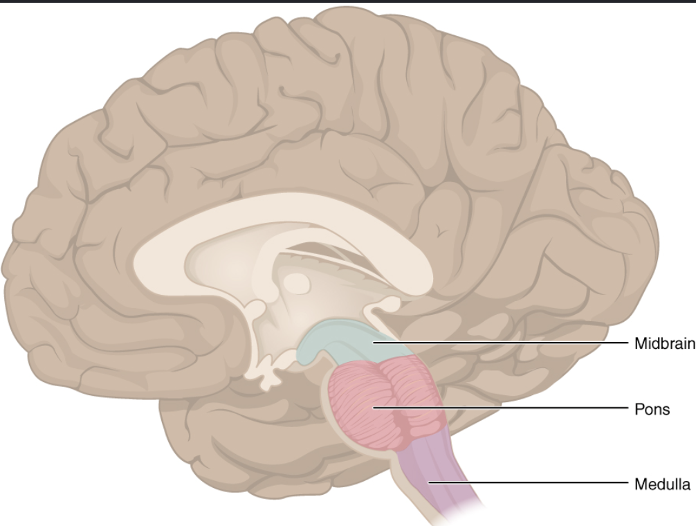
contains pons, medulla, reticular formation, midbrain

4 structures the brainstem contains
pons
medulla
reticular formation
midbrain
medulla
the hindbrain structure that is the brainstem’s base
slight swelling in the spinal cord just after it enters the skull
controls heartbeat + breathing
a conscious mind is not needed to orchestrate our hearts’ pumping + lungs breathing as seen in brain-damaged patients in vegetative state — brainstem handles that task

pons
helps coordinate movements + control sleep
just above the medulla
the largest part of brainstem
thalamus
egg-shaped structures that act as brain’s sensory control center
forebrain’s sensory control center
sits atop the brainstem
directs messages to sensory receiving areas in the cortex + transmits replies to the cerebellum + medulla
receives info from all sense EXCEPT smell, routes them to respective regions
similar to a train station (what Seoul is to SK’s trains)
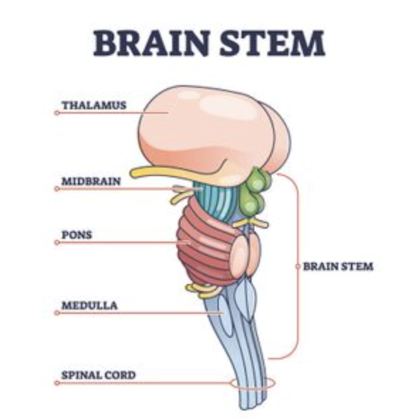
__ is what the thalamus being to sensory info is
Seoul to South Korea’s trains — a hub through which traffic passes en route to various locations
reticular formation
a nerve network that travels through the brainstem into thalamus
inside brainstem, between your ears
extends from spinal cord —> thalamus
filters info + pays an important role in controlling arousal / state of alertness
nerve network governed by reticular activating system
Giuseppe Moruzzi + Horace Magoun discovered that the reticular formation also controls arousal / state of alertness
as spinal cord’s sensory input flows up to thalamus, some travels through reticular formation, which filters incoming stimuli + relays important info to other brain areas
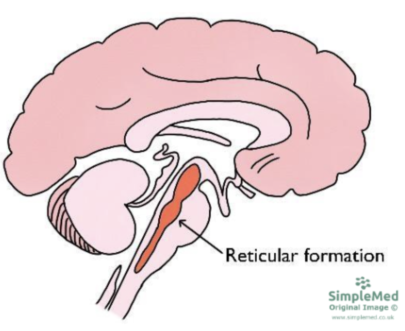
who discovered that the reticular formation also controls arousal / state of alertness? + how?
Giuseppe Moruzzi + Horace Magoun discovered that the reticular formation also controls arousal / state of alertness in 1949
electrically stimulated sleeping cat’s reticular formation, almost instantly became awake + alert
Magoun severed a cat’s reticular formation w/out damaging nearby sensory pathways — the cat lapsed into a coma + never woke up
cerebellum
the hindbrain’s “little brain” @ rear of brainstem
processes sensory input, coordinates movement output + balance, enables learning + nonverbal learning
contains over half of the brain’s neurons, operates just outside awareness
cerebellum + basal ganglia are both deep brain structures involved in motor movement
coordinates voluntary movement w/ pons
involved when a soccer player masterfully controls the ball
coordination suffers under alcohol’s influence

which brain structure contains over half of the brain’s neurons
cerebellum
T or F — injured cerebellum = difficulty walking, keeping balance, texting a friend
true
jerky / exaggerated
cannot be dancer/guitarist
T or F — our brain processes most information within our awareness
false
our brain processes most information outside our awareness
we are aware of the results of our brain’s labor, but not how we construct the visual image
limbic system
associated w/ emotions, drives, memory formations
neural system located mostly in forebrain — below cerebral hemispheres
consists of amygdala, hypothalamus, hippocampus, thalamus, pituitary gland
can thank limbic system for the emotion when we enjoy a joke

what 5 structures reside in the limbic system
amygdala
hypothalamus
hippocampus
thalamus
pituitary gland
amygdala
two lima-bean-sized neural clusters in limbic system
linked to emotion
enables aggression + fear
people with amygdala lesions displayed reduced arousal to fear + anger-arousing stimuli
animals’ electrically stimulated amygdala illicited fear + anger response
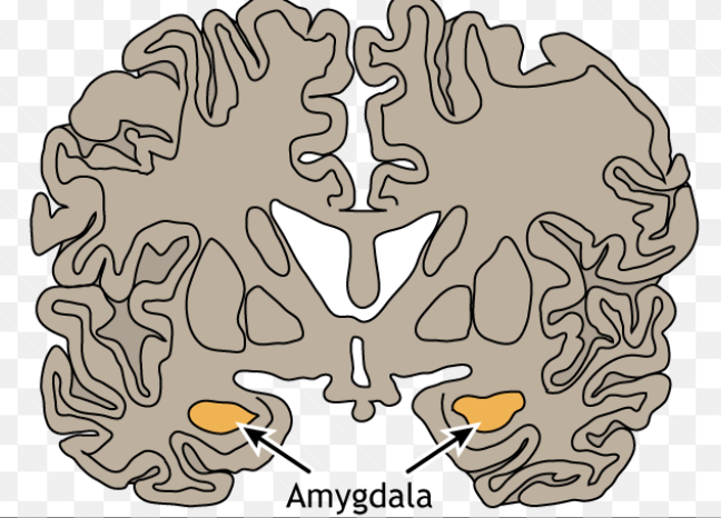
amygdala lesion + stimulation case studies
Heinrich Kluver + Paul Rucy made an ill-tempered rhesus monkey —> mellow creature after an amygdala lesion in 1939
people with amygdala lesions display reduced arousal to fear + anger arousing stimuli
patient S.M. (woman w/ lesion) — “the woman with no fear”, even when threatened w/ gun
stimulating one part of a cat’s amygdala illicits its attack response, while stimulating another part makes it cower in fear when locked in the same cage as a small mouse
electrically stimulating amygdala of normal animal illicits anger + fear responses
T or F — monkeys + humans w/ amygdala damage become less fearful of strangers
true
people w/ amygdala lesions display reduced arousal to fear + anger arousing stimuli
T or F — criminal behavior is linked to amygdala dysfunction
true
T or F — the brain is neatly organized into structures that correspond to behavior categories
false
the brain is not neatly organized into structures that correspond to behavior categories
ex: the amygdala is engaged with other mental phenomena
being afraid / aggressive occurs in many brain areas (not just amygdala)
ex: if you destroy a car battery, it won’t run; but battery is just one link in an integrated system
hypothalamus
linked to emotion + reward
helps govern endocrine system
directs several maintenance activities (eating, drinking, body temp)
important link in command chain governing bodily maintenance
a limbic system neural structure lying below (hypo) the thalamus
neural clusters in the hypothalamus influence different functions
together, they help maintain a steady (homostatic) internal state
later experiments locate other “pleasure centers” / reward centers
other limbic system reward centers in other species — ex: dolphins + monkeys have nucleus accumbens in front of hypothalamus
T or F — animals come equipped w/ built-in systems that reward activities essential to survival
true
animal research has revealed general dopamine-related award systems + specific centers associated w/ pleasures (eating, drinking, sex)
“if you were designing a robot vehicle to walk into future + survive, you’d wire it up so that behavior that ensured survival of the self or species — like sex and eating — would be naturally reinforcing”
T or F — stimulating brain’s hedonic hot spots (reward circuits) produces more desire than enjoyment
true
some evidence indicates that humans have limbic centers for pleasure
when electrodes implanted in violent patients’ reward center areas, they only reported mild pleasure
unlike Olds + Milners’ rats, patients are not driven to frenzy
T or F — disorders may stem from malfunctions in natural brain systems for pleasure + well being
true
people genetically predisposed to this reward deficiency syndrome may crave whatever provides missing pleasure / relieves negative feelings (ex: aggression, rich food, drugs, alcohol)
hippocampus
a neural center in limbic system that helps process explicit (conscious) memories (of facts + events) for storage
a curved brain structure
lost hippocampus = lost ability to form new memories of facts + events
those who survive hippocampal brain tumor in childhood struggle to remember new information as adult
NFL players to experiences over one loss-of-consciousness concussions may latter have shrunken hippocampus + poor memory
size + function of hippocampus decreases as we grow older, furthers cognitive decline

corpus callosum
the large band of neural fibers connecting the two brain hemispheres + carrying messages between them
allows left + right hemispheres to relay information with each other
people with severed corpus callosum don’t possess this communication

cerebral cortex
the surface layer / intricate fabric of interconnected neural cells covering the forebrain’s cerebral hemispheres
body’s ultimate control + information processing center
cortex means “bark” — scholars used Latin + Greek words as graphical descriptions
contains ~ 20-23 billion of the brain’s nerve cells + 300 trillion synaptic connections
mammals’ complex cerebral cortex offers high capacity for learning + thinking, enabling adaptations to ever changing environments
what makes humans distinct is the size + interconnectivity of our cerebral cortex

what distinguishes humans most from other animals?
our cerebral cortex
cerebrum
the 2 central hemispheres that contribute 85% of the brain’s weight
enables our perceiving, thinking, speaking
cerebral hemispheres come as a pair (like other brain structures — thalamus, hippocampus, amygdala)
covered by cerebral cortex

what contributes 85% of the brain’s weight
the cerebrum (brain’s 2 central hemispheres)
structure of cortex
makes brain appear as a wrinkled organ, shaped like oversized walnut
w/out wrinkles, flattened cerebral cortex would require 3x the area — around the size of large pizza
brain’s left + right hemispheres filled mainly w/ axons connecting cortex to brain’s other regions
many functions require the interplay of several lobes
frontal lobes, parietal lobes, occipital lobes, temporal lobes
names of 4 lobes
frontal lobes
parietal loes
occipital lobes
temporal lobes


name…
1) frontal lobe
2) parietal lobe
3) temporal lobe
4) occipital lobe
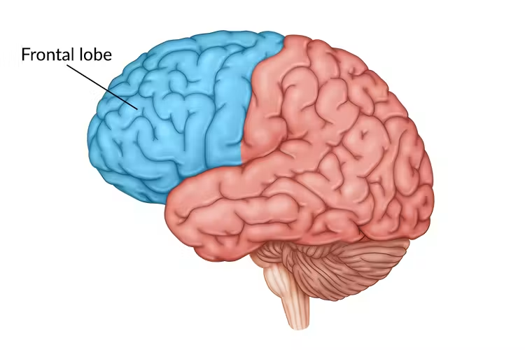
frontal lobe
enables linguistic processing, muscle movements, higher order thinking, executive function (ex: making plans + judgements)
the portion of the cerebral cortex lying just behind the forehead
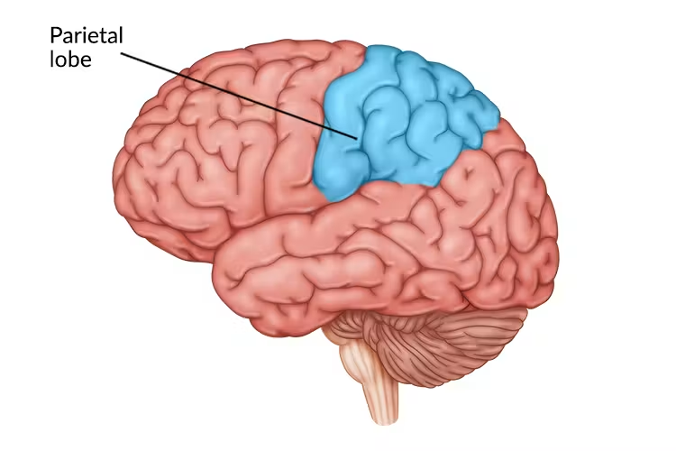
parietal lobes
receives sensory input for touch + body position
the portion of the cerebral cortex lying @ top of the head + toward the ear
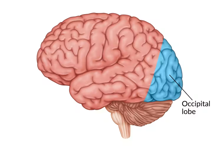
occipital lobes
includes areas that receive information from the visual fields
the portion of the cerebral cortex lying @ back of head
temporal lobes
enables language processing
includes the auditory areas, each of which receives information primarily from opposite ear
the portion of the cerebral cortex lying ~ above the ears

motor cortex
a cerebral cortex area @ the rear of the frontal lobes that controls voluntary movements
the brain devotes more tissue to sensitive areas + areas requiring precise control — finger have greater representation in cortex than your upper arm
discovered by German physicians Gustav Fritsch + Eduard Hitzig in 1870
used mild electrical stimulation to parts of an animal’s cortex to make parts of its body move — effects were selective
stimulation caused movement when applied to arch-shaped region @ back of frontal lobe, running ear-to-ear across top of brain
stimulating parts of this region in left / right hemisphere caused movements of specific body parts on opposite side of body

how can scientists predict a monkey’s arm motion just before it moves?
by repeatedly measuring motor cortex activity preceding specific arm movements
2 case studies of how scientists can determine if a person w/ paralysis can move a robotic limb by stimulation of the brain
100 tiny recording electrodes implanted into motor cortexes of 3 monkeys
gained rewards by using joystick to follow a moving red target —> matched brain signals w/ arm movement —> programmed a computer to monitor signals + operate joystick
resulted in mind-reading computer — monkey thinking about a move led to cursor moving w/ proficiency
Ian Burkhart’s legs + arms paralyzed; by learning his unique brain response patterns, the computer can predict his brain activity to help make movements
through implanting recording electrodes in his motor cortex
similarly, prosthetic voices created by reading the brain’s motor commands that direct vocal movement
somatosensory cortex
a cerebral cortex area @ the front of the parietal lobes that registers + processes body touch + movement sensations
parallel to + just behind the motor cortex
specializes in receiving information from skin senses (touch, temperature) + movement
the more sensitive the body region, the larger the somatosensory cortex area devoted to it
ex: humans’ supersensitive lips project to larger brain area than toes to kiss; rats’ whisker sensations; owls’ hearing sensations

motor cortex vs somatosensory cortex
motor cortex : output
somatosensory cortex: input

which lobe is the visual cortex located in
occipital lobes
visual info you receive —> visual cortex in occipital lobes
visual information travels from occipital lobes —> other areas that specialize in tasks (identifying words, detecting emotions, recognizing faces)
if stimulated in occipital lobes, might see flashes of light/dashes of color (w/ normal vision)
LSD increases communication between visual cortex + other regions
produced visual hallucinations

which lobe is the auditory cortex in
temporal lobes
auditory info you receive —> auditory cortex in temporal lobes
auditory info travels a circuitous route from one ear —> auditory receiving area above opposite ear
if stimulated in auditory cortex, you might hear a sound
tinnitus in one ear is associated w/ activity in temporal lobe on brain’s opposite side
fMRI scans of people with schizophrenia has active auditory areas in temporal lobes
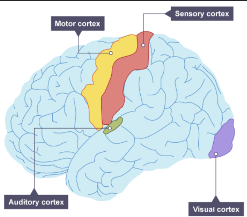
small cortical areas that either receive sensory input or direct muscular output occupy ~ __ of brain’s cerebral cortex
remaining vast regions of cortex are ___ areas
small cortical areas that either receive sensory input or direct muscular output occupy ~ ¼ of brain’s cerebral cortex
remaining vast regions of cortex are association areas
association areas
areas of the cerebral cortex that are not involved in primary motor / sensory functions
involved w/ higher mental functions (learning, remembering, thinking, speaking)
interprets, integrates, and acts on sensory info + links them w/ stored memories
electrically probing an association area will NOT trigger any observable response
cannot be neatly mapped
found in all 4 brain lobes
more intelligent animals have larger association areas
prefrontal cortex
frontal lobe
enables judgement, planning, social interactions, processing of new memories
in forward part of frontal lobes
people w/ damage to this area may have high intelligence test scores + great cake-baking skills; but wouldn’t be able to plan ahead to begin baking a cake for a party; if they did begin to bake, they might forget the recipe; if responsible for the absence of cake, may feel no regret

T or F — frontal lobe damage can alter personality, remove a person’s inhibitions, and leads to unrestrained moral judgements
true
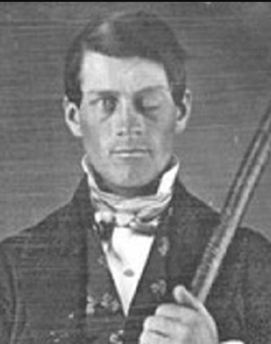
Phineas Gage, 1848: survived a rod up through his left cheek + out top of his skull, leaving frontal lobes damaged
quick recovery, HOWEVER, frontal lobes + brain regions that control emotion + decision making connections were damaged
drastic personality change, most mental abilities + memories intact
frontal lobes help w/ moral compass
T or F — parietal lobes (association area) enable math + spacial reasoning
true
large + unusually shaped in Einstein’s normal weight brain
when stimulated, patients had a desire to move their body part but didn’t
when further stimulated, patients thought they had moved their body part but didn’t
when stimulated in an association area in the frontal cortex near the motor area, they moved but didn’t know they had
suggests that our perception of moving flows not from the movement itself, but rather from our intention
what happens if association area on underside of right temporal lobe is damaged?
it enables face recognition
if damaged, you will be unable to identify the person even if they’re very familiar to you
T or F — our mental experiences + psychological health rely on coordinated brain activity
true
T or F — complex mental functions don’t reside in any single place
true
T or F — neuroplasticity is how our brain adapts to new situations
true
may occur after serious damage, especially in young children (whose undamaged hemisphere develops extra connection)
helps explain why some deaf people (who learned sign language before another language) may have enhanced peripheral motor detection vision
T or F — some neural tissue can reorganize in response to damage
true
T or F — blindness / deafness makes unused brain areas available for other uses, such as sound + smell
true
if a blind person uses one finger to read braille, the brain area dedicated to that finger expands as sense of touch invades the visual cortex that normally helps people see
when can brain function reassignment occur?
may occur when disease / damage frees up other brain areas normally dedicated to specific functions
how does brain often attempt self-repair? (2)
by reorganizing existing tissue
potentially though neurogenesis — producing new neurons
neurogenesis
the formation of new neurons
lateralization
the brain’s left + right hemispheres serving different functions
why did research spanning over a century conclude that the right hemisphere was significantly minor?
the left hemisphere’s accidents, strokes, and tumors impair reading, writing, speaking, math, and understanding
similar right-hemisphere injuries have less visibly dramatic effects, leading researchers to believe it was “minor”
what happened after 2 neurosurgeons speculated that severing the corpus callosum would stop major epileptic seizures — caused by amplification of abnormal brain activity bouncing back and forth between hemispheres?
seizures disappeared, surprisingly healthy, personality + intellect hardly affected
however, since communication lines between hemispheres severed, both hemispheres express themselves individually
information sharing doesn’t take place
split brain
a condition resulting from surgery that separates the brain’s 2 hemispheres by cutting the fibers (mainly those of the corpus callosum) connecting them
how sensory information is processed through 2 hemispheres
each eye receives sensory information from entire visual field
in each eye, information from left field of vision —> right hemisphere; information from right half of visual field —> left hemisphere (usually controls speech)
information received by either hemisphere is quickly transmitted to the other across the corpus callosum
explain the early experiment conducted by Gazzaniga (1967) on split-brain patients
asked patients to stare @ dot as he flashed HE o ART on a screen
HE appeared in left visual field —> transmitting to right hemisphere
ART appeared in right visual field —> transmitting to left hemisphere
asked patients to say (controlled by left hemisphere) what they had seen: reported ART
when asked to point w/ their left hand (controlled by right hemisphere) to what they had seen, they pointed to HE
roles of hemispheres
left hemisphere
in charge of speaking
skilled @ making quick, literal interpretations of language
right hemisphere
excels @ making inferences, helps modulate speech to make meaning clear, helps orchestrate our self-awareness